
Efficacy of Chitosan Nanoparticle Transplantation on Regeneration of Acute Spinal Cord Injury in Dogs Model


Efficacy of Chitosan Nanoparticle Transplantation on Regeneration of Acute Spinal Cord Injury in Dogs Model
Ahmed Kadhim Munahi1* , Hameed A. Al-Timmemi2

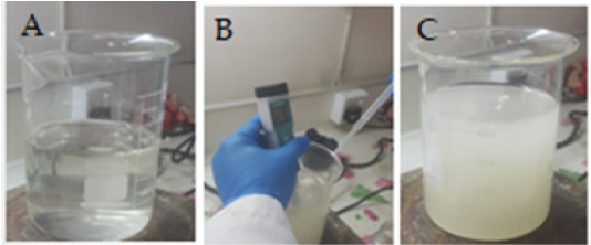
Photograph showing the steps of Fabrication of Chitosan nanoparticles A. Dissolving chitosan in deionized water and stirring. B. Adjusting to pH 7 by adding NaOH. C. filtering the formed semi-gel solution.

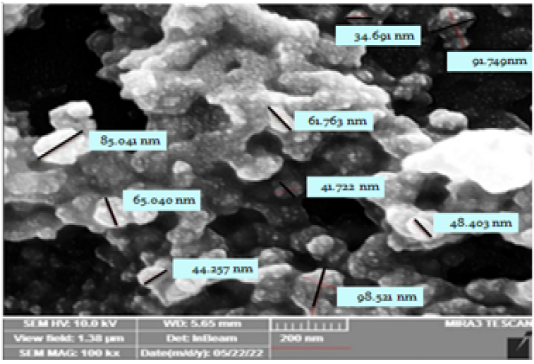
SEM image of synthesized nanoparticles shows spherical to elongated shape nanoparticles of about 62.065±7.632 nm.

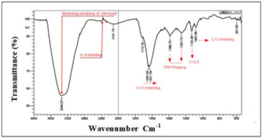
FT-IR spectrum of synthesized chitosan nanoparticles

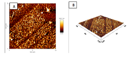
Atomic force microscopy images of chitosan nanoparticles indicate A. 2D and B. 3D

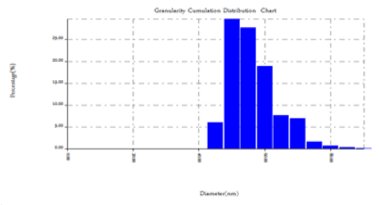
Atomic force microscopy image of chitosan nanoparticles shows the average particle diameter was 65.91 nm calculated in nanoscale size.
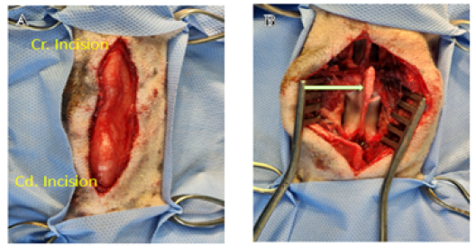
Photograph showing the initial steps of hemicordectomy. A. Shows the surgical incision over dorsal midline from L1 to L3. B. Elevated epaxial muscles from dorsal spinous processes, laminae, articular facets, and pedicles of L2 (arrow).
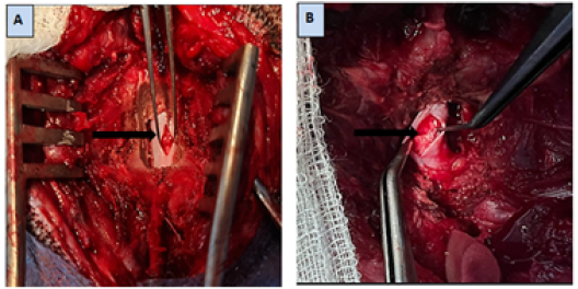
Photograph showing. A. A longitudinal incision is made through the meninges (arrow) B. Left lateral hemisection of the spinal cord (arrow).
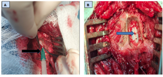
Photographs show. A. Implantation of chitosan nanoparticles at the injured spinal cord (arrow) B. Closed Dura matter (arrow).

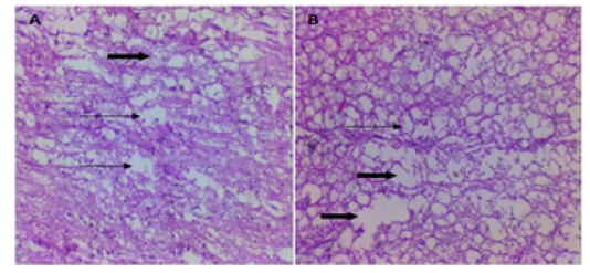
Micrograph of the longitudinal section of control group at the site of the spinal cord injuries A. 8 weeks PO shows multiple cystic cavity, containing granular cellular debris (thin arrows) surrounded by reactive gliosis and presented debris of necrotic with prominent vacuolization in white matter (thick arrow). B. 16 wks shows large cystic cavity (thick arrows) surrounded by glial scar tissue and vacuolated nerve fibers (thin arrow) H&EX10.

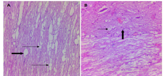
Micrograph of the longitudinal section of chitosan group at the site of the spinal cord injuries A. 8 weeks PO shows reduced cavities size and surround by glia cells (thin arrows) and moderate vacuolization (thick arrow). B. 16 weeks chitosan group shows small size cavity, injured area was filled with dense regenerative nerve fiber (thin arrow),

