Virulence Potential of Aspergillus flavus Isolated from Dogs
Virulence Potential of Aspergillus flavus Isolated from Dogs
Salma Ali Munshed*, Fadwa Abdul Razaq Jameel
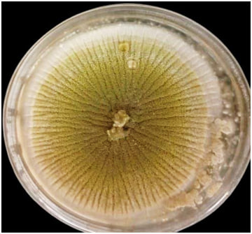
Macroscopic appearance of A. flavus on SDA at 25ºC for 7 days.
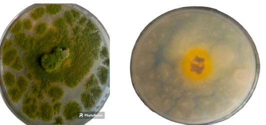
Aspergillus flavus on Czapek dox agar at 25 ºC for 4–5 days.
Microscopic appearance of A. flavu using stain lactophenol cotton blue (40x).
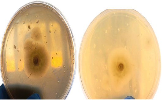
Hydrolysis of albumin by A. flavus in proteinase production medium.
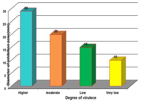
Measurement the diameter of inhibition zone in albumin medium.
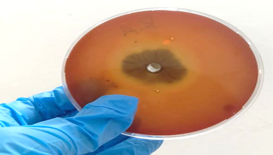
Hemolytic action by A. flavus on blood agar.
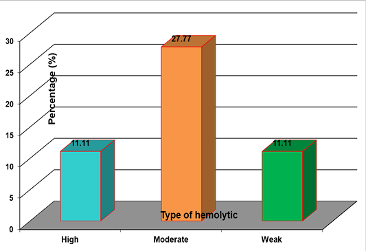
Results of hemolytic activity of A. flavus.
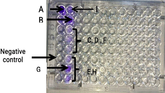
Microplate inoculated with nine A. flavus isolates with negative control for detection of bioflim formation result show high Biofilm formation in sample A, B and G, moderate in sample C, D, F and I and weak biofilm in sample E and H.
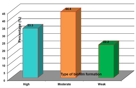
Biofilm formation by A. flavus.


