Histomorphological Development Study of the Heart of Local Awassi Sheep (Ovis aris) at Postnatal Stages
Histomorphological Development Study of the Heart of Local Awassi Sheep (Ovis aris) at Postnatal Stages
Ahmed Jamil Abid*, Ahlam Jaber Hamza
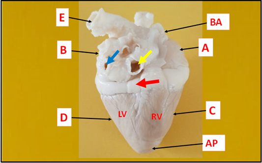
Anatomical photograph of heart in sheep at one month showing: (AP) apex of heart, (BA) base of heart, (A) right atrium, (B) left atrium, (RV) right ventricle, (LV) left ventricle, (C) cranial border, (D) caudal border, (E) aorta, (red arrow) coronary groove, (yellow arrow) cranial vena cava, (blue arrow) caudal vena cava.
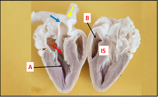
Anatomical photograph of heart in sheep at one month showing: (A) left ventricle, (B) right ventricle, (IS) interventricular septum, (red arrow) chordae tendinea, (yellow arrow) aorta, (blue arrow) brachiocephalic trunk.
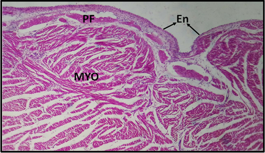
histological section of the heart (left ventricle) at one month in sheep showing: Endothelium (En), myocardium (MYO), purkinje fibers (PF), (H & E stain 10X).
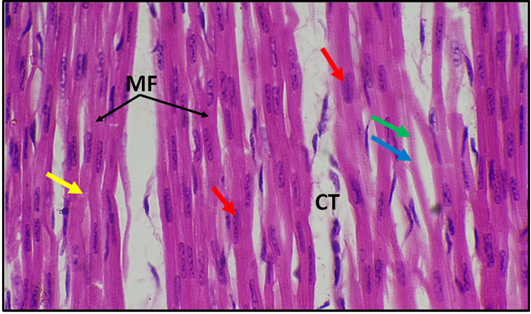
histological longitudinal section of the heart (right ventricle) in sheep at one month postnatal showing: muscle fibers (MB), nucleus (red arrow), A-band (green arrow), I-band (blue arrow), intercalated disks (yellow arrow), connective tissue (CT) (H & E stain 40X).
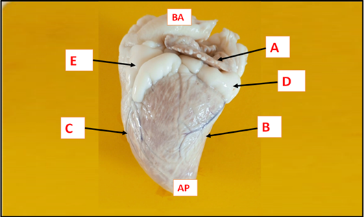
Anatomical photograph of heart in sheep at six months postnatal showing: (BA) base of heart, (AP) apex of heart, (A) right atrium, (B) cranial border, (C) caudal border, (D) coronary groove, (E) longitudinal groove.
Anatomical photograph of heart in sheep at six months postnatal showing: (A) left ventricle, (B) right ventricle, (C) right atrium, (D) aorta, (IS) interventricular septum (IS), (red arrow) chordae tendineae, (yellow arrow) moderator band.
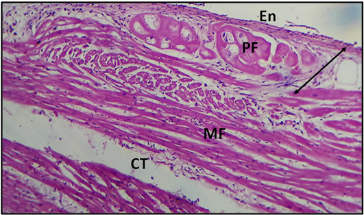
histological section of the heart (left ventricle) at six months in sheep showed: the endothelium layer (En), subendocardial (head double arrow), Purkinje fibres (PF), myocardium fibers (MF) (H & E stain 10X).
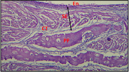
histological section of the heart (left ventricle) at six months in sheep showing the strongly PAS stain positive reactivity of the purkinje fibers (PF) in the subendocardium (SE) (PAS stain 10X).
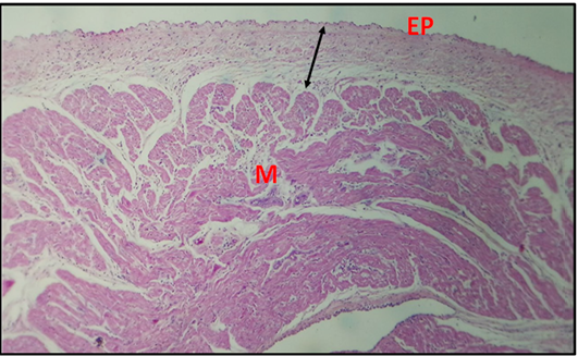
Histological cross-section of heart (right atrium) at (6) months postnatal showing: Epicardium (EP), subepicardial (double head arrow), myocardium (M) (H & E stain 4X).
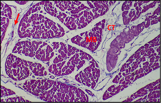
Histological cross-section of heart (left ventricle) at (6) months postnatal showing: Muscle bundles fibers (MB), connective tissue (CT), perimysium (red arrow) (Masson Trichrome stain 20X).


