
Brain Histopathological Changes After Treatment Using Calabash Fruit (Crescentia cujete L.) in Rat Model with Artificially Induced Ischemic Stroke


Brain Histopathological Changes After Treatment Using Calabash Fruit (Crescentia cujete L.) in Rat Model with Artificially Induced Ischemic Stroke
Jasir Hakim Hidayah1,2, Yos Adi Prakoso2, Sitarina Widyarini3*


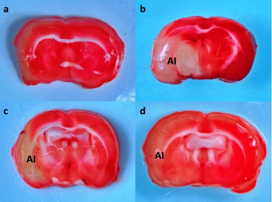
Brain macroscopy of rat model with artificially induced ischemic stroke after treatment. The sham-operated group showed there is infarct area was observed (a); ischemic stroke without treatment showed a large infarct area (AI) with white color (b); however, there is a decrease of infarct area (AI) from the group treated with 1.48 mg/kg BW of calabash (c); and pale infarct area (AI) from the group treated with 2.96 mg/kg BW of calabash (d). Macrophotograph, 2% TTC staining.
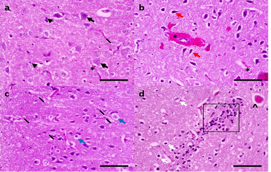
Brain histopathology of rat model with artificially induced ischemic stroke. Typical brain histology with an apparent nucleus and nucleolus are presented (black arrow), microglia (line), satellite cell (arrowhead), and intake blood vessels (*) from a sham-operated group (a); ischemic stroke group without treatment showed secondary haemorrhage (*) inside the Virchow Robin space and tissue, neuronal necrosis that marked by pyknosis neuron (red arrowhead) (b); most of the neuron suffered oedema (blue arrow) marked by pale cytoplasm and cleft that suspected by the crystalline inclusion, microgliosis (line) (c); moreover, the brain also showed perivascular oedema (^), oedema neuronal (white arrow) with neutrophilic and lymphocytic inflammation, and satellitosis (box) (d). H&E, 400×, skala bar: 50µm (a-d).

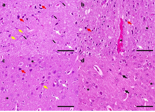
Brain histopathology of rat model with artificially induced ischemic stroke after calabash treatment. Group P3, treated with 0.74 mg/kg BW of calabash, showed moderate histopathological changes against neuronal necrosis (red arrow), microgliosis (line), and secondary haemorrhage (*) (a, b); group P4 that, treated with 1.48 mg/kg BW of calabash showed ghost cell (yellow arrow), oedema perineuronal (white arrow), pyknosis (red arrow), and secondary haemorrhage (*) (c); finally, the group P5 showed a dominate of typical neuron (black arrow), mild secondary haemorrhage (*), and mild oedema perineuronal (white arrow) (d). H&E, 400×, skala bar: 50µm (a-d).

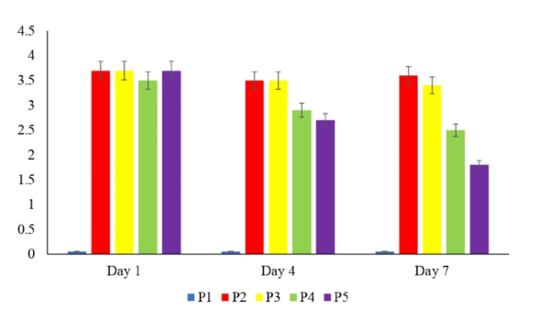
Neurodeficit score from rat model with artificially induced ischemic stroke.

