
Somatostatin (SS) DNA vaccine has been used to improve the growth and lactation of animals, however, the immune response is still unsatisfactory, especially in large animals. The present study aimed to investigate whether tissue plasminogen activator (tPA) could be applied as signal peptide for enhancing the effect of somatostatin DNA vaccine. A novel recombinant plasmid pEGS/2SS-tPA encoding two copies of somatostatin genes fused to tPA signal sequence was succesfully constructed and identified in MCF-7 cells. Subsequently, thirty female mice randomly were divided into three groups, treating with 50 μg pEGS/2SS-tPA (group T1), 50 μg pEGS/2SS (group T2), and 50 μg pEGFP-N1 (Control group) at weeks 0, 2, 4 and 6. Mating with male mice was performed at 2 weeks post final immunization. Results showed that mice both in T1 and T2 groups elicited significantly stronger anti-SS antibody level than that in control (p < 0.05), and mice in T1 group induced significantly higher serum GH and PRL concentration in comparison with T2 and control groups (p < 0.05). Expectantly, mice in T1 group also showed significantly higher average weight gain and offspring weaning weight than that in T2 and control groups, respectively (p < 0.05). Overall, these results suggested that novel somatostatin plasmid fused to tPA signal peptide obtained stronger immunogenicity, and better potentials of promoting growth and lactation, indicating that tPA is an effective signal peptide for enhancing the immune efficacy and performance of somatostatin DNA vaccine.
Research Article · Volume 50, Issue 5, 16 August 2018 · Open Access
Construction and Evaluation of Novel Plasmid DNA Encoding Somatostatin Fused to a Tissue Plasminogen Activator (tPA) in Mice
· · · ·
Affiliations & Notes ⌄
- 1Key Laboratory of Agricultural Animal Genetics, Breeding and Reproduction, (Huazhong Agricultural University), Ministry of Education, Wuhan 430070, PR China 2College of Animal Science and Technology &College of Veterinary Medicine, Huazhong Agricultural University, Wuhan 430070, PR China , Aixin Liang and Liguo Yang contributed equally in this article. , * Corresponding author: yangliguo889555@163.com
Article Info ⌄
- Journal:
- Pakistan Journal of Zoology
- Published:
- 16 August 2018

From the article
- PRL concentration and weaning weight of offspring in vaccinated group fused to tPA were significantly greater than that in the vaccinated group without tPA and control mice.
- These results indicated that SS DNA vaccine with tPA could effectively promote lactation of mice.
- In summary, the present report demonstrated that novel somatostatin DNA vaccine fused to tPA improved immunogenicity, weight gain as well as lactation by inducing improving higher secretion capacity of recombinant protein.
- Therefore, our data indicated that tPA can be used as a signal peptide for enhancing effect of SS DNA vaccine.
Selected from the article's own text.
Abstract
Introduction
Somatostatin-14 (SS-14) regulates the growth and lactation of animals by inhibiting the secretion of growth hormone (GH) ; and prolactin (PRL) (Gruszaka et al., 2012). Immunization against SS-14 DNA vaccines could improve the growth of mice , rat , swine and lambs and lactation in mice by affecting the secretion of GH and PRL. Although the use of DNA vaccines is one successful strategy comparing protein vaccine, synthetic peptide vaccine and genetic engineering vaccine for immunization with somatostatin, due to no purification of protein antigens and saving cost ; , the immunogenicity of these somatostatin DNA vaccines was still low, and the effects was also inadequate in improving growth and lactation of animals, especially in large animals. Therefore, we need to further optimize the somatostatin DNA vaccine to improve its immune efficacy.
Tissue-type plasminogen activator (tPA) signal peptide is a serine proteinase which could increase humoral immune responses of DNA vaccines . Many studies suggested that antigen of DNA vaccine secreted influenced effectiveness and magnitudes of the immune responses ; ; ; , which may be due to that the signal peptide enable recombinant antigen of DNA vaccine to be secreted and thus raised access to the major histocompatibility complex class II (MHC-II) pathway of antigen presentation. The tPA, a secretory signal peptide, was usually fused into the 5’-end of plasmid DNA expressing various antigens which enhanced various antigens’ surface expression and thus improved DNA vaccine’s humoral immune response (Goncalves et al., 2015; . Hepatitis B surface antigen S (HBsAg-S) with 226 residues has been widely applied to improve the immunogenicity of somatostatin DNA vaccines because of the somatostatin’s small molecular mass and then incomplete antigenicity ; ; Kotiw et al., 2012; . Though these somatostatin DNA vaccines fused to the HBsAg-S gene can lead to the surface expression, the surface expression efficiency of the somatostatin antigens may be relatively low, resulting in the weak humoral immune responses of somatostatin DNA vaccines.
The objective of this study is to verify this hypothesis whether tPA can be used as a signal peptide to enhance immune efficacy and performance of somatostatin DNA vaccine. The effect of novel somatostatin DNA vaccine in promoting mice growth and lactation was evaluated by measuring serum somatostatin antibody GH, PRL concentration, average weight gains and offspring weaning weight.
Materials and methods
Vaccine construction and identification
The recombinant vaccine (pEGS/2SS-tPA) was constructed by inserting the sequence of tissue plasminogen activator signal peptide (tPA) into previous somatostatin DNA vaccine (pEGS/2SS) . The pEGS/2SS-tPA was confirmed to be constructed successfully proved by double enzyme digestion and sequence comparison.
Human breast cancer cell MCF-7 (purchased from China Center for Type Culture Collection) was cultivated in incubator at 37°C in 5% CO2 and containing Dulbecco’s modified Eagles medium (DMEM, Gibco, Shanghai, China) with 15% fetal bovine serum (Gibco, Shanghai, China). These cells were then transfected with pEGS/2SS-tPA, pEGS/2SS and pEGFP-N1 vector by using LipofectamineTM 2000 Kit (Invitrogen, Shanghai, China). The pictures of green fluorescent were taken after transfection of 24 h and 48 h, respectively.
On the other hand, MCF-7 was lysed by conventional lysis buffer for 30 min on ice, and then the supernatant was prepared by centrifuging at 4°C. BCA Protein Assay Kit (Sangon Biotech, Shanghai, China) was used to detect protein concentration in supernatant. After 5 min for boiling, we used the sodium dodecyl sulphate polyacrylamide gel electrophoresis (SDS-PAGE) to separate the supernatant protein, and then electrophoretically transferred onto nitrocellulose polyvinylidene fluoride (PVDF) membrane. These samples were blocked in 1% BSA in tris-buffered saline (TBS) and then incubated with primary anti-SS antibody (Santa Cruz, Shanghai, China, dilutions at 1:300) and GAPDH antibody (Boster, Wuhan, China, dilutions at 1:1000), followed by incubation with secondary antibodies horseradish peroxidase- (HRP) conjugated goat anti-rabbit IgG (Boster, Wuhan, China, dilutions at 1:1000). The densities of bands were scanned by enhanced chemiluminescence reagent (Pierce, Rockford, IL, USA).
Real time PCR detection
At 48 h after cell transfection, total RNA from MCF-7 cells was extracted by TRIZOL (Invitrogen, Shanghai, China) and then reverse-transcribed using PrimeScriptTM RT reagent Kit (TaKaRa, Dalian, China) according to the manufacturer’s instructions. Real-time PCR reactions were run using QuantiFast® SYBR® Green PCR Kit (Qiagen, 204054, Germany) on BIORAD CFX Manager Machine. The specific SS, PRL receptor, JAK2, STAT5, DGAT1, as well as GAPDH primers (Table I) were designed and synthesized by Sangon Biotech (Shanghai, China). Samples were prepared according to the manufacturer’s instructions. The data were subsequently quantified according to the comparative cycle threshold (CT) method and relative expression of target genes was obtained by calculating 2-ΔΔCT.
Table I.- Primers used in this study for real time PCR.
| Gene (Size) | Sequence (5' to 3') | Accession No. |
| S/2SS (228bp) | F: CATCCTGCTGCTATGCCTCA | KT235604 |
| R: CCGTCCGAAGGTTTGGTACA | ||
| PRLR (225bp) | F: CGTCGCCTTCTAAAACTGGC | NM_000949 |
| R: AGAAAGCCCAGCCCAGAAAA | ||
| JAK2 (113bp) | F: GTGCCTTTGAAGACCGGGAT | AF005216 |
| R: TAGAGGGTCATACCGGCACA | ||
| STAT5 (156bp) | F: GGAACTCTTACGCCGACCAA | L41142 |
| R: AAACTCACAACACGACCGCT | ||
| DGAT1 (276bp) | F: GGAACTCCGAGTCTGTCACC | NM_012079 |
| R: CAGCTGCGTTGCCATAGTTG | ||
| GAPDH (259bp) | F: ACAGTCAGCCGCATCTTCTT | NM_002046 |
| R: GACAAGCTTCCCGTTCTCAG |
Mice immunization
Thirty Kunming mice (6-week-old, female) were purchased (Wuhan Institute of Biological Products Co., Ltd, Wuhan, China) and fed in the experimental animal house of Huazhong Agricultural University which following conforming to the National Institutes of Health guidelines in care and use of laboratory animals. One week after caging, the mice were randomly divided into three groups (Groups T1, T2 and control), which were respectively intramuscular injected with 50μg pEGS/2SS-tPA or pEGS/2SS or naked pEGFP-N1 plasmids dissolved in 100μl saline solution. The mice were vaccinated total four times with a two week interval. Tail blood at weeks 0, 2, 4 and 6 was collected in EDTA anticoagulant tube, then was centrifuged at 3,000 rpm for 10 min and stored at -20°C until analysis.
Specific SS antibody detection
An indirect ELISA method was used to detect the specific SS IgG antibodies. 96-well polystyrene flat-bottom plates were coated with 100 ng SS antigen dissolved in bicarbonate buffer and incubated overnight at 4°C. Then the plates were blocked with 1% BSA in phosphate-buffered saline (PBS) at 37°C for 1 h. We added 1:200 dilution of plasma samples dissolved in PBS containing 0.05% Tween-20 into the plates and then incubated them at 37°C for 1 h. Meanwhile, standard SS antibody was used as positive control for the drawing standard curve. Goat anti-mouse IgG secondary antibody (1:2000) with horseradish peroxidase-conjugated (HRP) labeled (Boster, Wuhan, China) in PBST were added to plates and incubated for 1 h at 37°C. 150 μl tetramethylbenzidine (TMB) substrate were added to plates and incubated them at 37°C for 25 min. The reaction was stopped with 2 M H2SO4 and the optical density (OD) absorbance was read at 450 nm wavelength filter. According to the standard SS antibody concentration and OD450 values, the standard curve was drawn. Then SS antibody concentrations of these samples were calculated by the SS standard curve and the OD450 values of examined samples. Results were evaluated using the ratio of known positive sample’s OD450 (P) to known negative sample’s OD450 (N) . If the P/N ratio is greater than 2.0 and the OD450 value is greater than 0.2, the sample will be judged to be positive, otherwise it will be seen as negative one .
Serum GH and PRL assay
Serum GH and PRL concentration was detected by double antibody sandwich ELISA from purchasing commercial kits (Laibio, Shanghai, China). All the treatment was simultaneously run for avoiding inter-assay variation. The assay sensitivity of GH and PRL were respectively ≤ 0.1 ng/ml. The coefficients of variation for GH and PRL assays were respectively < 7.46% and < 11.58%.
Records of weight gain and weaning weight
After the mice were fasted for 12 h, their body weight were recorded at weeks 0, 4 and 8 after immunization and then their average weight gains, was calculated. At week 2 after the final immunization, each cage was put one male mouse for mating, the successful breeding was checked by observing the female mice vaginal suppository in the next day. After that, the mice’s pregnancy was observed at any time, and offspring birth weight and weaning weight (3 weeks old) were recorded.
Statistical analysis
Statistically significant (p < 0.05) differences between groups in terms of anti-SS antibody, serum GH and PRL concentration, average weight gains, offspring weaning weight were assessed using one-way ANOVA and unpaired Student’s t-test from the SAS 8.1 analytical software program. These data were showed as Mean±SD.
Results
Vaccine construction and detection
We engineered a recombinant plasmid pEGS/2SS-tPA, in which the sequence of tissue plasminogen activator signal peptide (tPA) was inserted into previously described somatostatin DNA vaccine (pEGS/2SS) (Fig. 1A). The fusion gene S/2SS and tPA gene insertion sites, sequence, and the direction were correctly detected by restriction endonuclease digestion and sequencing methods (Fig. 1A, B). Intensity of green fluorescence displayed that the expression level of target gene was higher at 48 h after transfection (Fig. 2). Additionally, the green fluorescence treated with pEGS/2SS-tPA distributed dispersedly in the cytoplasm (Fig. 2), which was different from the distribution of green fluorescence in pEGFP-N1 transfected cells. Result of real-time PCR also showed the highly significant increase in the relative expression of SS (Fig. 3). In order to evaluate whether the SS fusion protein was secreted by pEGS/2SS-tPA transfected cells, samples collected from culture supernatants were analyzed by Western blot. Result analysis showed that both of the MCF-7 cells transfected with pEGS/2SS-tPA and pEGS/2SS plasmid contained 28 kDa fusion protein bands which was consistent with the size of target protein (Fig. 4). The negative control did not show specific band (Fig. 4). These results showed that the novel recombinant SS vaccine constructed can correctly express the fusion protein in transfected MCF-7 cells and secreted more SS fusion protein into extracellular space.
Fig. 4. S/2SS fusion protein detection in cell supernatants by Western blot. Lane 1, cells transfected with pEGS/2SS-tPA; Lane 2, cells transfected with pEGS/2SS; Lane 3, cells transfected with negative control pEGFP-N1. GAPDH protein was selected as the standardized correction of S/2SS fusion protein expression quantity.
mRNA level of lactation-related genes
As the result of SS over-expression, the mRNA levels of some genes related to lactation changed in MCF-7 cells. Compared with cells transfected with negative vector, the relative expression levels of PRLR (PRL receptor), JAK2, STAT5 and DGAT1 in cells treated with pEGS/2SS-tPA or pEGS/2SS were dramatically decreased (Fig. 5; p < 0.05), suggesting SS could regulate expression the lactation related genes in vitro.
Table II.- Positive rate and concentration of SS antibody in different vaccine groups.
| Group | Positive rate | Antibody concentration (pg/mL) |
| T1 | 66.67% | 5.45±2.27a |
| T2 | 50.00% | 4.78±1.94a |
| C | 0.00 | - |
Values with different lowercases represented significantly different in the same column (p < 0.05).
SS antibody response
Specific anti-SS- antibody was detected in T1 and T2 groups at week 8 after primary immunization. Anti-SS antibody concentration in T1 and T2 groups was both significantly higher than control group (Table II; p < 0.05). Anti-SS antibody concentration in T1 group is higher than that in T2 group; however, there was no significantly difference. The ratio of positive antibodies in T1 group (66.67%) was obviously higher than that one in T2 group (50.00%).
Serum GH and PRL concentration
Mice in T1 group showed significantly higher serum GH concentration than that in T2 and control at week 8 after primary immunization (Fig. 6; p < 0.05) and we did not find significantly difference between T2 and control. The serum PRL concentration of T1 group was extremely significantly higher than that of T2 and control groups at week 8 after primary immunization (Fig. 6; p < 0.01), and we did not find significantly difference between T2 and control.
Table III.- Average weight gain of mice in different vaccine groups (g).
| Group | Week 4 after vaccinated | Week 8 after vaccinated |
| T1 | 23.61±1.82a | 29.76±2.30a |
| T2 | 23.28±1.47a | 28.82±2.13a |
| C | 21.64±1.40b | 26.14±1.82b |
Values with different lowercases represented significantly different in the same column (p < 0.05).
Table IV.- Average weaning weight of offspring.
| Group | Weaning weight (g) |
| T1 | 12.20±2.19a |
| T2 | 11.02±1.89b |
| C | 9.54±1.87c |
Values with different lowercases represented significantly different in the same column (p < 0.05).
Effect on the growth performance
The body weight was monitored in the whole experimental period. The average weight gains of mice in T1 and T2 groups were significantly higher than those in control group at week 4 and week 8 after primary vaccination (p < 0.05; Table III); however, there is no significant difference between T1 and T2 groups.
Effect on the weaning weight of offspring
Discussion
Somatostatin immuno-neutralization technology has been proved to be a potent strategy in promoting growth and lactation of animal ; ; , 2014;; ; . Although our previous somatostatin DNA vaccines fused to HBsAg-S gene were used to enhance animal growth and lactation performance, the immune effects of these somatostatin DNA vaccines was relatively unsatisfactory ; ; ; , which may be due to the weak secretion capacity of encoded antigen. The tissue plasminogen activator signal sequence (tPA) is a specific targeting signal that targets the expressed antigen directly to the plasmic reticulum (ER), and therefore obviates the need for the antigen to be processed and translocated to the cytoplasm ; . The tPA signal peptide has been widely used in improving the secreting levels of the expressed antigen ; , resulting in stronger humoral and cellular immune ; . Therefore, a novel somatostatin DNA vaccine fused to tPA signal peptide was successfully constructed and identified the fusion protein expression in MCF-7 cells in this study. Compared to pEGS/2SS-transfected cells, pEGS/2SS-tPA-transfected cells secreted more fusion protein, suggesting tPA drived more secretion of the recombinant SS protein.
Several studies indicate that the efficiency and magnitude of immune responses elicited by DNA vaccines are affected by the ability of the expressed antigen to be secreted ; . Subsequently, the immune response and hormone level of such novel SS vaccine was evaluated. Compared to T2 and control groups, recombinant SS protein fused to tPA was able to induce strong SS specific antibodies and obtain more positive mice (66.67% vs. 50.00%, 66.67% vs. 0.00%), resulting in the higher GH concentration at week 8 post immunization. The possible reason is that tPA signal peptide could guide a large number of recombinant antigens to transport to extracellular which elevated uptake by APC, and thus activate the immune system . Based on the higher GH secretion at week 8 after immunization, the average weight gain of mice in Group T1 and T2 are higher than that in control group.
In this research, the lactation related gene expression was detected in MCF-7 cells by Real time PCR. Compared with pEGS-N1 transfected cells, the relative expression levels of PRLR, JAK2, STAT5 and DGAT1 in cells treated with pEGS/2SS-tPA or pEGS/2SS were dramaticlly decreased, suggesting overexpression of SS fusion gene are able to decline the expression of PRLR, JAK2, STAT5 and DGAT1 genes. PRLR, JAK2 and STAT5 are key components in the mammary JAK/STAT signaling pathway, which advances milk synthesis. On the surface of mammary epithelial cells, binding with PRL, long isoform PRLR is activated and undergoes the dimerization and the conformational change, followed by JAK2 recruitment and STAT5 phosphorylation; and then activated STAT5 targets the promoter of genes (e.g. β-casein), triggering the transcription associated with milk component synthesis . The down-regulation of PRLR and STAT5 is accompanied by a decrease in PRL signaling and mammary involution . Accordingly, abundant SS is more likely to play a negative role in lactation through hindering JAK/STAT signaling. Bratthauer et al. (2008) found that the expression level of PRLR tended to be opposite to that of STAT5a in common breast epithelial cells. Our result is consistent with Bratthauer’s research. The mRNA level of PRLR in pEGS/2SS-tPA transfected cells was higher than that in pEGS/2SS transfected cells, while the STAT5 level in pEGS/2SS-tPA treated cells was relatively lower. Subsequently, we detected that immune effect of novel SS DNA vaccine fused to tPA signal peptide in promoting mice lactation performance. PRL concentration and weaning weight of offspring in vaccinated group fused to tPA were significantly greater than that in the vaccinated group without tPA and control mice. These results indicated that SS DNA vaccine with tPA could effectively promote lactation of mice. Interestingly, although there was no significant difference in the concentration of PRL between the immunized group without tPA and control group, weaning weight of offspring in the immunized group without tPA was significantly greater than that in control mice, which may be due to that the testing time was at the descending period of prolactin. Previous study showed that after female mice were treated with the SS DNA vaccine, the weight of their offspring during lactation increased faster compared with control group, especially at the first two weeks of lactation . Similarly, results in this experiment also displayed an obviously rise in weight gain of offspring after SS DNA immunization; however, the significant distinction existed until the end of lactation. It might due to diverse adjuvant and vectors have an influence on milk quality and functioning time of SS DNA vaccine.
In summary, the present report demonstrated that novel somatostatin DNA vaccine fused to tPA improved immunogenicity, weight gain as well as lactation by inducing improving higher secretion capacity of recombinant protein. Vaccination of mice with the pEGS/2SS-tPA induced higher levels of the specific SS antibody, resulting in higher concentration of GH and PRL. Moreover, pEGS/2SS-tPA was able to increase the weight gain in vaccinated mice and weaning weight in offsprings. Therefore, our data indicated that tPA can be used as a signal peptide for enhancing effect of SS DNA vaccine.
Acknowledgements
This study was financially supported by the National Natural Science Foundation of China (No. 31402078), and the Earmarked Fund for Modern Agro-industry Technology Research System (CARS-37-04B).
Statement of conflict of interest
All authors declare that there is no conflict of interest in this study.
Figures
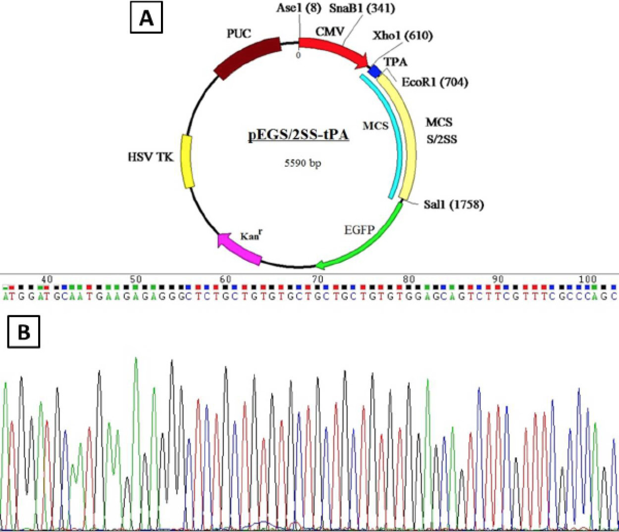
Construction of plasmid pEGS/2SS-tPA. The sequence of tissue plasminogen activator signal peptide (tPA) was inserted into previous somatostatin DNA vaccine (pEGS/2SS). A, the plasmid profile of pEGS/2SS-tPA; B, The sequencing analysis of pEGS/2SS-tPA.
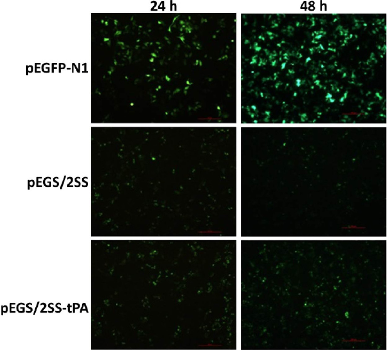
Expression and distribution of green fluorescent in MCF-7 cells at 24 h and 48 h after transfection. Scale bar = 200μm.
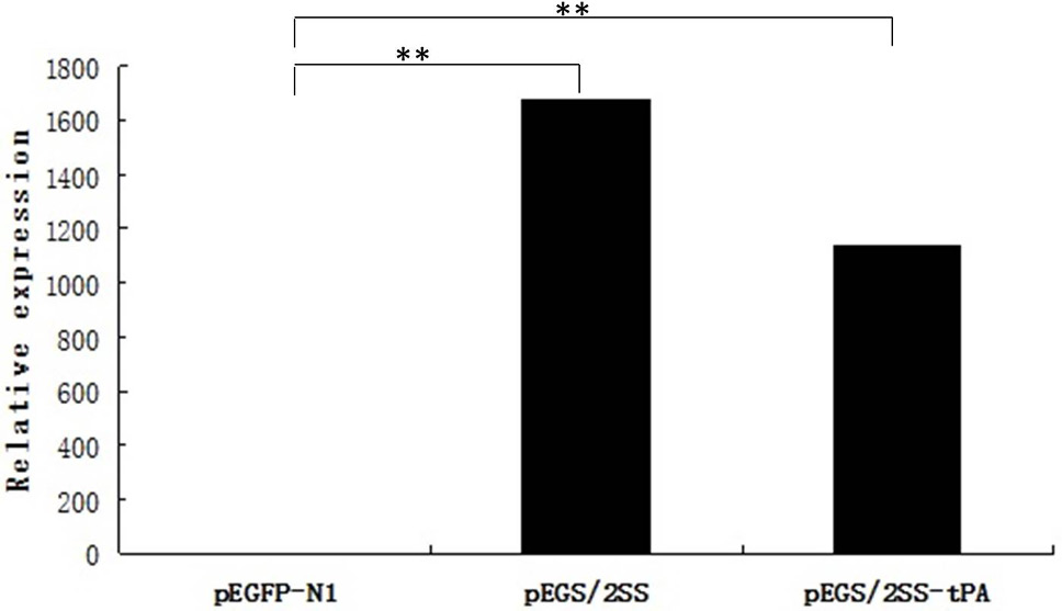
Relative expression of SS mRNA by real-time PCR. The Ct value of SS was normalized to that of GAPDH, and the SS expression level in cells transfected with pEGS/2SS-tPA or pEGS/2SS was calculated relative to cells transfected with pEGFP-N1. **P<0.01.
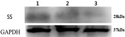
S/2SS fusion protein detection in cell supernatants by Western blot. Lane 1, cells transfected with pEGS/2SS-tPA; Lane 2, cells transfected with pEGS/2SS; Lane 3, cells transfected with negative control pEGFP-N1. GAPDH protein was selected as the standardized correction of S/2SS fusion protein expression quantity.
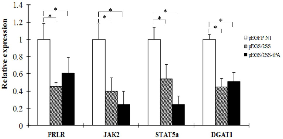
Relative expression of PRLR, JAK2, STAT5a and DGAT1 by Real-time PCR. The Ct value of SS was normalized to that of GAPDH, and the expression level of targets genes in cells transfected with pEGS/2SS-tPA or pEGS/2SS was calculated relative to cells transfected with pEGFP-N1. *P<0.05.
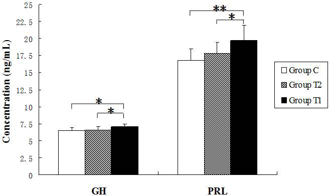
Serum GH concentration (ng/mL) and serum PRL concentration (ng/mL) in T1, T2 and C groups vaccinated with pEGS/2SS-tPA, pEGS/2SS and pEGFP-N1 at 8 week after immunization. *P<0.05 and **P<0.01.
References
- Adams, J.M., Otero-Corchon, V., Hammond, G.L., Veldhuis, J.D., Qi, N. and Low, M.J., 2015. Somatostatin is essential for the sexual dimorphism of GH secretion, corticosteroid-binding globulin production, and corticosterone levels in mice. Endocrinology, 156: 1052-1065. https://doi.org/10.1210/en.2014-1429[CrossRef]
- Ashok, M.S. and Rangarajan, P.N., 2002. Protective efficacy of a plasmid DNA encoding Japanese encephalitis virus envelope protein fused to tissue plasminogen activator signal sequences: studies in a murine intracerebral virus challenge model. Vaccine, 20: 563-570. https://doi.org/10.1016/S0264-410X(01)00492-3[CrossRef]
- Bai, L.Y., Liang, A.X., Zhang, J., Yang, F.F., Han, L., Huo, L.J. and Yang, L.G., 2011. Effects of immunization against a DNA vaccine encoding somatostatin gene (pGM-CSF/SS) by attenuated Salmonella typhimurium on growth, reproduction and lactation in female mice. Theriogenology, 75: 155-163. https://doi.org/10.1016/j.theriogenology.2010.08.001[CrossRef]
- Bratthauer, G.L., Strauss, B.L. and Barner, R., 2008. Reversed expression of the JAK/STAT pathway related proteins prolactin receptor and STAT5a in normal and abnormal breast epithelial cells. Breast Cancer (Auckl), 1: 7-14. https://doi.org/10.4137/BCBCR.S549[CrossRef]
- Campo, V., Arbocco, F., Sasso, C.V., Actis, E.A., Carón, R.W., Hapon, M.B. and Jahn, G.A., 2016. Hypothyroidism advances mammary involution in lactating rats through inhibition of PRL signaling and induction of LIF/STAT3 mRNAs. Mol. Cell. Endocrinol., 419: 18-28. https://doi.org/10.1016/j.mce.2015.09.023[CrossRef]
- Cao, S.X., Yang, L.G., Zhang, W.W., Mao, D.G. and He, X.H., 2005. Construction and expression of somatostatin DNA vaccine pEGS/2SS in HeLa. Chin. J. Vet. Sci., 25: 499-502.
- Ciernik, I.F., Berzofsky, J.A. and Carbone, D.P., 1996. Induction of cytotoxic T lymphocytes and antitumor immunity with DNA vaccines expressing single T cell epitopes. J. Immunol., 156: 2369-2375.
- Costa, S.M., Yorio, A.P., Gonçalves, A.J., Vidale, M.M., Costa, E.C., Mohana-Borges, R., Motta, M.A., Freire, M.S. and Alves, A.M., 2011. Induction of a protective response in mice by the dengue virus NS3 protein using DNA vaccines. PLoS One, 6: e25685. https://doi.org/10.1371/journal.pone.0025685[CrossRef]
- Farshadpour, F., Makvandi, M. and Taherkhani, R., 2015. Design, construction and cloning of truncated ORF2 and tPAsp-PADRE-truncated ORF2 gene cassette from hepatitis E virus in the pVAX1 expression vector. Jundishapur J. Microbiol., 8: e26035. https://doi.org/10.5812/jjm.26035[CrossRef]
- Golden, J.W., Josleyn, M.D. and Hooper, J.W., 2008. Targeting the vaccinia virus L1 protein to the cell surface enhances production of neutralizing antibodies. Vaccine, 26: 3507-3515. https://doi.org/10.1016/j.vaccine.2008.04.017[CrossRef]
- Gonçalves, A.J., Oliveira, E.R., Costa, S.M., Paes, M.V., Silva, J.F., Azevedo, A.S., Mantuano-Barradas, M., Nogueira, A.C., Almeida, C.J. and Alves, A.M., 2015. Cooperation between CD4+ T cells and humoral immunity is critical for protection against dengue using a DNA vaccine based on the NS1 antigen. PLoS Negl. Trop. Dis., 9: e0004277. https://doi.org/10.1371/journal.pntd.0004277[CrossRef]
- Gonzalez, M.C., Kostrzak, A., Guetard, D., Pniewski, T. and Sala, M., 2009. HIV-1 derived peptides fused to HBs Ag affect its immunogenicity. Virus Res., 146: 107-114. https://doi.org/10.1016/j.virusres.2009.09.007[CrossRef]
- Gruszka, A., Culler, M.D., Melmed, S., 2012. Somatostatin analogs and chimericsomatostatin-dopamine molecules differentially regulate human growth hormone and prolactin gene expression and secretion in vitro. Mol. Cell. Endocrinol., 362: 104-109. https://doi.org/10.1016/j.mce.2012.05.020[CrossRef]
- Han, Y., Liang, A., Han, L., Guo, A., Jiang, X. and Yang, L., 2014. Efficacy and safety of an oral somatostatin DNA vaccine without antibiotic resistance gene in promoting growth of piglets. Scand. J. Immunol., 79: 244-250. https://doi.org/10.1111/sji.12155[CrossRef]
- Jahn, GA., Daniel, N., Jolivet, G., Belair, L., Bole-Feysot, C., Kelly, P.A. and Djiane, J., 1997. In vivo study of prolactin (PRL) intracellular signalling during lactogenesis in the rat: JAK/STAT pathway is activated by PRL in the mammary gland but not in the liver. Biol. Reprod., 57: 894-900. https://doi.org/10.1095/biolreprod57.4.894[CrossRef]
- Jorritsm, S.H., Gowans, E.J., Grubor-Bauk, B. and Wijesundara, D.K., 2016. Delivery methods to increase cellular uptake and immunogenicity of DNA vaccines. Vaccine, 348: 5488-5494. https://doi.org/10.1016/j.vaccine.2016.09.062[CrossRef]
- Kotiw, M., Johnson, M., Pandey, M., Fry, S., Hazell, S.L., Netter, H.J., Good, M.F. and Olive, C., 2012. Immunological response to parenteral vaccination with recombinant hepatitis B virus surface antigen virus-like particles expressing Helicobacter pylori KatA epitopes in a murine H. pylori challenge model. Clin. Vaccine Immunol., 19: 268-276. https://doi.org/10.1128/CVI.05295-11[CrossRef]
- Li, Z., Howard, A., Kelley, C., Delogu, G., Collins, F. and Morris, S., 1999. Immunogenicity of DNA vaccines expressing tuberculosis proteins fused to tissue plasminogen activator signal sequences. Infect. Immunol., 67: 780-4786.
- Liang, A., Cao, S., Han, L., Yao, Y., Moaeen-Ud-Din, M. and Yang, L., 2008. Construction and evaluation of the eukaryotic expression plasmid encoding two copies of somatostatin genes fused with hepatitis B surface antigen gene S. Vaccine, 26: 2935-2941. https://doi.org/10.1016/j.vaccine.2008.03.036[CrossRef]
- Liang, A., Riaz, H., Dong, F., Luo, X., Yu, X., Han, Y., Chong, Z., Han, L., Guo, A. and Yang, L., 2014.Evaluation of efficacy, biodistribution and safety of antibiotic-free plasmid encoding somatostatin genes delivered by attenuated Salmonella enterica serovar Choleraesuis, Vaccine, 32: 1368-1374. https://doi.org/10.1016/j.vaccine.2014.01.026[CrossRef]
- Liang, R., van den Hurk, J.V., Zheng, C., Yu, H., Pontarollo, R.A., Babiuk, L.A. and van Drunen Littel-van den Hurk, S., 2005. Immunization with plasmid DNA encoding a truncated, secreted form of the bovine viral diarrhea virus E2 protein elicits strong humoral and cellular immune responses. Vaccine, 23: 5252-5262. https://doi.org/10.1016/j.vaccine.2005.06.025[CrossRef]
- Luo, M., Tao, P., Li, J., Zhou, S., Guo, D. and Pan, Z., 2008. Immunization with plasmid DNA encoding influenza A virus nucleoprotein fused to a tissue plasminogen activator signal peptide elicits strong immune responses and protection against H5N1 challenge in mice. J. Virol. Meth., 154: 121-127. https://doi.org/10.1016/j.jviromet.2008.08.011[CrossRef]
- Midha, S. and Bhatnagar, R., 2009. Anthrax protective antigen administered by DNA vaccination to distinct subcellular locations potentiates humoral and cellular immune responses. Eur. J. Immunol., 39: 159-177. https://doi.org/10.1002/eji.200838058[CrossRef]
- Redding, L. and Weiner, D.B., 2009. DNA vaccines in veterinary use. Expert Rev. Vaccines, 8: 1251-1276. https://doi.org/10.1586/erv.09.77[CrossRef]
- Sun, Y., Sinclair, S., Wynn, P. and McDowell, G.H., 1990. Immunization against somatotropin release inhibiting factor increases milk yield in ewes. Crop Pasture Sci., 41: 393-400. https://doi.org/10.1071/AR9900393[CrossRef]
- Wang, J.Y, Song, W.T, Li, Y., Chen, W.J., Yang, D., Zhong, G.C., Zhou, H.Z., Ren, C.Y., Yu, H.T. and Ling, H., 2011. Improved expression of secretory and trimeric proteins in mammalian cells via the introduction of a new trimer motif and a mutant of the tPA signal peptide. Appl. Microbiol. Biotechnol., 91: 731-740. https://doi.org/10.1007/s00253-011-3297-0[CrossRef]
- Watanabe, I., Ross, T.M., Tamura, S., Ichinohe, T., Ito, S., Takahashi, H., Sawa, H., Chiba, J., Kurata, T., Sata, T. and Hasegawa, H., 2003. Protection against influenza virus infection by intranasal administration of C3d-fused hemagglutinin. Vaccine, 21: 4532-4538. https://doi.org/10.1016/S0264-410X(03)00510-3[CrossRef]
- Woo, W.P., Doan, T., Herd, K.A., Netter, H.J. and Tindle, R.W., 2006. Hepatitis B surface antigen vector delivers protective cytotoxic T-lymphocyte responses to disease relevant foreign epitopes. J. Virol., 80: 3975-3984. https://doi.org/10.1128/JVI.80.8.3975-3984.2006[CrossRef]
- Xue, C.L., Liang, A.X., Mao, D.G., Han, L., Liu, X.B., Zhang, J. and Yang, L.G., 2010. Effect of genetic adjuvants on immune respondance, growth and hormone levels in somatostatin DNA vaccination-induced Hu lambs. Vaccine, 28: 1541-1546. https://doi.org/10.1016/j.vaccine.2009.11.058[CrossRef]
- Yang, S., Wang, C., Fang, X., Zhai, L., Dong, C., Ding, L., Meng, J. and Wang, L., 2010. Fusion of C3d molecule with neutralization epitope(s) of hepatitis E virus enhances antibody avidity maturation and neutralizing activity following DNA immunization. Virus Res., 151: 162-169. https://doi.org/10.1016/j.virusres.2010.04.011[CrossRef]
- Yang, S.Y., Du, F.K. and Pao, X., 2017. Characterization, tissue-specific and developmental stage expression of somatostatin in Coilia nasus. Pakistan J. Zool., 49: 257-265. http://dx.doi.org/10.17582/journal.pjz/2017.49.1.257.265[CrossRef]
- Yi, I., Lim, D., Kim, J., Chung, M., Choi, Y., Kim, Y. and Kim, J., 1999. Effects of active immunization against somatostatin or somatostatin analogues on milk production in rats. Nutr. Res., 19: 1061-1072. https://doi.org/10.1016/S0271-5317(99)00066-4[CrossRef]
Cited by 1 article
In journals published by ResearchersLinks.
Services
Article usage statistics
Loading…