
Our previous study has indicated that Somatostatin type-2 receptor (SSTR2) antibody enhances mechanical hyperalgesia in dorsal root ganglion neurons (DRGs) based on Pinch-nerve injury painful model. However, more details on the role of SSTR2 involved should have been clearly elucidated. Here in this study, we focused on the early phase of painful process; detected the painful hyperalgesia and allodynia induced by Pinch-nerve injury firstly; and then we analysed the expression of SSTR2 protein in DRGs after injecting Carrageenan, the inflammation-induced reagent into the mouse left-hind paw(Ipsilateral-side). Compared with the SSTR2 in normal right -hind paw(contralateral-side) DRGs. The variation of SSTR2 protein expression is fast, because about 15 min after injection the significant up-regulated expression of the SSTR2 proteins are found in inflammatory DRG neurons compared to that of in control DRG neurons. The process is also time-dependent, because no difference is seen after one-day injection. Meanwhile, we also analysed the SSTR2 expression in other types of painful models (i.e., DR and SNI); Thus, our data suggested that changes of SSTR2 expression at the short time painful stage which has influenced in the painful signal transduction maintain and may be the clues and evidence for Somatostatin inhibits neuronpathic pain based on our previous work.
Research Article · Volume 54, Issue 5, 30 May 2022 · Open Access
Expression of Somatostatin Type-2 Receptors in Mouse Dorsal Root Ganglion at Early Stage of Pain Models: Evidence for the Inhibitory Role of Somatostatin in Pain
· · · ·
Affiliations & Notes ⌄
- 1Institute of Medicine, Medical Research Center, Jishou University, Jishou, Hunan, 416000, China 2Hunan Engineering Laboratory for Analyse and Drugs Development of Ethnomedicine in Wuling Mountains, Jishou University, Jishou, China 3Center Hospital of Hunan Province, Changsha, Hunan, China. Qiong Xiang and Jing-Jing Li contributed equally. * Corresponding author: lxh_surgeon@yeah.net
Article Info ⌄
- Journal:
- Pakistan Journal of Zoology
- Published:
- 30 May 2022

From the article
- After 4 days injection, however the SSTR2 decreased significantly compared to any other time point before.
- To determine the role of protein in the evaluate whether the variation of SSTR2 expression at the early phase of painful process based on differents pain models, we chose the SSTR2 subtype of somatostatin receptors for the evaluation of protein expessed in DRG neurons.
- Taken together, our data indicate that the process of SSTR2 expression in DRGs after Carrageenan injection is extremely quick, and also the function-related plasticity was affected as well as plastic signal was generated and transmited to the spinal cord.
- Here, the data provided evidence that this molecular process may be the link which is responsible for that inhibition of Somatostatin- SSTR2 treatment of neuropathic and inflammatory pain.
Selected from the article's own text.
Abstract
INTRODUCTION
Somatostatin is a regulatory peptide that takes widely actions in endocrine, neuroendocrine, neuronal, smooth muscle, and immune cells ; . These actions include modulation of neurotransmission in the central and peripheral nervous system , inhibition of hormone secretion by the pancreas and the pituitary, inhibition of exocrine secretion in the pancreas and the gastrointestinal tract, and regulation of smooth muscle contraction . Moreover, somatostatin has been shown to inhibit secretion and growth by a number of neuroendocrine tumors . The biological actions of somatostatin are mediated by its five G protein-coupled receptors, named SSTR1 through SSTR5 . The SSTR2 receptor is widely distributed in the central and peripheral nervous system ; . Recently, some papers reported that administration of octreotide (OCT); the agonist of SSTR2 receptor provided analgesic effects not only in different experimental animal pain models but also applied in some clinical cases ; ; . On the other hand, The recent study demonstrated that local administration of OCT significantly regulate pain in both central and peripheral nervous system ; .
Meanwhile, our group in a recent study suggested that administration of SSTR2 antibody significantly attenuated mechanical hyperalgesia in the Pinch nerve injury-induced pain model in mice. However, to date, little information has been available on expression of SSTR2 in DRGs in mouse based on the early stage of painful process in animal models. In the present study, we analysed the time couse of painful behavior in Pinch-nerve injury pain model we used previously and then detected the expression of the SSTR2 in short time phase after Carrageenan injection, focusing on the mouse dorsal root ganglion by western blot and also comparing the difference among other two types of pain models as well.
MATERIALS AND METHODS
Animals tissue
The experiments were performed on male Institute of Cancer Research (ICR) mice weighing 25-30g. All animals were kept in Animal Center of Jishou University and also under standard conditions on a 12 h day/night cycle with free access to food and water. All experiments were performed in accordance to with the NIH’s Guide for the Care and Use of Laboratory Animals. The study has been approved by the local Ethical Committee for animal experiments (Jishou University).
Sciatic nerve pinch (SNP)-induced nerve injury model
Pain behavior was induced by pinch of the sciatic nerve in experimental animals as performed according to the method described previously by Xiang et al. (2016). Briefly, the mice were anesthetized with chloral hydrate (2%, 10μl/g, i.p.) and then their left sciatic nerves were pressed one time with a pair of tweezers for 3-5s, and pinching was stopped if the mice showed cramps in its hind paw. After surgery, the muscle and skin was closed through standard procedure and the animals were allowed to recover for painful behavior testing. The mice were grouped randomly as followed: (i) pinch-operated group, in which pinch injury was infected in the left sciatic nerves of 10 mice, (ii) sham-operated mouse, in which the left sciatic nerves were exposed, but the sciatic nerves were not pinched in another set of 10 mice.
To induce inflammatory pain, 10 µl Carrageenan (Sigma, USA) was injected into the plantar surfaces of hindpaws. The same volume of saline was injected in control animals.
Dorsal rhizotomy (DR) and spared nerve injury (SNI)
DR and SNI operations were performed on mice (25-30g) under anesthesia with i.p. administration of chloral hydrate. Briefly, a half-sided laminectomy was performed at the lumbar level, approximately at the L4 to S1 segments. Axotomy of two or three of the central processes of the dorsal roots was made with microsiccors. The mice were allowed to survive for 1 week after axotomy and were sacrificed. The procedure of SNI surgury was done as described by Decosterd and Woolf (2000). Mice were kept 3 weeks after SNI surgery. For analysis of SSTR2 expression in DRG neurons, DRGs tissues were rapidly dissected out and frozen on dry ice.
Behavioral test for pain evaluation
Mechanical hyperalgesia by von frey test
At one time point before pinch induction (baseline), as well as everyday of pinch, mechanical hyperalgesia was determined on both ipsilateral and contralateral hind paws. Animals were placed on a mesh floor and allowed to acclimate to the testing device. The filaments were applied in ascending order beginning with the lowest filament (0.07 g) to the central region of the plantar surface of a hind paw until a filament was found from which a hind paw withdrawal response was observed. Measurements were performed in triplicate, and means were calculated as mechanical hyperalgesia thresholds. Baseline tactile sensitivity of both hind paws was determined before surgery (Pre). A statistically significant decrease in the hind paw withdrawal threshold was considered indicative of tactile allodynia.
Thermal hyperalgesia by hargreaves device
Briefly, the thermal hyperalgesia was assessed in the hind paws according to Boettger, . After accommodation of the animals to the testing device, paw withdrawal latencies (PWLs) to radiant heat were measured. A digital timer automatically read the duration between the start of heat stimulation and paw withdrawal. Temperature of the glass plate was adjusted so that the baseline PWLs of normal mice were 6-10 s, and a cut off time of 15 s was used to avoid any tissue damage. Mean latencies were calculated and used as a measure of the threshold of withdrawal to heat stimulus.
Assessment of cold allodynia
Cold allodynia was measured by applying 100 μl of acetone on the plantar surface of the ipsilateral hind paw and the responses were observed for 20 s and graded to 5-point scale: Score 0, no response; score 1-2, quick withdraw; score 3, prolonged withdraw or repeated flicking; score 4, continued withdraw and flicking; score 5, repeated flicking with the licking of paw. Acetone was applied on the skin of hind paw with an interval of 5 min and the individual scores noted in 20 s interval were added to get the single value over a cumulative period of 60s .
Time course of SST2R expression and tissues preparations
Tissues were obtained immediately for western blot after the painful behavior testing at 15 min, 90 min, 1 day, 4 days. Both ipsilateral and contralateral L4-L5 DRGs were removed, kept on dry ice and and lysed in RIPA buffer containing 150 mM NaF, 2 mM sodium orthovanadate and protease inhibitors (protease inhibitor mixture; Roche). An equal amount of protein was loaded and separated on sodium dodecylsulphate polyacrylamide gel; the separated proteins were transferred onto polyvinyl difluoride (PVDF) membrane followed by blocking with BSA for 1 h. Primary antibodies against SSTR2 (Santa Cruz Biotechnology) and GAPDH (Cell signal) were used. Then the membranes were incubated overnight at 4°C with primary antibodies of SSTR2, (Santa Cruz Biotechnology, USA), GAPDH (Cell Signaling Technology, MA, USA). The relative band densities were quantified using software (Image J 1.36; NIH, USA). Equal loading of protein was confirmed by measuring GAPDH expression.
Quantitative evaluations
Data was presented as the mean (%)±standard error of mean (S.E.M). Data were subjected to statistical evaluation using Student T-tests. Statistically significant differences between groups were expressed as p values less than 0.05.
RESULTS
Sciatic nerve pinch (SNP) induced mechanical hyperalgesia
The mechanical allodynia noted by paw withdrawal threshold (PWT) are shown in Figure 2 by von Frey filament testing. Before SNP surgery, the baseline of PWT showed no significant difference (P > 0.05) among all the animals (Data are not shown here). One days after surgery, the mechanical allodynia induced by SNP presented a significant decrease compared with the sham group (P < 0.05); the painful behavior in SNP group remained low consistently until 2 weeks-day (14 day). However, not very sharply significant variation was observed after day 8 to day14 compared with the sham-operated group.
Sciatic nerve pinch (SNP) induced thermal hyperalgesia
The thermal hyperalgesis caused by SNP is shown in Figure 3A. The baselines of thermal withdrawal threshold (TWT) among all mice were similar. The SNP group mice showed the development of thermal hyperalgesia significantly according to the reduced TWL values (P < 0.05) from the 15min during the day 1 to day 7 compared to the sham group.
Sciatic nerve pinch (SNP) induced cold allodynia
Figure 3B shows the cold allodynia tested by actone. Mice subjected to SNP showed no significant increase in response to cold stimulus compared to sham animals, which is evidented from the values of paw withdrawal.
DRG SSTR2 expression following carrageenan injection
Figure 4A shows SSTR2 expression in both contralateral-side (R) and ipsilateral-side(L) DRGs. In ipsilateral DRG neurons, the expression of SSTR2 protein was significantly increased after 90min Carrageenan injection (no significant difference) compared with the contralateral DRG neurons. Likewise, there was a sharp increase in DRGs at 90min as well as a slight increase after 1day Carrageenan injection compared with 15min this time point in the total DRG neurons from pinched animals. There was however a significant reduction in either contralateral (R) or ipsilateral (L) DRGs on day 4 (Fig. 4A, * p < 0.05 vs day 1; ** p < 0.01 vs 90 min).
DRG SSTR2 expression after DR and SNI
In order to detect the SSTR2 expression levels after the short time or long-time injury treatment, DR and SNI painful model were executed. As shown in Figure 4B, expression level of SSTR2 in ipsilateral-side (L) DRGs was increased after RN as compared to the contralateral-side(R). In contrast, SSTR2 expression in ipsilateral-side (L) DRGs was decreased when compared to the contralateral-side(R) based on the 3 weeks SNI model, However, no significant difference was detected in these two groups.
DISCUSSION
Although the somatostatin receptors, in particular the SSTR2 subtype, are well established and most successful among the peptide receptor–targeting candidates ; ; , information on SSTR2 functional role is limited and obscure at the early phase of painful process in several models. The present results distinctly show that SSTR2, expressed at the ipsilatral-side of DRGs, is functional and up-regulated within 90 min after immflamation induced by Carrageenan injection from the mice left paw. This observation is similar to other group’s findings ; ; however, the up-regulation tendency is significantly reversed after 1 day immflamation process. After 4 days injection, however the SSTR2 decreased significantly compared to any other time point before. Alternatively, DR and SNI are used to determine the variation of SSTR2 expression monitored by western blot as complementary tests.
To determine the role of protein in the evaluate whether the variation of SSTR2 expression at the early phase of painful process based on differents pain models, we chose the SSTR2 subtype of somatostatin receptors for the evaluation of protein expessed in DRG neurons. Indeed, somatostatin receptors family have been found to be expressed widely and be excellent targets ; . Focused on the DRGs, we have, therefore, evaluated the differences of SSTR2 protein expression under imflamationary conditions at different time points. In addition, another two widely used pain models- DR as well as SNI were evaluated at the same time ; ; ; . We investigated painful hyperalgesia and allodynia behavior on the basis of different models, in animals DRG tissue dissected. Western blot methods were used for this purpose, with specific and well-established SSTR2 antibodies.
In addition, the experssion of SSTR2 in DRGs is most likely adjustable along the painful process. One day after Carrageenan injection, the once up-regulated SSTR2 receptors appear to slow down, accompanied by the alleviated inflammatory behavor and after four days, there is little SSTR2 neither in ipsi-side nor in contral-side DRGs. The pattern of pain model used in the present study is well compatible with an SSTR2 expression at the early stage of painful process. Meanwhile, the data from DR and SNI models suggest that, 1 week DR surgery, SSTR2 are, which they are detected in a amounts comparable to the levels after 3 weeks SNI surgery; which are consistent with several published papers ; ; .
CONCLUSION
Taken together, our data indicate that the process of SSTR2 expression in DRGs after Carrageenan injection is extremely quick, and also the function-related plasticity was affected as well as plastic signal was generated and transmited to the spinal cord. It is also known to express SSTR2 under this inflammatory conditions. A physiologic somatostatin target organ was evaluated under the same conditions in our further study. Our previous data also found that p38MEAK, one of the SSTR2 effectors in signal transduction pathway in cell was down-regulated in octreotide. The specific SSTR2 agonist treated mice compared with antagonist treated and normal mice and showed high pain threshold in 2-weeks SNI pain model . Here, the data provided evidence that this molecular process may be the link which is responsible for that inhibition of Somatostatin- SSTR2 treatment of neuropathic and inflammatory pain.
ACKNOWLEDGEMENTS
The work was supported by the National Natural Science Foundation of China (No.82060681; No.81560609), Excellent Youth of the Education Department of Hunan Province (No.18B315) and OCPC program funding (No. 2018022).
Statement on the welfare of animals
All procedures performed in studies involving animals were approved by the local Ethical Committee for animal experiments (Jishou University)
Statement of conflict of interest
The authors have declared no conflict of interest.
Figures
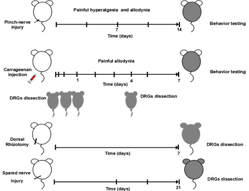
Procedures for establishing pain-related mouse model and behavioral tests. Following the Pinch-nerve injury, mice in the Pinched group, and sham group were housed and subjected to thermal hyperalgesia behavior and cold allodynia tests consecutively within 7days and prolonged subsequently for 14days for Von-Frey tests. n = 5 per group. After Carrageenan injection (Carrageenan-treated group), and sham group were respectively injected with saline, mice were sacrificed after 15min , 90min, 1day and 4days and prolonged on the 7th day. After Dorsal Rhizotomy (DR) was completed, mice were housed for 7 days and subjected to DRGs dissection. n = 5 per group. The mice were performed Spared nerve injury (SNI) surgery; kept for 21days (3 weeks) and also subjected to DRGs dissection. n = 5 per group.
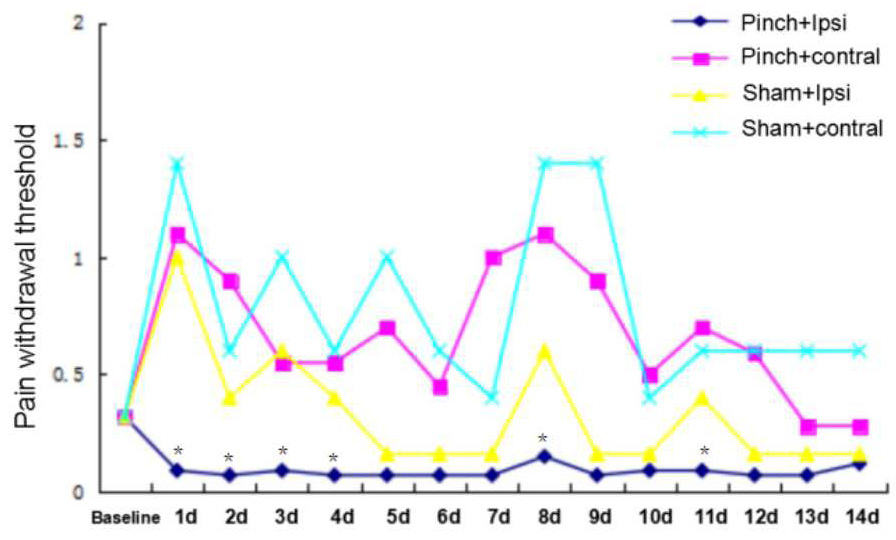
Mechanical hyperalgesia induced after Pinch nerve injury. Time course of mechanical pain withdrawal threshold measured by the von Frey filaments after unilateral Pinch (n=5). Ipsilateral hind paw displays a strongly painful behavior measured by pain withdrawal threshold compared with sham group animals (n=5); This mechanical hyperalgesia is not seen in the contralateral paw compared with sham group animals (n=5). Data are expressed as mean±S.EM. *, P <0.05 compared with the sham group.
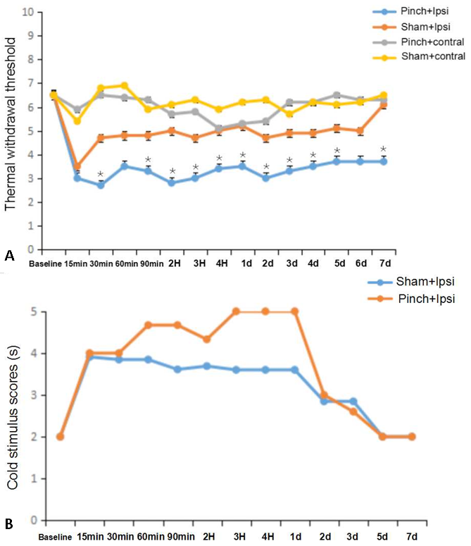
Thermal hyperalgesia and cold allodynia induced after Pinch nerve injury. (A)Time course of thermal hyperalgesia tests after Pinch. The paw withdrawal latency (Seconds) after nociceptive hot stimulation significantly decreased at 30min time point in Pinch group (n=5) compared with sham group (n=5); This thermal hyperalgesia sustained for 7days. The contralateral paw has not shown this mechanical hyperalgesia compared with sham group animals (n=5). (B)cold allodynia tests after Pinch.Time course of cold stimulus scores measured by the Actone after Pinch-nerve injury (n=5). Ipsilateral hind paw displays a strongly but not significantly painful behavior measured by pain scores compared with sham group animals (n=5); This cold allodynia is not done in the contralateral paw compared with sham group animals (n=5). Data are expressed as mean±S.EM. *, P <0.05 compared with the sham group.
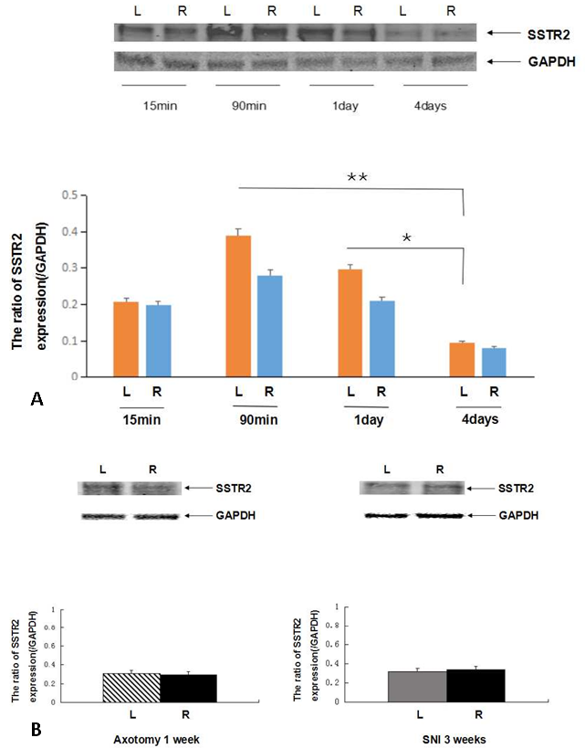
The expression of SSTR2 protein in DRGs by western bolt. (A) SSTR2 expression in DRGs after Carrageenan injection. Mice sacrificed at 15min, 90 min, 1day and 4 day after Carrageenan injected in left- hind paw(L), compared with right-side untreated hind paw, showed up-regulated SSTR2 expression in DRG neurons at 15min and 90 min these two time points (no significant differences existed, respectively); However, 1day after injection of Carrageenan, SSTR2 protein showed down-regulated in left-side DRGs(L), after 4 day, The SSTR2 protein sharply reduced in left-side DRGs(L) compared to right-side DRGs(R). Error bars represent standard error of the mean±SEM. Significant differences are indicated by *, P<0.05; **. P<0.01 (n=5). (B) The expression of SSTR2 in DRGs on the basis of DR and SNI models. Tissues from ipsilateral hind paw DRGs (L) are compared with tissues from contralateral hind paw DRGs(R) after DR 1 week and SNI 3weeks surgery. SSTR2 expression are not showed significant variations in neither of these two models (n=5).
References
- Benya, R.V., Kusui, T., Shikado, F., Battey, J.F. and Jensen, R.T., 1994. Desensitization of neuromedin B receptors (NMB-R) on native and NMB-R-transfected cells involves down-regulation and internalization. J. biol. Chem., 269: 11721-11728.
- Blevins, G.T., Vandewesterlo, E.A. and Williams, J.A., 1994. Nucleoside diphosphate kinase associated with rat pancreatic membranes regulates CCK receptor activity. Am. J. Physiol., 267: 886-874. https://doi.org/10.1152/ajpgi.1994.267.5.G866[CrossRef]
- Cescato, R., Loesch, K.A., Waser, B., Mäcke, H.R., Rivier, J.E. and Reubi, J.C., 2010. Agonist-biased signaling at the sst2A receptor: The multi-somatostatin analogs KE108 and SOM230 activate and antagonize distinct signaling pathways. Mol. Endocrinol., 24: 240-249. https://doi.org/10.1210/me.2009-0321[CrossRef]
- De Martino, M.C., Hofland, L.J. and Lamberts, S.W., 2010. Somatostatin and somatostatin receptors: From basic concepts to clinical applications. Prog. Brain Res., 182: 255-280. https://doi.org/10.1016/S0079-6123(10)82011-4[CrossRef]
- Decosterd, I. and Woolf, C.J., 2000. Spared nerve injury: an animal model of persistent peripheral neuropathic pain. Pain, 87: 149-158. https://doi.org/10.1016/S0304-3959(00)00276-1[CrossRef]
- Epelbaum, J., Dournaud, P., Fodor, M. and Viollet, C., 1994. The neurobiology of somatostatin. Crit. Rev. Neurobiol., 8: 25-44.
- Flatters, S.J.L. and Bennett, G.J., 2004. Ethosuximide reverses paclitaxel-and vincristine-induced painful peripheral neuropathy. Pain, 109: 150-161. https://doi.org/10.1016/j.pain.2004.01.029[CrossRef]
- Liu, Q., Cescato, R., Dewi, D.A., Rivier, J., Reubi, J.C. and Schonbrunn, A., 2005. Receptor signaling and endocytosis are differentially regulated by somatostatin analogs. Mol. Pharmacol., 68: 90-101. https://doi.org/10.1124/mol.105.011767[CrossRef]
- Lynds, R., Chuang, L., Gong-Wei, L., Xie-Qi, S. Annika, R., Kamal, M. and Sten, S.T-M., 2017. Neuronal plasticity of trigeminal ganglia in mice following nerve injury. J. Pain Res., 10: 349-357. https://doi.org/10.2147/JPR.S120092[CrossRef]
- Moore, S.D., Madamba, S.G., Joels, M. and Siggins, G.R., 1988. Somatostatin augments the M-current in hippocampal neurons. Science, 239: 278-280. https://doi.org/10.1126/science.2892268[CrossRef]
- Mulak, A., Larauche, M., Biraud, M., Million, M., Rivier, J. and Taché, Y., 2015. Selective agonists of somatostatin receptor subtype 1 or 2 injected peripherally induce antihyperalgesic effect in two models of visceral hypersensitivity in mice. Peptides, 63: 71-80. https://doi.org/10.1016/j.peptides.2014.10.013[CrossRef]
- Mariana, M., Leiguarda, C., Gastón, G., McCarthy, C. and Brumovsky, P., 2017. Spinal activation of the NPY Y1 receptor reduces mechanical and cold allodynia in rats with chronic constriction injury. Peptides, 92: 38-45. https://doi.org/10.1016/j.peptides.2017.04.005[CrossRef]
- Olias, G., Viollet, C., Kusserow, H., Epelbaum, J. and Meyerhof, W., 2004. Regulation and function of somatostatin receptors. J. Neurochem., 89: 1057-1091. https://doi.org/10.1111/j.1471-4159.2004.02402.x[CrossRef]
- Patel, Y.C., 1999. Somatostatin and its receptor family. Front. Neuroendocrinol., 20: 157-198. https://doi.org/10.1006/frne.1999.0183[CrossRef]
- Prasoon, P., Kumar, R., Gautam, M., Sebastian, E.K., Reeta, K.H. and Ray, S.B., 2015. Role of somatostatin and somatostatin receptor type 2 in postincisional nociception in rats. Neuropeptides, 49: 47-54. https://doi.org/10.1016/j.npep.2014.12.002[CrossRef]
- Qu, C.L., Dang, Y.H. and Tang, J.S., 2015. Administration of somatostatin analog octreotide in the ventrolateral orbital cortex produces sex-related antinociceptive effects on acute and formalin-induced nociceptive behavior in rats. Neurochem. Int., 87: 77-84. https://doi.org/10.1016/j.neuint.2015.06.002[CrossRef]
- Rai, U., Thrimawithana, T.R., Valery, C. and Young, S.A., 2015. Therapeutic uses of somatostatin and its analogues: Current view and potential applications. Pharmacol. Ther., 152: 98-110. https://doi.org/10.1016/j.pharmthera.2015.05.007[CrossRef]
- Reubi, J.C., 1997. Regulatory peptide receptors as molecular targets for cancer diagnosis and therapy. J. Nucl. Med., 41: 63-70.
- Schottelius, M., Poethko, T. and Herz, M., 2004. Featured article: first-(18) F-labeled tracer suitable for routine clinical imaging of sst receptor-expressing tumors using positron emission tomography. Clin. Cancer Res., 10: 3593–3606. https://doi.org/10.1158/1078-0432.CCR-03-0359[CrossRef]
- Shi, T.J., Xiang, Q., Zhang, M.D., Barde, S., Kai-Larsen, Y., Fried, K., Josephson, A., Glück, L., Deyev, S.M., Zvyagin, A.V., Schulz, S. and Hökfelt, T., 2014. Somatostatin and its 2A receptor in dorsal root ganglia and dorsal horn of mouse and human: expression, trafficking and possible role in pain. Mol. Pain, 10: 12. https://doi.org/10.1186/1744-8069-10-12[CrossRef]
- Shi, T.J., Xiang, Q., Zhang, M.D., Tortoriello, G., Hammarberg, H., Mulder, J., Fried, K., Wagner, L., Josephson, A., Uhlén, M., Harkany, T. and Hökfelt, T., 2012. Secretagogin is expressed in sensory CGRP neurons and in spinal cord of mouse and complements other calcium-binding proteins, with a note on rat and human. Mol. Pain, 8: 80. https://doi.org/10.1186/1744-8069-8-80[CrossRef]
- Shunsuke, Y., Matsuda, M., Yamaguchi, Y., Sawa, T. and Amaya, F., 2017. Dexmedetomidine prolongs levobupivacaine analgesia via inhibition of inflammation and p38 MAPK phosphorylation in rat dorsal root ganglion. Neuroscience, 361: 58-68. https://doi.org/10.1016/j.neuroscience.2017.08.011[CrossRef]
- Song, P., Hu, J.Y. and Zhao, Z.Q., 2002. Spinal somatostatin SSTR2A receptors are preferentially up-regulated and involved in thermonociception but not mechanonociception. Exp. Neurol., 178: 280-287. https://doi.org/10.1006/exnr.2002.8025[CrossRef]
- Thacker, M., Rivera, L.R., Cho, H.J. and Furness, J.B., 2011. The relationship between glial distortion and neuronal changes following intestinal ischemia and reperfusion. Neurogastroenterol. Motil., 23: e500-9. https://doi.org/10.1111/j.1365-2982.2011.01696.x[CrossRef]
- Tsai, Y.C., So, E.C., Chen, H.H., Wang, L.K. and Chien, C.H., 2002. Effect of intrathecal octreotide on thermal hyperalgesia and evoked spinal cFos expression in rats with sciatic constriction injury. Pain, 99: 407-413. https://doi.org/10.1016/S0304-3959(02)00107-0[CrossRef]
- Wild, D., Schmitt, J.S. and Ginj, M., 2003. DOTA-NOC, a high-affinity ligand of somatostatin receptor subtypes 2, 3 and 5 for labelling with various radiometals. Eur. J. Nucl. Med. mol. Imag., 30: 1338-1347. https://doi.org/10.1007/s00259-003-1255-5[CrossRef]
- Xiang, Q., Yu, C., Zhu, Y.F., Li, C.Y., Tian, R.B. and Li, X.H., 2016. Nuclear factor erythroid 2-related factor 2 antibody attenuates thermal hyperalgesia in the dorsal root ganglion: Neurochemical changes and behavioral studies after sciatic nerve-pinch injury. Injury, 47: 1647-1654. https://doi.org/10.1016/j.injury.2016.06.006[CrossRef]
Services
Article usage statistics
Loading…