
Immunopathological and Comparative Study of Concanavalin- A and Streptococcus lyophilized Antigen in Immunized Rats


Immunopathological and Comparative Study of Concanavalin- A and Streptococcus lyophilized Antigen in Immunized Rats
Sura Ayed Radam*, Inam Badr Falih
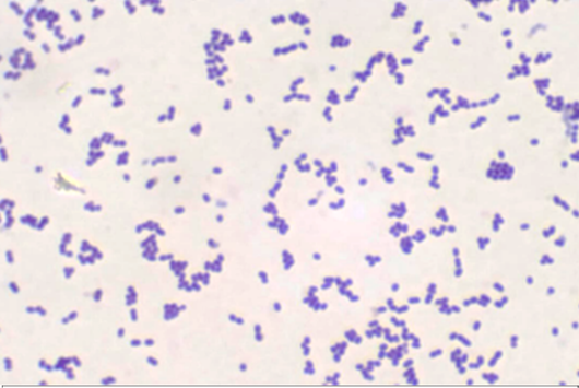
Gram positive S. pyogenes cocci (X100).
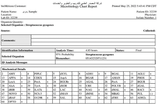
Microbiological chart report of vitek-2 test.
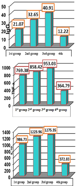
Mean and standard error of (IgG, TNF-a and IL-10) titers of the 1st, 2nd and 3rd immunized groups and 4th control negative groups at 28 day postimmunization.
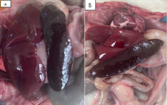
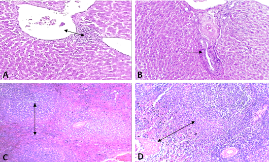
Gross appearance of hepatosplenomegaly in 3rd group (A) and 2nd group (B).
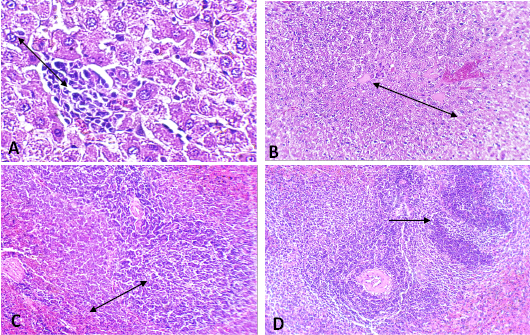
A histopathological slices in rat immunized with Con-A at 28 day post immunization. (A) Liver showed perivenular MNCs infiltration with ductal dilation. (B) Liver showed slight portal MNCs infiltration with ductal dilation. (C) Spleen showed reactive lymphoid hyperplasia with slight splenic vessels congestion. (D) Spleen showed lymphoid hyperplasia with vascular fibromuscular hypertrophy.
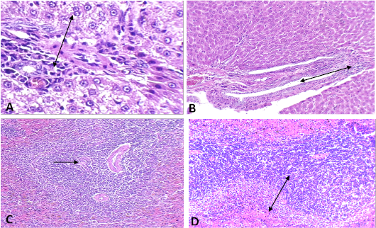
A histopathological slices in rat immunized with mixed Ag. at 28 day post immunization. (A) Liver showed perivascular and periductiolar MNCs infiltration with nuclear pyknosis and hyper eosinophilic of hepatocyte cytoplasm. (B) Liver showed mild periductal MNCs infiltration with obvious ductal dilation. (C) Spleen showed periarteriolar lymphoid hyperplasia. (D) Spleen showed great lymphoid hyperplasia with red pulp congestion.


