Comparison of Photochemical Tissue Bonding Using Rose Bengal Dye and Conventional Suturing for Closure of Incisional Cutaneous Wounds in Canine Model
Comparison of Photochemical Tissue Bonding Using Rose Bengal Dye and Conventional Suturing for Closure of Incisional Cutaneous Wounds in Canine Model
Saad Ahmad1, Shahbaz ul Haq1, Shujaat Hussain2, Khurram Ashfaq1, Shahrood Ahmed Siddiqui3,4, Arsalan Khan5*, Abubakar Yameen1, Muhammad Wasim Usmani6, Rafiq Ullah7 and Muhammad Arslan Aslam1*
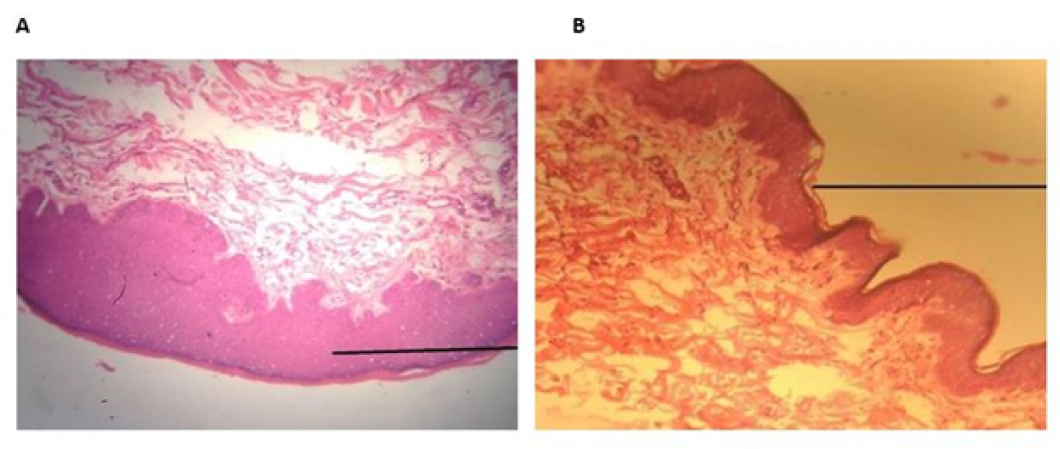
Histopathological evaluation of wound healed with PTB. The photomicrograph showed maximum leucocytic infiltration. More prominent and regular re-epithelization was seen. Prominent keratinization was seen, thickness of epidermis and dermis was more. H & E; 10X. (B) Histopathological evaluation of wound healed with silk. The silk treated photomicrograph showed less thickness of epithelium as compared to PTB. Thickness of epidermis and dermis was less. No inflammatory cells were seen. H & E; 10X.
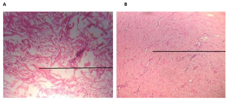
Histopathological Evaluation of Wound Healing Treatments. (A) Treated with PTB, showing loose connective tissue with thick collagen fiber deposition at 10X magnification, H&E stain. (B) Treated with silk, displaying dense connective tissue with thin collagen fibers.
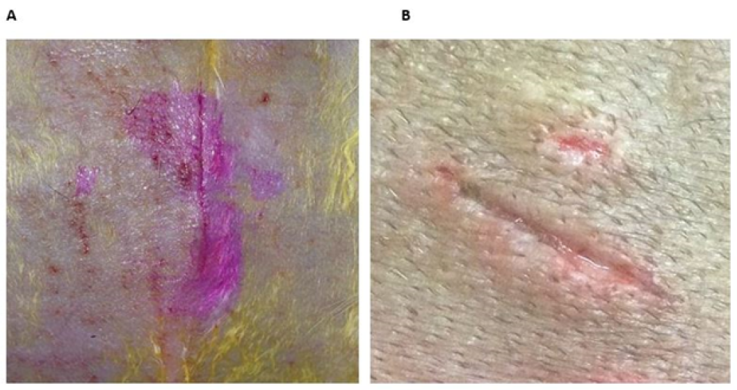
Wound appearance after PTB treatment. Full thickness cutaneous incisional wound showing complete tissue bonding just after the treatment applied. (B): Healing score. Wound appearance healed after 3.2 ±0.86 days showing no suture marks and no inflammation over the incision site held under post-operative care.


