
Adverse Histo-Physiological Damages of Increasing Consummation of Puma (Super Fat) on Female Rats


Adverse Histo-Physiological Damages of Increasing Consummation of Puma (Super Fat) on Female Rats
Fatima Aziz Mahdi Al-Badry
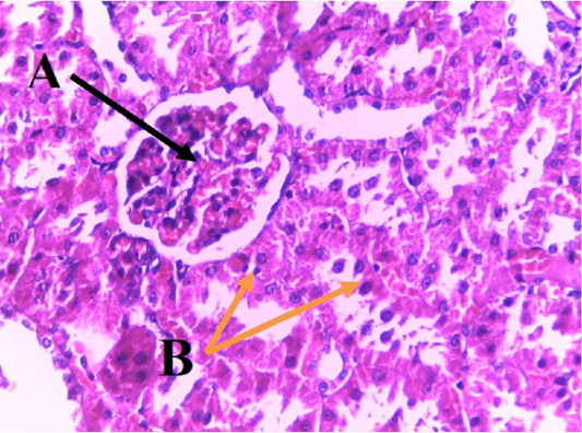
Section in kidney of control group showing cortex of kidney consist of glomerulus (A) renal tubules in medulla (B) (H&E) (100 X).
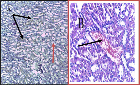
Section in kidney of Puma super fat group (One month) showing, (A): hemorrhage (black arrow) natural renal tubules (red arrow). (B): Section in kidney of Puma group (Two months) showing congestion of blood vessel (black arrow) stained by H&E, (A) (40 X) (B) (100 X).
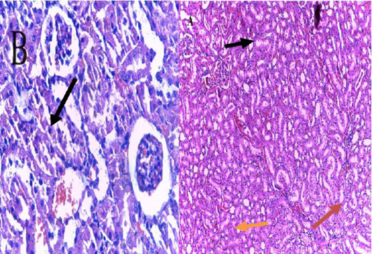
Section in kidney of Puma group (Two months) showing (A): hemorrhage among renal tubules (black arrow). (B): glomerular atrophy (black arrow) hemorrhage (red arrow) dilatation of Bowman’s capsule (orange arrow), stained by H & E, (A) (40 X), (B) (100 X).
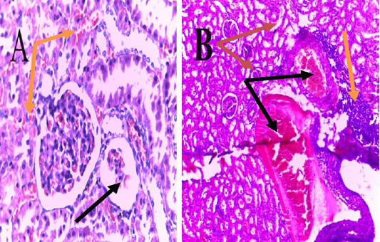
Section in kidney of Puma group (Three month) showing (A): absence of glomerulus (black arrow) severs bleeding (orange arrow). (B) sever congestion (black arrow) infiltration of inflammatory cells (orange arrow) shrinkage of glomerulus (red arrow) with autolysis stained by H&E, (A) (100 X) (B) (40 X).
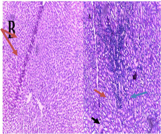
Section in the kidney of Puma group (Three months) showing (A): sever infiltration of inflammatory cells (red arrow). (B): Section in the liver of control group showing central vein (red arrow) hepatocytes (blue arrow) sinusoids (black arrow), stained by H & E, (A), (B) (40 X).

Section in the liver of Puma (Super fat) group (One month) showing (A): simple enlargement of sinusoids (Black arrow) natural tissue (orange arrow). (B) Puma group (Two months) showing congestion (black arrow) hypertrophy of hepatocytes (orange arrow) enlargement of sinusoids (red arrow) stained by H&E, (A) (100 X) (B) (40 X).
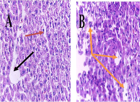
Section in the liver of Puma group (Two months) showing (A) dilation of central vein (black arrow) sinusoids (red arrow). (B): necrosis (orange arrow) stained by H&E, (A) (100 X) (B)(40X).
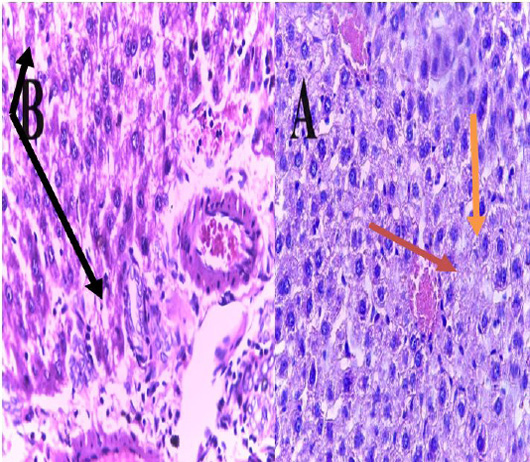
Section in the liver of Puma group (Three months) showing (A): congestion of central vein (black arrow). (B): fibrosis (red arrow) congestion (orange arrow) stained by H&E, (A), (B) (100X).

Section in the liver of Puma group (Three months) showing inflammation (red arrow) (H&E) (100X).

