
Placental maladaptation due to defective trophoblast invasion, vascular remodeling and molecular signaling defects are the primary factors that cause intrauterine growth restriction (IUGR). Besides its classical endocrine functions, human chorionic gonadotropin (hCG) plays a vital role in the regulation of trophoblast differentiation, angiogenesis and immune adaptation during early placentation. New data indicates that the malfunction of hCG-regulation, , is the cause of abnormal placental development and decreased uteroplacental perfusion. This review discusses the mechanistic role of hCG in pathogenesis of IUGR, highlighting its role in trophoblast invasion, angiogenic signaling, and hypoxia-related feedback pathways. Clinical evidence shows that abnormal levels of maternal serum hCG are predictive of adverse pregnancy outcomes; but its predictive capacity as an isolated biomarker is limited. Incorporating hCG into multimarker screening models and investigating isoform-specific functions can enhance its clinical use. Better comprehension of hCG dysregulation can aid in earlier identification and improved control of at-risk pregnancies.
Review · Volume 72, Issue 2, 14 September 2026 · Open Access
Role of Human Chorionic Gonadotropin Dysregulation in Intrauterine Growth Restriction: Mechanisms and Clinical Implications
·
Affiliations & Notes ⌄
- 1Department of Medical Imaging Technology, University of Sialkot, Sialkot, Pakistan. *Correspondence | Amna Bibi, Department of Medical Imaging Technology, University of Sialkot, Sialkot, Pakistan
- 2Email: amna.bibi@uskt.edu.pk
Article Info ⌄
- Journal:
- Biologia (Lahore)
- Published:
- 14 September 2026
From the article
- Human chorionic gonadotropin (hCG) assumes a central role in the regulation of placental development that goes beyond its traditional endocrine role and extends to the regulation of trophoblast behaviour, vascular adaptation and immune modulation.
- Synthesized evidence on the association of dysregulation of hCG, specifically at the level of its biologically active isoforms, with the pathogenesis of intrauterine growth restriction has been found to exert coordinated effects in response to placental hypoxia.
- One of the most important revelations of the existing research significant insights can be obtained to gain an understanding of the active regulatory role of hCG in placental activity.
- Finally, hCG is a crusade between molecular signaling and clinical pathology in IUGR.
Selected from the article's own text.
Abstract
Introduction
Intrauterine growth restriction (IUGR) is a significant obstetric complication, which is caused by the inability of the fetus to reach its genetically determined growth potential, mainly due to compromised placental function. It is linked with high perinatal morbidity and mortality, and long-term metabolic and cardiovascular outcomes in adulthood ; ; . The pathophysiological pathway of IUGR is closely associated with abnormal placentation, especially when there is defective trophoblast invasion and insufficient remodeling of the uterine spiral arteries, leading to a compromised uteroplacental perfusion and chronic fetal hypoxia ; . Latest evidence suggests that early disruption in molecular signaling pathways that control placental development is a critical factor in the development of this condition ; .
Human chorionic gonadotropin (hCG) is a glycoprotein hormone that is mainly synthesized by trophoblastic cells and is essential for the successful early pregnancy by promoting placental development and establishment. Apart from its classical endocrine role to sustain the corpus luteum, hCG has autocrine and paracrine properties which play a role in trophoblast differentiation, invasion, angiogenesis, and maternal immune tolerance ; d’Hauterive et al., 2022). Different isoforms of hCG, such as intact hCG, free β-hCG and hyperglycosylated hCG (hCG-H), have different biological functions, with hCG-H playing a particularly critical role in promoting extravillous trophoblast invasion and placental anchoring ; . These functions play a crucial role in creating appropriate placental vascularization and fetal development. Alterations in the concentrations of the protein β-hCG during the first and second trimesters have been associated with impaired fetal development and placental dysfunction ; ; . Moreover, longitudinal research has shown that disruptions in hCG dynamics are linked to changes in the growth pathways in the fetus, indicating that hCG can be used as an early warning of hysteresis of the placental functions ; . Although the evidence has been mounting, the exact role of hCG in IUGR is yet to be completely comprehended. Most studies have focused on the total or free β-hCG concentrations without differentiating between the biologically important isoforms, and therefore, limiting the use of mechanisms. In addition, the utilization of hCG as a predictive biomarker of IUGR has not been completely validated. It is this ambiguity that highlights the need to critically assess hCG dysregulation in the broader context of placental pathophysiology.
There have been several reviews summarizing the general endocrine role of hCG in pregnancy, but a few studies have focused on the endocrine role of hCG as a possible mechanism underlying IUGR. Although there are distinct roles for intact hCG, free β-hCG and hyperglycosylated hCG in placentation, most previous reviews have focused on hCG as a single entity.
Therefore, the review below will focus on the role of human chorionic gonadotropin, specifically on its unique isoforms, in IUGR. It emphasizes isoform-specific dysregulation in relation to the invasiveness of the trophoblast, angiogenic signaling and feedback mechanisms in the setting of hypoxia, critically reviews the clinical evidence that supports the association of hCG with IUGR outcomes and explores its role as a tool for early identification of at-risk pregnancies.
Materials and Methods
The identification of literature was carried out by searching the PubMed and Google Scholar databases for articles published from 2000 to 2026, focusing on those published in the past five years. The search terms used were combinations of human chorionic gonadotropin, hCG, hyperglycosylated hCG, intrauterine growth restriction, fetal growth restriction, trophoblast invasion, placental dysfunction, and angiogenic imbalance. Only studies that presented original clinical, biochemical or mechanistic data on hCG or its isoform(s) in relation to placental function or IUGR, or relevant background information on placental biology were included. Case reports, abstracts and articles not written in English were not used. The reference lists of key articles were also examined to look for other relevant articles not found in the initial search.
Functional biology of human chorionic gonadotropin in placentation
Human chorionic gonadotropin (hCG) is a structurally heterogeneous glycoprotein hormone that can only be active when its concentration exceeds a threshold and when its molecular isoforms are present in the appropriate proportion. They include intact hCG, free α-hCG, free β-hCG, nicked hCG, and hyperglycosylated hCG, which are modified in different ways and have undergone different proteolytic degradation ; . This means that each isoform has different receptor affinities and biological activities, allowing hCG to control both local and endocrine placental functions. Intact hCG can mediate classical luteotropic activity via the luteinizing hormone/choriogonadotropin receptor (LHCGR), while free β-hCG and hyperglycosylated hCG are more involved in autocrine and paracrine regulation of trophoblast proliferation, differentiation and invasion ; .
Of these variants, hyperglycosylated hCG is a form of functional specialization that is predominant during early gestation and is critical in the invasive phenotype of trophoblast cells. The molecular conformation changes in hyperglycosylated hCG increase its capacity to act on trophoblast proliferation and invasion in the absence of its classical endocrine actions ; . It is a predominant isoform found to be produced by extravillous cytotrophoblasts and mediates the metamorphosis of such cells into an invasive phenotype that is needed to achieve successful implantation and anchoring of the placenta ; . The distinct biological functions of hCG isoforms highlight the importance of their tightly regulated expression for normal placental development and maturation.
In the background of placentation, hCG is a pivotal controlling element in the differentiation and invasion of trophoblasts. To respond to early implantation, there is coordinated signaling between the embryo and part of the maternal endometrium, which is mediated by hCG. It facilitates the differentiation of the cytotrophoblasts into syncytiotrophoblasts and extravillous trophoblasts, thus supporting the production of hormones and their invasive ability ; . Human chorionic gonadotropin (hCG) stimulates trophoblast motility and matrix degradation, which are crucial in penetration into the decidua and remodeling of maternal spiral arteries. Such activities are strictly controlled, as very high or low invasion may lead to disruptions in the placenta and impair the development of the fetus. Its contribution to placental development is further emphasized by the interaction of hCG with key molecular regulators of trophoblast invasion. Empirical evidence suggests that hCG can act in signaling pathways linked to the transforming growth factor beta superfamily that play key roles in determining trophoblast differentiation and invasive gene behaviour . hCG promotes a balance in proliferation and invasion in the population of trophoblasts through the modulation of these pathways. Moreover, it has also been involved in the regulation of extracellular matrix remodeling, and the penetration of the trophoblast cells into maternal tissues can be remodeled and controlled .
Another vital procedure that hCG affects and that happens during the development of the placenta is known as angiogenesis. Proper vascularization provides adequate nutrient and oxygen delivery between the maternal and fetal circulations. It is known that hCG promotes vascular growth and adaptation in the first trimester of pregnant women by stimulating the expression of angiogenic factors, their receptors in the placental bed, and subsequent vascular development . This effect is especially pertinent in the first trimester, when the rapid growth of the placenta vascular network is essential in order to meet the growing metabolic needs. Human chorionic gonadotropin (hCG) plays a role in the formation of a normal uteroplacental circulation by increasing angiogenic signaling.
Besides the direct effects on trophoblasts and endothelial cells, hCG has a great impact in modulating the maternal immune response to favour pregnancy. Its immune tolerance is necessary to prevent the rejection of the semi-allogeneic fetus by its maternal and fetal immune systems and therefore to allow the implantation to succeed. Human chorionic gonadotropin (hCG) has been established to have a role in modulating the population of immune cells in the decidua, including natural killer
Table 1: Functional roles of human chorionic gonadotropin (hCG) isoforms in placentation with supporting evidence from key studies.
|
hCG Isoform |
Primary source |
Biological function |
Mechanistic role in placentation |
Evidence (Key Studies) |
|
Intact hCG |
Syncytiotrophoblast |
Maintains corpus luteum, progesterone secretion |
Supports early pregnancy maintenance and endometrial receptivity |
Cole, 2010; Stenman et al., 2006 |
|
Free β-hCG |
Placental trophoblasts |
Circulating subunit measurable in serum |
Reflects trophoblast activity and placental function |
Dugoff et al., 2004; Yaron et al., 2002 |
|
Hyperglycosylated hCG (hCG-H) |
Extravillous cytotrophoblasts |
Promotes trophoblast invasion and proliferation |
Regulates extravillous trophoblast invasion and spiral artery remodelling |
Cole, 2007; Evans, 2016; Herghelegiu et al., 2023 |
cells that are present in the uterus . By inducing the production and regulation of the production of cytokines, positively affecting immune cell activity, hCG promotes an environment that allows tolerance to take place and yet maintains the controlled production of inflammatory signals required to promote tissue remodeling.
Notably, these pathways are interdependent, and the role of one aspect can affect the others; there is a need to strictly regulate the expression and activity of hCG during the initial gestation. It is thus crucial to balance the various hCG forms and their downstream consequences to achieve normal placental architecture and functionality.
Pathophysiology of intrauterine growth restriction: A placental perspective
Intrauterine growth restriction is essentially a disease of placental insufficiency in which structural and functional abnormalities in the placenta impede the transfer of oxygen and nutrients to the developing fetus. The etiologic pathology of this condition has its genesis early in gestation and is intricately linked to failure in trophoblast behaviour, vascular remodeling, and molecular signaling within the placental microenvironment. These processes are closely coordinated when under normal circumstances, and are increasingly suggested to be involved in their pathogenesis.
Defective trophoblast invasion and spiral artery remodeling
One of the characteristic features of IUGR is insufficient invasion of extravillous trophoblast cells into the maternal decidua and myometrium. During normal placentation, these cells undergo a tightly regulated differentiation into an invasive phenotype that is capable of maintaining an increased uteroplacental blood flow. The failure of such a process leads to the retention of narrow vessels with high resistance to perfusion; these vessels are limited to perfusion of the intervillous space ; .
On a molecular scale, trophoblast invasion is controlled by a combination of signaling pathways that control and regulate cell proliferation, differentiation, and degradation of the extracellular matrix. The modulator of the balance between trophoblast proliferation and invasive capacity is played by members of the transforming growth factor beta superfamily . Failure to regulate these pathways may result in impairment in trophoblast migration, as well as inadequate vascular remodeling. Notably, local regulatory factors in the placental environment, such as hCG, have been found to exert control over these signaling cascades. Signaling defects by hCG may thus play a role in the impaired invasion caused by IUGR by destabilizing the molecular pathways regulating the actions of trophoblasts.
Placental hypoxia and oxidative stress
Abnormal remodeling of the spiral artery leads to lower uteroplacental perfusion, which puts the placenta in a hypoxic environment. A core element of IUGR, and a precursor of a cascade of adaptive and pathological responses that further impair the functional capabilities of the placenta. Hypoxic conditions change the metabolism of cells, enhance the formation of reactive oxygen species, and cause oxidative stress in cells of both trophoblast and endothelium . The disruption of cellular integrity, cellular differentiation, and cellular signaling pathways is one of the effects of oxidative stress. The alterations impair the exchange landscape of the placenta and diminish its ability to propel the development of the fetus. Moreover, the current evidence suggests that cellular damage also occurs due to hypoxia, including the promotion of ferroptosis-related pathways, which contribute to vascular injury in the placenta, further enhancing the perfusion deficit . Hypoxia may alter hCG production, indicating a feedback interaction.
Angiogenic imbalance and vascular dysfunction
Placental efficiency hinges on the growth and development of a well-structured vascular network, which makes effective maternal-fetal exchange possible. This is disrupted in IUGR by an imbalance between pro-angiogenic and anti-angiogenic factors, failing this process. Changes in angiogenic signaling have been consistently linked to adverse pregnancy outcomes, such as fetal growth restriction ; .
In the placental bed, the expression of angiogenic growth factors and their receptors is closely regulated to encourage coordinated vascular expansion. The imbalance in these two leads to decreased vessel formation, dysfunction of endothelial cells and augmented vascular resistance . These alterations combine to cause hemodynamic abnormalities typical of IUGR as well as further reduce the availability of nutrients and oxygen to the fetus.
hCG-immune cell interactions, especially the uterine natural killer cells, contribute to the vascular restructuring and immune-mediated control of angiogenesis . The instability of these interactions can also impair the adaptive compensatory vascular responses in IUGR.
This combined view explains why considering IUGR not as a clinical context but as the product of multifaceted molecular and cellular aberrations in the placenta. An understanding of the interaction of hCG with these pathways is an essential stepping stone in establishing the mechanism and clinical relevance involved in the hormonal regulation and structural abnormality observed in IUGR.
Mechanisms of hCG dysregulation in intrauterine growth restriction
As discussed in Section 2, trophoblast invasion, angiogenesis and immune adaptation during placentation are regulated by hCG. Based on recent findings, deregulation of hCG and its isoforms in IUGR pregnancies can interfere with the same processes, underlining the importance of understanding the molecular, cellular and clinical interactions between these hormones to elucidate the pathogenesis of IUGR ; .
Isoform-specific dysregulation and functional consequences
One of the hallmarks of the biology of hCG is the existence of structurally distinct hCG isoforms that exhibit distinct activities ; . These include hyperglycosylated hCG, the most abundant in early pregnancy, which is made by the invasive extravillous cytotrophoblasts ; . In addition to endocrine activity, hyperglycosylated hCG also performs autocrine and paracrine effects to stimulate trophoblast proliferation, differentiation
and invasion ; . Because of its extensive glycosylation, it can change the interactions with receptors and intracellular signaling, which contributes to the increased invasiveness of the phenotype necessary for successful development of the placenta ; ; .
Reduced production or functional impairment of hyperglycosylated hCG has been associated with insufficient trophoblast invasion into the maternal decidua, resulting in shallow placentation . In contrast, intact hCG primarily functions to maintain the corpus luteum and support progesterone production during early pregnancy, whereas hyperglycosylated hCG directly regulates trophoblast proliferation, differentiation, and invasion ; . These functional differences indicate that dysregulation of specific hCG isoforms has distinct pathophysiological consequences for placental development and may contribute to the pathogenesis of IUGR .
Trophoblast differentiation affects the relative abundance of the hCG variants. Any form of disruption in these pathways of normal differentiation can alter the balance between the endocrine and autocrine versions of hCG and affect local signaling within the placental microenvironment . This disproportion may play a role in the failure of the trophoblast subtypes to communicate with each other and provide suitable coordination of the process of placental development. Moreover, experimental research has shown that hyperglycosylated hCG stimulates the ability of cellular processes to invade another cell by choosing an appropriate cellular strategy ; .
hCG-mediated regulation of trophoblast invasion
Invasion by Trophoblast is a highly regulated process where the activation of signaling pathways that regulate cellular adhesion, migration, and proteolytic activity is coordinated. Human chorionic gonadotropin (hCG) has an important modulatory role in this process and regulates intracellular signaling pathways that influence trophoblast phenotype and invasive potential, in turn, affecting spiral artery remodeling. Members of the transforming growth factor beta superfamily participate in the regulation of the balance between trophoblast proliferation and differentiation as well as in the disruption of these pathways. Human chorionic gonadotropin also regulates cytoskeletal remodeling, extracellular matrix degradation, and the expression of proteolytic enzymes required for trophoblast invasion through its interaction with multiple signaling pathways ; ; . In pregnancies complicated by IUGR, the signaling of hCG deregulates these processes, resulting in a decreased invasion depth, impaired matrix remodeling and an inadequate spiral artery transformation, ultimately leading to loss of uteroplacental perfusion. This association has further been supported by clinical evidence, whereby distorted maternal serum β-hCG dynamics during early pregnancy have been associated with adverse outcomes, including fetal growth restriction, an indicator of underlying trophoblast dysfunction ; . These findings indicate that the hCG dysregulation is no longer a secondary effect but may also work towards the pathogenesis of placental insufficiency.
Trophoblasts express the LH/CG receptor (LHCGR) that is activated by hCG. After binding this receptor, hCG can stimulate PI3K/Akt and MAPK pathways. PI3K/Akt prolongs the survival of trophoblasts and promotes vascular remodeling. Their proliferation, differentiation and migration are controlled by MAPK signaling. Disruption of hCG-LHCGR signaling could lead to inadequate function of these pathways, and shallow trophoblast invasion is associated with IUGR .
The invasion of the trophoblast must also involve a breakdown of the extracellular matrix. This is primarily carried out by two enzymes, MMP-2 and MMP-9. MMP-2 is more active in early first trimester trophoblasts, while MMP-9 is more active later . Both enzymes are influenced by hCG, and decreased activity of MMP-2/MMP-9 has been associated with insufficient remodeling of the spiral arteries and poor trophoblast invasion, both of which occur in IUGR.
Disruption of angiogenic signaling and vascular development
The development of an effective, tightly regulated angiogenic signaling pathway that coordinates the proliferation, migration and differentiation of endothelial cells relies upon well-coordinated angiogenic signaling, particularly via intercellular adhesion molecules, maintaining the development of a properly functioning placental vascular network. Human chorionic gonadotropin has been found to play a role in the regulation of these pathways, affecting the expression of angiogenic factors and endothelial receptors in the placental bed. In this way, it helps to develop a low-resistance uteroplacental circulation that is adequate to sustain fetal growth. Angiogenic imbalance is observed in IUGR due to reduced pro-angiogenic signaling and poor vascular development. Research has shown changes in maternal serum markers, such as hCG, to relate to changes in angiogenic profiles and adverse pregnancy outcomes ; . The relation between hCG and the angiogenic pathways is multifaceted, and it goes both directly through the direct effects of the angiogenic pathways on the endothelial cells and indirectly through the modulation of trophoblast-derived factors.
Here are two pathways that are of particular importance: HIF-1α and VEGF. Placental tissue is hypoxic, and HIF-1α accumulates, which aids trophoblast survival. However, continuous exposure to high levels of HIF-1α can inhibit normal trophoblast differentiation and spiral artery remodeling . HIF-1α also regulates VEGF and its receptors that promote blood vessel growth in the placenta. hCG and HIF-1α are both oxygen-sensitive, so hypoxic signaling of hCG may exacerbate the angiogenic imbalance associated with abnormal HIF-1α/VEGF activity. Another factor regulating the behavior of trophoblast stem cells is Wnt/β-catenin signaling, which is known to affect preeclampsia and IUGR . This is still an unanswered question because no study has yet demonstrated a direct relationship between hCG and Wnt/β-catenin in the human placenta.
Angiogenic signaling in response to hCG dysregulation may be impaired, resulting in compromised function of endothelial cells and formation of vessels. Moreover, hCG affects the activities of uterine natural killer cells, which are also critical in the regulation of vascular remodeling by the secretion of angiogenic mediators . This interaction might be crucial to defective vascular adaptation in IUGR.
Together, dysfunctional trophoblast invasion and angiogenic imbalance cause structural and functional defects of the placental vasculature, which leads to decreased uterine placental perfusion and placental insufficiency ; . These vascular malformations cause increased vascular resistance, reduced maternal–fetal blood flow and decreased delivery of oxygen and nutrients necessary for normal fetal growth ; . It is possible that dysregulation of hCG can contribute to progression of placental dysfunction in IUGR, involving disrupted trophoblast function and angiogenic signaling, as previously described ; ; .
Hypoxia-driven feedback and hCG dysregulation
Placental hypoxia is one of the outcomes and initiators of pathological processes in IUGR. The defective invasion of the trophoblast results in reduced perfusion, an alteration to the cellular metabolism and signaling in the placenta. Hypoxia leads to the development of reactive oxygen species and facilitates oxidative stress, which further influences the viability and functioning of trophoblasts .
Placental hypoxia is a hallmark of placental dysfunction in intrauterine growth restriction (IUGR) and profoundly affects trophoblast function and placental development. Continued hypoxia leads to mitochondrial dysfunction and oxidative stress, which leads to poor trophoblast invasion, poor remodeling of spiral arteries, and placental insufficiency. Furthermore, hypoxia also inhibits the synthesis of placental hormones such as chorionic gonadotropin (CG), which is crucial for normal placental development and trophoblast activity. All of these changes lead to placental dysfunction and fetal growth restriction (FGR) ; .
Clinical evidence linking hCG and intrauterine growth restriction
Human chorionic gonadotropin (hCG) has been widely studied in maternal serum from routine prenatal testing as a potential placental function biomarker and a marker of fetal growth restriction (FGR). The clinical value of each molecular form of hCG has not been fully studied, and most clinical studies have focused on either total hCG or free β-subunit of human chorionic gonadotropin (free β-hCG). There have been various reported associations of hCG with FGR, but these vary due to demographic differences, gestational timing for sampling, hCG assays used, thresholds for hCG, and definitions of FGR. Therefore, it is recommended that hCG could be considered as a marker of placental function rather than a reliable sole marker of IUGR ; .
First trimester hCG and early prediction of IUGR
First trimester screening cohorts suggest that maternal hCG levels could be related to subsequent fetal growth, though the direction of the relationships and significance of the findings differ between studies. In an analysis of 7,987 mother–child pairs from the Generation R Study, a population-based prospective cohort in Rotterdam, the Netherlands, maternal total hCG concentrations measured predominantly during the late first trimester were associated with fetal growth and birth weight. These associations differed depending on gestational age at hCG measurement and fetal sex, suggesting that these should be taken into account when interpreting maternal hCG concentrations .
In addition, clinical screening studies have been conducted to evaluate free β-human chorionic gonadotropin. The First- and Second-Trimester Evaluation of Risk Trial is a large population-based screening study in the United States that evaluated the relationship of extreme levels of free β-hCG, pregnancy-associated plasma protein-A, and nuchal translucency to obstetric complications. The correlation between reduced pregnancy-associated plasma protein-A and FGR was more clearly established, and free β-hCG was mildly decreased in pregnancies that later were complicated by FGR. The results indicate that the level of free β-hCG alone does not have a strong predictive value when compared to pregnancy-associated plasma protein-A .
In a similar study, Goetzinger et al. measured the pregnancy-associated plasma protein-A, free β-hCG, and maternal factors in association with the development of fetal growth abnormalities. After their analysis, they concluded that high free β-hCG, low pregnancy-associated plasma protein-A, along with certain maternal characteristics, were statistically significant predictors of fetal growth restriction, but that the overall predictive power of the model was modest .
However, not every study has shown a relationship between free β-hCG and fetal growth restriction. Yaron et al., in a cohort of 1,622 singleton pregnancies, noted that very low free β-hCG was linked to a higher risk of spontaneous miscarriage, but not related to fetal growth restriction or most other adverse pregnancy outcomes. The authors therefore concluded that free β-hCG was a poor predictor of these complications .
More recent evidence is conflicting. In a Spanish case–cohort study, the 119 pregnancies affected by IUGR were compared with a random subcohort of 383 pregnancies, carried out in 2025. First-trimester pregnancy-associated plasma protein-A and free β-hCG were tested as continuous and percentile values. It found associations among these screening markers and subsequent IUGR, but the study design and population restricted to a single centre do not allow the findings to be extrapolated or taken as evidence of good predictive accuracy .
The clinical literature suggests that abnormal concentrations of hCG in the first trimester may be linked to fetal growth in some populations, but the link is inconsistent and is generally less strong than the links suggested for pregnancy-associated plasma protein-A. This variation may be due to differences in the type of hCG assays, the values used to define abnormal levels of hCG, the gestational timing of measurements, fetal sex, mother-to-child characteristics, and the definitions of fetal growth restriction. Therefore, for the time being, hCG cannot be considered as an independent reliable marker of IUGR.
Second trimester hCG and established placental dysfunction
Second-trimester maternal serum biomarkers provide additional information on placental function and the risk of adverse pregnancy outcomes. During this stage of pregnancy, maternal serum hCG is routinely evaluated as part of second-trimester prenatal screening, in combination with other biochemical markers such as alpha-fetoprotein (AFP) and unconjugated estriol (uE3), and, in quadruple screening, inhibin A. Several studies have investigated whether abnormal maternal hCG concentrations during the second trimester are associated with subsequent fetal growth restriction (FGR) and other placental complications ; .
These findings have been reinforced by subsequent studies that have demonstrated that second-trimester levels of hCG, especially when added to the first-trimester level, are associated with poor fetal growth. Indicatively, ‘active’ predictive analysis using a combination of first-trimester free β-hCG into a single test alongside second-trimester intact hCG into a single test has been demonstrated to be correlated with both the risk of IUGR as well as the risk of hypertensive pregnancy disorders . The correlation of the values implies that consistently abnormal values in the level of hCG were only due to continuous malfunction in the placenta, not transient change.
The combined predictive value of second-trimester markers has also been assessed in conjunction with clinical parameters and other biochemical markers. Research that has evaluated multi- marker models has shown better prediction of poor outcomes when hCG is incorporated in the broader screening systems . The role of hCG in these models can, however, be low compared to the other markers, especially those that are directly related to angiogenesis and placental vascular activity.
Notably, hCG levels in the second trimester seem to be an indicator of known placental pathology, as opposed to the presence of early mechanistic changes. Consequently, their usefulness might be found not in being an early predictive aid but in identifying already-at-risk pregnancies. This difference is important to acknowledge the need to pay attention to the time when the biomarker will be measured, with respect to the disease pathogenesis.
Evidence from cohort studies and meta-analyses
Many cohort studies and meta-analyses make it possible to view the clinical significance of hCG in predicting negative pregnancy outcomes from a broader perspective. Systematic reviews have shown that abnormal levels of hCG can predict a higher risk of complications associated with abnormal levels of hCG, including preeclampsia and fetal growth restriction; but the predictive power of hCG alone is weak . Equally, meta-analyses assessing the effectiveness of β-hCG in screening situations have reported moderate effectiveness with substantial variations, since it depends on the study design and population characteristics .
More recent meta-analytical evidence on assisted reproductive environments has also illuminated the issue of early hCG dynamics and their relationships with pregnancy outcomes. These studies imply that early β-hCG levels after embryo transfer are linked to later pregnancy viability and pregnancy complications, such as growth restriction, although their predictive value already depends on their context ;. Although these findings may bring the applicability of hCG beyond natural conception, it also highlights how the clinical context may influence the interpretation of biomarkers.
Comparative studies provide some themes which are consistent across the studies. First, abnormal hCG values can be reliably correlated with adverse outcomes, and thus, it can be concluded that hCG is a reflection of underlying placental dysfunction. Second, the direction of association varies; that is, there are both low and high directions associated with the risk factor, indicating that there are optimum ranges of physiological parameters that are crucial
Table 2: Summary of clinical studies evaluating the association between maternal serum hCG levels and intrauterine growth restriction (IUGR).
|
Study / Year |
Trimester |
Marker(s) |
Key Findings |
Clinical Interpretation |
|
Dugoff et al., 2004 |
1st |
Free β-hCG, PAPP-A |
Abnormal levels associated with adverse outcomes, including IUGR |
Useful in early risk screening (limited alone) |
|
Yaron et al., 2002 |
1st |
Free β-hCG |
Both high and low levels are linked to adverse outcomes |
Non-linear relationship with placental function |
|
Barjaktarovic et al., 2017 |
1st |
Total hCG |
Associated with fetal growth patterns |
Reflects early placental development |
|
Sharony et al., 2018 |
1st + 2nd |
Free β-hCG + intact hCG |
Combined abnormal levels linked to IUGR and preeclampsia |
Persistent abnormal levels reflect placental dysfunction |
|
Huang et al., 2022 |
Meta analysis |
β-hCG |
Moderate predictive value for adverse outcomes |
Not sufficient as a standalone biomarker |
|
Zhang et al., 2021 |
Meta analysis |
β-hCG |
Significant association but variable accuracy |
Requires integration with other markers |
in normal placentation. Third, hCG plus other biomarkers, especially the angiogenesis and placental perfusion-associated ones, can improve the predictive performance of hCG.
In general, the available clinical data suggest that abnormal levels of hCG in maternal blood are linked to placental dysfunction and a higher risk of intrauterine growth restriction; but this association is not consistent in all studies and can be affected by the differences in the study populations, the gestational age at which the blood is sampled, the assay method, and the definition of abnormal. Thus, the use of hCG alone is of limited value, but could be used with other biochemical markers, ultrasound markers, and maternal clinical markers to assist in risk assessment. The clinical value of hCG could further be enhanced in future studies, which could include evaluation of individual hCG isoforms and standardization of prediction models using multiple markers.
Human chorionic gonadotropin as a predictive biomarker in intrauterine growth restriction
Maternal serum human chorionic gonadotropin is used in the usual prenatal screening and might be useful as an indirect marker for placental function. Total hCG or free β-hCG levels are abnormal in cases of fetal growth restriction and other complications. The direction and magnitude of these relationships do, however, depend on gestational age, maternal factors, assay techniques, study population and definition of fetal growth restriction. Thus, the use of hCG alone is insufficient in determining the reliability of IUGR prediction ; .
Diagnostic limitations
The clinical value of hCG is limited by substantial physiological and analytical variability. Both elevated and reduced hCG levels have been associated with adverse outcomes, making it difficult to establish universally applicable cutoff values. In addition, most routine assays measure total hCG or free β-hCG without distinguishing biologically different isoforms. This may reduce diagnostic accuracy because isoforms such as hyperglycosylated hCG have distinct roles in trophoblast invasion and placental development ; . Consequently, hCG should be considered an indicator of possible placental dysfunction rather than an independent diagnostic marker of IUGR.
Role in combined screening models
The predictive value of hCG improves when it is incorporated into multimarker screening models. Combining hCG with pregnancy-associated plasma protein-A, maternal characteristics, Doppler ultrasound findings, and markers of placental angiogenesis provides a broader assessment of placental function than any single biomarker ; ; . However, the additional contribution of hCG may remain modest compared with markers that more directly represent placental vascular function.
Therefore, the most appropriate clinical role of hCG is as a supportive component of integrated risk-assessment models. Future studies should determine whether isoform-specific measurements and standardized longitudinal assessment can improve its usefulness in the early identification of pregnancies at risk of IUGR.
Clinical and translational implications
Human chorionic gonadotropin has a proven application in prenatal screening, wherein it helps assess placental functioning and risk stratification against developing adverse pregnancy outcomes. In the scenario of intrauterine growth restriction, it has clinical implications associated with its linkage to early placental development as opposed to its diagnostic power. The presence of abnormal maternal serum β-hCG levels, when interpreted together with other biochemical and clinical measurements, is an indirect sign of placental dysfunction and thus is included in a screening algorithm ; .
Despite this research interest, current international clinical guidelines do not recommend hCG measurement as part of formal IUGR screening. The 2019 FIGO guideline states that PAPP-A can be included in preeclampsia risk models if already obtained from aneuploidy screening, but is not recommended for prediction of IUGR. First-trimester biochemical markers such as hCG are not included in the Doppler assessment and clinical risk factors used to monitor IUGR in the 2024 updated version of RCOG Green-top Guideline No. 31. Also, ISUOG recommendation regarding first trimester ultrasound does not mention growth restriction screening, either using hCG or PAPP-A ; ; . This disconnect between research data and clinical recommendations may be related to cost and availability of assays: isoform-specific tests for hCG, especially hyperglycosylated hCG, are not widely available outside the research laboratory setting, and this may restrict their use in antenatal care. Future trials should be conducted on the efficacy of the integration of hCG into existing screening platforms to see if it is necessary to enhance the value of the screening to justify the additional cost.
Nevertheless, it only plays a supportive role and not the final role. Introduction of hCG into combined screening models is the most important clinical use of hCG. Multimarker methods, which incorporate hCG, angiogenic factors and Doppler index, have been shown to improve the identification of pregnancies at risk of growth restriction when compared to the single-marker strategy ; . These models will allow us to identify placental insufficiency earlier and to additionally monitor the conditions, which is a key fact of fetal outcomes maximization. In this context, hCG indicates something related to the functions and the endocrine action of the trophoblast, complementing the vascular and hemodynamic indicators. From a translational perspective, there is an emerging body of evidence on the isoform-specific functions, which may allow current clinical practice involving total or free β-hCG to underestimate its potential. Hyperglycosylated hCG (which is directly involved in trophoblast invasion) is a more biologically relevant marker of placental development . Increased sensitivity of screening strategy could be achieved by improving the development of assays which could differentiate between HCG isoforms. A clinical application of such would later on be likely to involve the incorporation of isoform-specific measurements of personalised risk models, enhancing the accuracy of early detection and management of pregnancies complicated by intrauterine growth restriction.
Limitations of current evidence
The hCG interpretation of intrauterine growth restriction is limited by important methodological and biological issues, which not only constrain research outcomes but also the clinical applicability of research results. The heterogeneity of the study designs, the dissimilarity in characteristics of the population when the biomarkers are assessed, and how fetal growth restriction is defined are some of the major limitations. Such differences lead to inconsistent findings between the studies and restrict the ability to compare the findings reported . As such, the orientation and the strength of the association between hCG and IUGR are variable. Analytical variability is also a problem that makes interpretation problematic. The discrepancies in the measured concentrations of hCG in different laboratories are due to differences in assay methodologies, such as antibody specificity and calibration standards, among others . It is this lack of uniformity that makes developing a set of standard reference ranges difficult and makes establishing the reproducibility of results difficult. In addition, the majority of clinical assays do not differentiate between the biologically different isoforms, although it has been demonstrated that such variants do possess biologically distinct functions in placenta development. Another important factor is the biological variability. Gestational age, maternal factors, and fetal characteristics influence birth hCG maternal serum levels, resulting in a wide physiological variation . Such variability means that hCG cannot be considered a standalone biomarker and has a high chance of false classification in case prediction is targeted. Besides that, hCG is also linked to several complications of pregnancy, such as preeclampsia and miscarriage and, therefore, cannot specifically detect IUGR .
These constraints underscore the importance of having uniform methodologies, isoform-specific analysis and integrated biomarker approaches to enhance the reliability and clinical usefulness of hCG in predicting intrauterine growth restriction.
Future directions
Future studies should concentrate on the improvement of the role of human chorionic gonadotropin in intrauterine growth restriction based on mechanistically informed and clinically applicable approaches. The research of hCG isoforms, in particular, hyperglycosylated hCG, which has been involved in regulating trophoblast invasion and placental development, is a priority area. The tools used as a means of clinical assessment are mostly based on the total or free β-hCG measurements, which fail to reflect the functional diversity of the hormone. It could be more advantageous to develop isoform-specific assays, as this would help better understand the differences in placental biology and detect abnormal placentation earlier ; .
An increment in the multimarker predictive framework is another urgent direction. Research has shown that the combination of hCG, angiogenic markers and Doppler indices has been shown to predict better than the individual biomarkers ; . Molecular and biochemical data should be integrated into future models within complex integrative frameworks that are reflective of the complex pathophysiology of IUGR. Implementation of the principles of the personalized medicine is also necessary to be able to improve clinical outcomes. The changes in the dynamics of hCG indicate that the population-based thresholds may not be the best for all people. Integrating patient-specific factors into predictive algorithms, as well as longitudinal monitoring of changes in biomarkers, can help better diagnostics and provide an opportunity to conduct clinical management individually.
New technologies are opening additional directions for hCG-related research in IUGR. The transcriptome profiling of IUGR-affected placenta has already identified differentially expressed genes and altered splicing patterns, indicating that the gene expression changes in placenta are more complex than single-marker studies suggest . Similar sequencing strategies could be used, specifically in hCG-producing trophoblast populations, to better understand the mechanisms of isoform expression at the cellular level. Machine learning algorithms are also being created to forecast fetal growth restriction based on clinical, biochemical, and imaging data, independent of each other ; . Although they have not yet been developed especially for hCG or its isoforms, they demonstrate that using several types of data enhances prediction over any single one of these. The inclusion of isoform-specific hCG data in such models is a logical extension and is little explored.
Lastly, research has to be carried out across different disciplines in a bid to close the gap between mechanistic knowledge and clinical practice. Combining findings of experiments on trophoblast biology with vast clinical data will be crucial in establishing cause and effect, as well as coming up with targeted interventions. These measures can lead to hCG turning away from being a general measure of pregnancy to a specific tool allowing intrauterine growth restriction to be identified and controlled early.
Conclusion
Human chorionic gonadotropin (hCG) assumes a central role in the regulation of placental development that goes beyond its traditional endocrine role and extends to the regulation of trophoblast behaviour, vascular adaptation and immune modulation. Synthesized evidence on the association of dysregulation of hCG, specifically at the level of its biologically active isoforms, with the pathogenesis of intrauterine growth restriction has been found to exert coordinated effects in response to placental hypoxia. One of the most important revelations of the existing research significant insights can be obtained to gain an understanding of the active regulatory role of hCG in placental activity. The changes in its expression and isoform ratio could influence several processes at the same time, which contribute to the aggravation of placental dysfunction and deteriorated growth of the fetus. In this mechanistic viewpoint, molecular disturbances that occur in the placenta relate to clinically apparent changes that occur later in pregnancy. Although the association between abnormal hCG levels and adverse outcomes has been consistent for many years, its predictive power is still limited as it is to be used in isolation. Their complexity in terms of regulation and the impact of physiological variability causes their inclusion in larger and multi-parameter models, which are more appropriate in terms of reflecting the multifactorial nature of placental disease. The increased clinical relevance of the technique can be further achieved through developments in using isoform-specific analysis and longitudinal evaluation. Finally, hCG is a crusade between molecular signaling and clinical pathology in IUGR. It is imperative to shift towards mechanistically informed and integrated solutions that will permit full utilization of its potential in terms of great help in early detection, risk stratification and targeted management of pregnancies affected by placental insufficiency.
Acknowledgments
The authors acknowledge the academic support and resources provided by their respective institutions. No specific funding was received for this work.
Novelty Statement
This review provides an updated and focused overview of the role of human chorionic gonadotropin (hCG), particularly its isoform-specific dysregulation, in the pathogenesis of intrauterine growth restriction (IUGR). It integrates current evidence on trophoblast invasion, angiogenic signaling, hypoxia-related pathways, and the emerging clinical relevance of hCG as a potential biomarker for early identification of IUGR.
Authors’ Contribution
All authors contributed substantially to the conception and design of the study, literature review, drafting of the manuscript, and critical revision of the content. All authors reviewed and approved the final version of the manuscript.
Generative AI and AI assisted technology statement
The authors declare that no generative AI and AI assisted technology was used in the creation of this manuscript.
Conflict of interest
The authors have declared no conflict of interest.
Figures
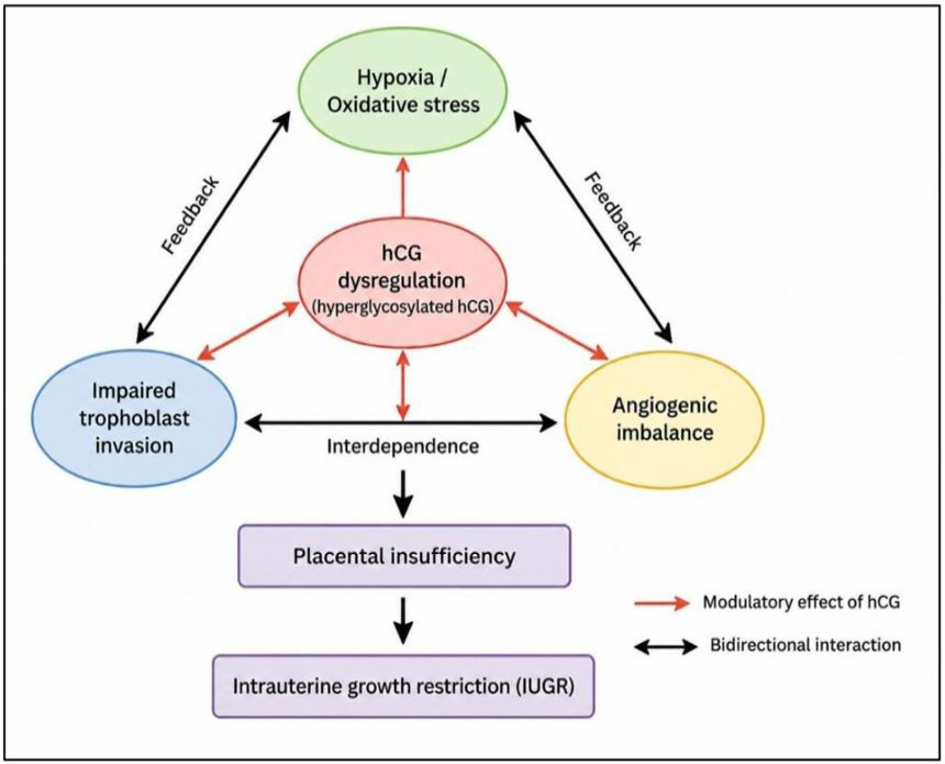
Integrated network of placental dysfunction in intrauterine growth restriction (IUGR). Impaired trophoblast invasion, hypoxia, and angiogenic imbalance interact in a bidirectional manner, with hCG dysregulation acting as a central modulator. These interconnected processes collectively contribute to placental insufficiency and fetal growth restriction
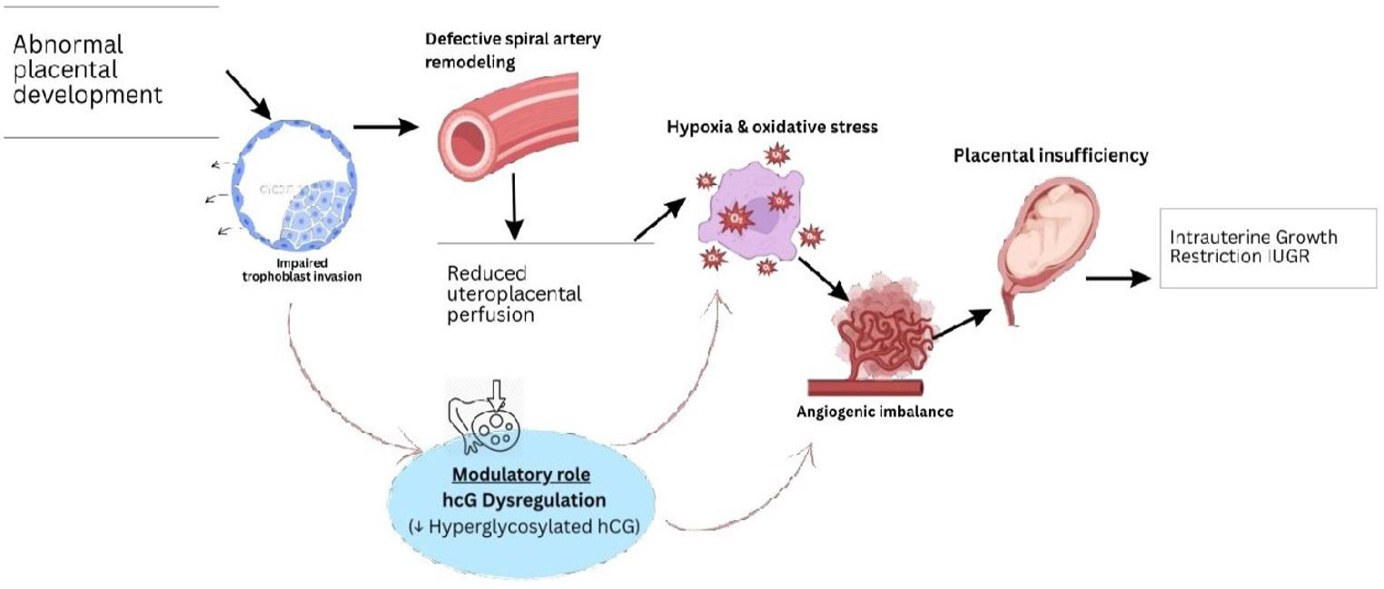
Mechanistic model of placental dysfunction and the modulatory role of hCG dysregulation in IUGR.
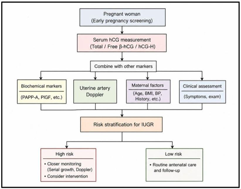
Clinical integration of hCG in multimarker screening for intrauterine growth restriction (IUGR). Maternal serum hCG, when combined with biochemical markers, Doppler assessment, and maternal factors, contributes to risk stratification and guides clinical management.
References
- Barjaktarovic, M., Korevaar, T.I., Jaddoe, V.W., de Rijke, Y.B., Visser, T.J., Peeters, R.P. and Steegers, E.A., 2017. Human chorionic gonadotropin (hCG) concentrations during the late first trimester are associated with fetal growth in a fetal sex-specific manner. Eur. J. Epidemiol., 32(2): 135-144. https://doi.org/10.1007/s10654-016-0201-3[CrossRef]
- Baschat, A.A., 2004. Pathophysiology of fetal growth restriction: implications for diagnosis and surveillance. Obstet. Gynecol. Surv., 59(8): 617-627. https://doi.org/10.1097/01.OGX.0000133943.54530.76[CrossRef]
- Beune, I.M., Damhuis, S.E., Ganzevoort, W., Hutchinson, J.C., Khong, T.Y., Mooney, E.E. and Gordijn, S.J., 2021. Consensus definition of fetal growth restriction in intrauterine fetal death: A Delphi procedure. Arch. Pathol. Lab. Med., 145(4): 428-436. https://doi.org/10.5858/arpa.2020-0027-OA[CrossRef]
- Birken, S., Kovalevskaya, G. and O’Connor, J., 2001. Immunochemical measurement of early pregnancy isoforms of HCG: Potential applications to fertility research, prenatal diagnosis, and cancer. Arch. Med. Res., 32(6): 635-643. https://doi.org/10.1016/S0188-4409(01)00329-0[CrossRef]
- Bischof, P. and Campana, A., 2000. Molecular mediators of implantation. Best Pract. Res. Clin. Obstet. Gynaecol., 14(5): 801-814. https://doi.org/10.1053/beog.2000.0120[CrossRef]
- Butler, S.A., Luttoo, J., Freire, M.O., Abban, T.K., Borrelli, P.T. and Iles, R.K., 2013. Human chorionic gonadotropin (hCG) in the secretome of cultured embryos: hyperglycosylated hCG and hCG-free beta subunit are potential markers for infertility management and treatment. Reprod. Sci., 20(9): 1038-1045. https://doi.org/10.1177/1933719112472739[CrossRef]
- Ceballos, M.A., Gómez-Acebo, I., Gallego L.C.C., Alonso-Molero, J., Vilares, C.S., Odriozola, F.J.M. and Dierssen-Sotos, T., 2025. Study of first-trimester serum levels of β-hCG and PAPP-A as a screening test for fetal development of intrauterine growth restriction. BMC Pregn. Childb., 25(1): 655. https://doi.org/10.1186/s12884-025-07787-7[CrossRef]
- Cole, L.A., 2007. Hyperglycosylated hcg. Placenta, 28(10): 977-986. https://doi.org/10.1016/j.placenta.2007.01.011[CrossRef]
- Cole, L.A., 2010. Biological functions of hCG and hCG-related molecules. Reprod. Biol. Endocrinol., 8(1): 102. https://doi.org/10.1186/1477-7827-8-102[CrossRef]
- Conde-Agudelo, A., Papageorghiou, A., Kennedy, S. and Villar, J., 2013. Novel biomarkers for predicting intrauterine growth restriction: A systematic review and meta-analysis. BJOG Int. J. Obstet. Gynaecol., 120(6): 681-694. https://doi.org/10.1111/1471-0528.12172[CrossRef]
- Cuckle, H., 2014. Prenatal screening using maternal markers. J. Clin. Med., 3(2): 504-520. https://doi.org/10.3390/jcm3020504[CrossRef]
- d’Hauterive, S.P., Close, R., Gridelet, V., Mawet, M., Nisolle, M. and Geenen, V., 2022. Human chorionic gonadotropin and early embryogenesis. Int. J. Mol. Sci., 23(3): 1380. https://doi.org/10.3390/ijms23031380[CrossRef]
- Dugoff, L., 2010. First-and second-trimester maternal serum markers for aneuploidy and adverse obstetric outcomes. Obstet. Gynecol., 115(5): 1052-1061. https://doi.org/10.1097/AOG.0b013e3181da93da[CrossRef]
- Dugoff, L., Hobbins, J.C., Malone, F.D., Porter, T.F., Luthy, D., Comstock, C.H. and Craigo, S.D., 2004. First-trimester maternal serum PAPP-A and free-beta subunit human chorionic gonadotropin concentrations and nuchal translucency are associated with obstetric complications: A population-based screening study (the FASTER Trial). Am. J. Obstet. Gynecol., 191(4): 1446-1451. https://doi.org/10.1016/j.ajog.2004.06.052[CrossRef]
- Evans, J., 2016. Hyperglycosylated hCG: A unique human implantation and invasion factor. Am. J. Reprod. Immunol., 75(3): 333-340. https://doi.org/10.1111/aji.12459[CrossRef]
- Figueras, F. and Gardosi, J., 2011. Intrauterine growth restriction: New concepts in antenatal surveillance, diagnosis, and management. Am. J. Obstet. Gynecol., 204(4): 288-300. https://doi.org/10.1016/j.ajog.2010.08.055[CrossRef]
- Goetzinger, K.R., Singla, A., Gerkowicz, S., Dicke, J.M., Gray, D.L. and Odibo, A.O., 2009. The efficiency of first-trimester serum analytes and maternal characteristics in predicting fetal growth disorders. Am. J. Obstet. Gynecol., 201(4): 412. e411-412. e416. https://doi.org/10.1016/j.ajog.2009.07.016[CrossRef]
- Herghelegiu, C.G., Veduta, A., Stefan, M.F., Magda, S.L., Ionascu, I., Radoi, V.E. and Calin, A.M., 2023. Hyperglycosylated-hCG: Its role in trophoblast invasion and intrauterine growth restriction. Cells, 12(12): 1647. https://doi.org/10.3390/cells12121647[CrossRef]
- Hu, X.Q. and Zhang, L., 2021. Hypoxia and mitochondrial dysfunction in pregnancy complications. Antioxidants, 10(3): 405. https://doi.org/10.3390/antiox10030405[CrossRef]
- Huang, J., Liu, Y., Yang, H., Xu, Y. and Lv, W., 2022. The effect of serum β-human chorionic gonadotropin on pregnancy complications and adverse pregnancy outcomes: A systematic review and meta-analysis. Comput. Mathemat. Methods Med., 2022(1): 8315519. https://doi.org/10.1155/2022/8315519[CrossRef]
- Huppertz, B., 2018. The critical role of abnormal trophoblast development in the etiology of preeclampsia. Curr. Pharma. Biotechnol., 19(10): 771-780. https://doi.org/10.2174/1389201019666180427110547[CrossRef]
- Kabodmehri, R., Ghanami, G.N., Zahiri, S.Z., Sharami, S.H., Milani, F., Hasanpour, M. and Rafiei, S.Z., 2024. Levels of serum β-human chorionic gonadotropin after embryo transfer and subsequent miscarriage, pre-eclampsia, and intrauterine growth restriction. Health Sci. Rep., 7(4): e2015. https://doi.org/10.1002/hsr2.2015[CrossRef]
- Kalkunte, S.S., Mselle, T.F., Norris, W.E., Wira, C.R., Sentman, C.L. and Sharma, S., 2009. Vascular endothelial growth factor C facilitates immune tolerance and endovascular activity of human uterine NK cells at the maternal-fetal interface. J. Immunol., 182(7): 4085-4092. https://doi.org/10.4049/jimmunol.0803769[CrossRef]
- Korevaar, T.I., Steegers, E.A., de Rijke, Y.B., Schalekamp-Timmermans, S., Visser, W.E., Hofman, A. and Medici, M., 2015. Reference ranges and determinants of total hCG levels during pregnancy: The generation R study. Eur. J. Epidemiol., 30(9): 1057-1066. https://doi.org/10.1007/s10654-015-0039-0[CrossRef]
- Kovalevskaya, G., Kakuma, T., Schlatterer, J. and O’connor, J., 2007. Hyperglycosylated HCG expression in pregnancy: Cellular origin and clinical applications. Mol. Cell. Endocrinol., 260: 237-243. https://doi.org/10.1016/j.mce.2006.02.021[CrossRef]
- Lee, S.U., Choi, S.K., Jo, Y.S., Wie, J.H., Shin, J.E., Kim, Y.H. and Ko, H.S., 2024. Prediction model of late fetal growth restriction with machine learning algorithms. Life, 14(11): 1521. https://doi.org/10.3390/life14111521[CrossRef]
- Li, Y., Yan, J., Chang, H.M., Chen, Z.J. and Leung, P.C., 2021. Roles of TGF-β superfamily proteins in extravillous trophoblast invasion. Trends Endocrinol. Metab., 32(3): 170-189. https://doi.org/10.1016/j.tem.2020.12.005[CrossRef]
- Licht, P., Russu, V. and Wildt, L., 2001. On the role of human chorionic gonadotropin (hCG) in the embryo-endometrial microenvironment: implications for differentiation and implantation. Semin. Reprod. Med., (Vol and Pages?). https://doi.org/10.1055/s-2001-13909[CrossRef]
- Lin, Z., Wu, S., Jiang, Y., Chen, Z., Huang, X., Wen, Z. and Yuan, Y., 2024. Unraveling the molecular mechanisms driving enhanced invasion capability of extravillous trophoblast cells: A comprehensive review. J. Assist. Reprod. Genet., 41(3): 591-608. https://doi.org/10.1007/s10815-024-03036-6[CrossRef]
- Liu, L., Tang, L., Chen, S., Zheng, L. and Ma, X., 2024. Decoding the molecular pathways governing trophoblast migration and placental development; A literature review. Front. Endocrinol., 15: 1486608. https://doi.org/10.3389/fendo.2024.1486608[CrossRef]
- Majewska, M., Lipka, A., Paukszto, L., Jastrzebski, J.P., Szeszko, K., Gowkielewicz, M. and Majewski, M.K., 2019. Placenta transcriptome profiling in intrauterine growth restriction (IUGR). Int. J. Mol. Sci., 20(6): 1510. https://doi.org/10.3390/ijms20061510[CrossRef]
- Melamed, N., Baschat, A., Yinon, Y., Athanasiadis, A., Mecacci, F., Figueras, F. and McIntyre, H.D., 2021. FIGO (international federation of gynecology and obstetrics) initiative on fetal growth: Best practice advice for screening, diagnosis, and management of fetal growth restriction. Int. J. Gynaecol. Obstet., 152(Suppl 1): 3. https://doi.org/10.1002/ijgo.13522[CrossRef]
- Morris, R.K., Johnstone, E., Lees, C., Morton, V., Smith, G., Obstetricians, T.R.C.O. and Gynaecologists, 2024. Investigation and care of a small-for-gestational-age fetus and a growth restricted fetus (green-top guideline No. 31). BJOG: Int. J. Obstet. Gynaecol., 131(9): e31-e80. https://doi.org/10.1111/1471-0528.17814[CrossRef]
- Ong, C.Y., Liao, A.W., Cacho, A.M., Spencer, K. and Nicolaides, K.H., 2001. First-trimester maternal serum levels of placenta growth factor as predictor of preeclampsia and fetal growth restriction. Obstet. Gynecol., 98(4): 608-611. https://doi.org/10.1097/00006250-200110000-00015[CrossRef]
- Park, C., Alahari, S., Ausman, J., Liu, R., Nguyen, F., Sallais, J. and Caniggia, I., 2025. Placental hypoxia-induced ferroptosis drives vascular damage in preeclampsia. Circ. Res., 136(4): 361-378. https://doi.org/10.1161/CIRCRESAHA.124.325119[CrossRef]
- Pijnenborg, R. and Brosens, I., 2010. Deep trophoblast invasion and spiral artery remodelling. Cambridge University Press Cambridge. https://doi.org/10.1017/CBO9780511750847.012[CrossRef]
- Prast, J., Saleh, L., Husslein, H., Sonderegger, S., Helmer, H. and Knöfler, M., 2008. Human chorionic gonadotropin stimulates trophoblast invasion through extracellularly regulated kinase and AKT signaling. Endocrinology, 149(3): 979-987. https://doi.org/10.1210/en.2007-1282[CrossRef]
- Schiessl, B., Innes, B., Bulmer, J., Otun, H., Chadwick, T., Robson, S. and Lash, G., 2009. Localization of angiogenic growth factors and their receptors in the human placental bed throughout normal human pregnancy. Placenta, 30(1): 79-87. https://doi.org/10.1016/j.placenta.2008.10.004[CrossRef]
- Sharony, R., Sharon-Weiner, M., Kidron, D., Sukenik-Halevy, R., Biron-Shental, T., Manor, M. and Maymon, R., 2018. The association between maternal serum first trimester free βhCG, second trimester intact hCG levels and foetal growth restriction and preeclampsia. J. Obstet. Gynaecol., 38(3): 363-366. https://doi.org/10.1080/01443615.2017.1340441[CrossRef]
- Sovio, U., Gaccioli, F., Cook, E., Charnock-Jones, D.S. and Smith, G.C., 2021. Slowing of fetal growth and elevated maternal serum sFLT1: PlGF are associated with early term spontaneous labor. Am. J. Obstet. Gynecol., 225(5): 520. e521-520. e510. https://doi.org/10.1016/j.ajog.2021.04.232[CrossRef]
- Spencer, R., Maksym, K., Hecher, K., Maršál, K., Figueras, F., Ambler, G. and Hansson, S.R., 2023. Maternal PlGF and umbilical Dopplers predict pregnancy outcomes at diagnosis of early-onset fetal growth restriction. J. Clin. Invest., 133(18). https://doi.org/10.1172/JCI169199[CrossRef]
- Staun-Ram, E., Goldman, S., Gabarin, D. and Shalev, E., 2004. Expression and importance of matrix metalloproteinase 2 and 9 (MMP-2 and -9) in human trophoblast invasion. Reprod. Biol. Endocrinol., 2(1): 59. https://doi.org/10.1186/1477-7827-2-59[CrossRef]
- Stenman, U.H. and Alfthan, H., 2013. Determination of human chorionic gonadotropin. Best Pract. Res. Clin. Endocrinol. Metab., 27(6): 783-793. https://doi.org/10.1016/j.beem.2013.10.005[CrossRef]
- Stenman, U.H., Tiitinen, A., Alfthan, H. and Valmu, L., 2006. The classification, functions and clinical use of different isoforms of HCG. Hum. Reprod. Update, 12(6): 769-784. https://doi.org/10.1093/humupd/dml029[CrossRef]
- Sultana, Z., Qiao, Y., Maiti, K. and Smith, R., 2023. Involvement of oxidative stress in placental dysfunction, the pathophysiology of fetal death and pregnancy disorders. Reproduction, 166(2): R25-R38. https://doi.org/10.1530/REP-22-0278[CrossRef]
- Taeidi, E., Ranjbar, A., Montazeri, F., Mehrnoush, V. and Darsareh, F., 2023. Machine learning-based approach to predict intrauterine growth restriction. Cureus, 15(7): e41448. https://doi.org/10.7759/cureus.41448[CrossRef]
- Umapathy, A., Clark, A., Sehgal, A., Karanam, V., Rajaraman, G., Kalionis, B. and Murthi, P., 2024. Molecular regulators of defective placental and cardiovascular development in fetal growth restriction. Clin. Sci., 138(13): 761-775. https://doi.org/10.1042/CS20220428[CrossRef]
- Wald, N., Densem, J., Smith, D. and Klee, G., 1994. Four-marker serum screening for Down’s syndrome. Prenat. Diagn., 14(8): 707-716. https://doi.org/10.1002/pd.1970140810[CrossRef]
- Wen, Q., Zhang, R., Zhu, Y., Ling, Y. and Xiong, D., 2026. Diagnostic accuracy of initial serum β-hCG in predicting pregnancy outcomes post-SET in IVF/ICSI cycles: A systematic review and meta-analysis. Front. Endocrinol., 17: 1636981. https://doi.org/10.3389/fendo.2026.1636981[CrossRef]
- Yaron, Y., Ochshorn, Y., Heifetz, S., Lehavi, O., Sapir, Y. and Orr-Urtreger, A., 2002. First trimester maternal serum free human chorionic gonadotropin as a predictor of adverse pregnancy outcome. Fetal Diagn. Therapy, 17(6): 352-356. https://doi.org/10.1159/000065384[CrossRef]
- Yefet, E., Kuzmin, O., Schwartz, N., Basson, F. and Nachum, Z., 2017. Predictive value of second-trimester biomarkers and maternal features for adverse pregnancy outcomes. Fetal Diagn. Therapy, 42(4): 285-293. https://doi.org/10.1159/000458409[CrossRef]
- Zhang, X., Huangfu, Z., Shi, F. and Xiao, Z., 2021. Predictive performance of serum β-hCG MoM levels for preeclampsia screening: A meta-analysis. Front. Endocrinol., 12: 619530. https://doi.org/10.3389/fendo.2021.619530[CrossRef]
- Zhou, H., Zhao, C., Wang, P., Yang, W., Zhu, H. and Zhang, S., 2023. Regulators involved in trophoblast syncytialization in the placenta of intrauterine growth restriction. Front. Endocrinol., 14: 1107182. https://doi.org/10.3389/fendo.2023.1107182[CrossRef]
Services
Article usage statistics
Loading…