
Superovulation techniques in dogs are not well established because of the unique reproductive physiology of this species and the paucity of information on factors regulating postovulatory oocyte maturation. With the objective of rapidly obtaining mature oocytes in dogs regardless of their estrous period, we injected three healthy and anestrus females (~5 kg of weight each) with pregnant mare serum gonadotropin (PMSG) and human chorionic gonadotropin (hCG). From days 1 to 8 of the experiment, they received continuous PMSG intramuscular injections at a dosage of 200 IU/day. On day 12, they received an intramuscular injection of hCG at a dosage of 2000 IU. Then, on day 23, they underwent surgery for the collection of oocytes. The three females showed estrus symptoms and ovulation. The mean luteal number and follicular number in the ovaries were 21.7 and 7, respectively. An average of 21 oocytes were collected, and oocyte collection rate was 96.9%, including 4 at the germinal vesicle stage, 2.3 at metaphase I, and 14.7 at metaphase II. The mean oocyte maturation rate was 69.8%. This study is the first to show that multiple repeat administrations of PMSG and hCG can induce superovulation in female dogs, and it provides basic data for future studies on the reproductive physiology of dogs.
Research Article · Volume 58, Issue 3, 31 March 2026 · Open Access
Superovulation Induced by Multiple-Dose Administration of Pregnant Mare Serum Gonadotropin in Bitches
· ·
Affiliations & Notes ⌄
- 1Jilin Provincial Key Laboratory of Transgenic Animal and Embryo Engineering, Department of Animal Science, Yanbian University, Yanji, Jilin, 133002, China. 2College of Veterinary Science, The University of Agriculture, Peshawar, Pakistan. * Corresponding author: nkhanbuneri@gmail.com
Article Info ⌄
- Journal:
- Pakistan Journal of Zoology
- Published:
- 31 March 2026

From the article
- We injected the dogs with 2000 IU of hCG on day 11 to simulate the LH peak and ovulation.
- In summary, dogs can obtain a large number of high-quality mature oocytes by means of superovulation method; thus, this is an effective tool to understand the unique reproductive characteristics of dogs.
- In this study, multiple repeat administrations of PMSG were, for the first time, used to induce superovulation in dogs.
- This study provides basic data for future studies on the reproductive physiology of dogs.
Selected from the article's own text.
Abstract
Introduction
Superovulation is the most common technique used in assisted reproduction, and it is important for improving the efficiency of oocyte and animal production and reducing the number of oocyte donors . Canine reproductive physiology has unique characteristics that make extrapolation from farm animals (horses, cows, sheep, goats, pigs) unsuccessful in this species . Domestic bitches are non-seasonal monoestrus; they ovulate only once or twice per year with a few exceptions (Fuller). In most mammals, oocyte maturation occurs within the ovarian follicle, and mature oocytes are ovulated and fertilized within the oviduct. However, canine oocytes are ovulated at an immature state that requires a further 48 to 72 h for completion of meiosis within the oviduct ; . Because of this unique reproductive physiological characteristic and the paucity of information concerning factors regulating postovulatory oocyte maturation, superovulation techniques for dogs are not well established .
Canine embryos are scarce biological material, because of the inefficiency of superovulation and cycle induction/synchronization protocols . Difficulties encountered in collecting in vivo produced embryos and current the impossibility to produce canine embryos in vitro are other limiting factors. Currently, in vitro oocytes surgically obtained from female dogs in natural rut . However, as dogs are monoesters, and have estrous cycle of ~6 months, information on the duration of estrus is usually missing . Following natural estrus, without superovulation, an average of only six to eight good-quality embryos can be collected from each female dog . For livestock, such as cattle , pigs and sheep , the superovulation method is well established. However, because the reproductive biology of dogs is unique, the superovulation method is not yet well established for this species . Consequently, the availability of canine oocytes and knowledge on the reproductive physiology of bitches remain severely limited.
In this study, our aim is to collect mature oocytes by continuous injection of PMSG to superovulation the bitches. These findings may contribute to basic technologies such as in vitro maturation, in-vitro fertilization, and somatic cell nuclear transfer that are used in studies on reproductive aspects of dogs.
Materials and Methods
Animals and management
Three mixed-breed bitches aged 1.5 years and weighing 5 kg in anestrus were used in the study. They were provided by the Animal Hospital of The College of Agriculture, Yanbian University. The bitches were confirmed to be no abnormity of the reproductive system by ultrasonography and no symptom of estrus by vaginal smears and vulvar observation. They were fed standard commercial dog food once a day and given water ad libitum. Through vaginal cytological examination and vulvar observation, it was confirmed that the bitch was in a state of anestrus.
Design of superovulation protocols and estrus observation
All bitches were administered 200 IU pregnant mare serum gonadotropin (PMSG, Deasung Microbiological Labs. Co, Ltd, Seoul, South Korea) intramuscularly for 8 consecutive days, followed by a 3-day interval. Then on the 12th day, they received intramuscular injection of human chorionic gonadotropin (hCG; Deasung Microbiological Labs. Co, Ltd, Seoul, South Korea) at a dosage of 2000 IU (Fig. 1). During the entire trial, all animals were examined vaginal cytology and ultrasonic testing every day. After laparotomy, the dogs were sent back to the Animal Hospital. None of the dogs were used repeatedly. The first day of treatment was considered as day 1 of the experiment.
Estrus detection
After the superovulation treatment, the preoestrus and estrus stages, as well as the expected ovulation period, are determined by sexual behavior records, vaginal cytology and related evaluation criteria are based on those by Jurczak et al. (2016). Observe the normal ovary in anestrus and the follicles and corpora lutea by .
Laparotomy and oocyte collection
The dogs were fasted for 1 day before the experiment. All surgeries were performed under general anesthesia. Atropine sulfate (0.05 mg/kg; ShengDa, China) and acepromazine maleate (0.025 mg/kg; ShengDa, China) were administered as a pretreatment, and ketamine (5 mg/kg; ShengDa, China) was administered to induce anesthesia. Anesthesia was maintained using isoflurane. The laparotomy was performed on the 23rd day of the experiment.
An approximately 5 cm incision was made at 2 cm of the midline above the dogs umbilicus. The skin and subcutaneous tissue were cut to expose the abdominal cavity. A bending hemostatic forceps was used to extend the incision backward along the abdominal wall to 2 to-3 cm from the kidney, hook the uterine angle on one side, pull toward the incision, find and remove the ovarian suspension ligament, and then remove the ovary from the abdominal cavity. The same procedure was performed on the other ovary. The removed uterus was quickly placed in a vacuum flask and taken to the laboratory. Finally, the abdominal incision was closed using a two-layer closure followed by a surgical adhesive applied along the skin incision. Standard post-operative care of the bitches was performed by veterinarians.
After removal of the uterus, the entire bursal compartment around an ovary, including a small portion of the uterine horn immediately distal to the uterotubal junction, was removed. the excised part was transferred to a Petri dish on a warming pad and kept moist. Bursal window was identified, then made an opening in this region large enough to invert the bursal sac without cutting into the oviduct running through. corpora lutea and follicles on each ovary were recorded. oviduct was isolated from bursa. The infusion needle is linked to the fallopian tube. A hemostatic forceps can be used to place the fallopian tube over the needle. Flush the fallopian tube with a syringe containing 5 ml, and oocytes were collected and observed at the count under microscope.
Evaluation of oocyte maturation
Evaluation of oocyte maturation of the collected oocytes was performed under stereomicroscope. Cumulus-Oocyte Complexes (COCs) were denuded with 0.1% hyaluronidase by gentle pipetting and they were observed without fixing and mounting and oocytes with nucleus and extruded first polar body (metaphase II) was counted and look under fluorescence microscope. The remaining oocytes denuded were fixed in 4% formaldehyde solution. For assessing in vitro maturation, oocytes were stained with 5μg/mL Hoechst 33342 for 10 min. The stained oocytes were mounted on a glass slide with a 100% of glycerol drops. Chromatin state and position as well as spindle formation of oocytes were evaluated under UV light to determine the stage of meiosis as follows : germinal vesicle (GV) stage: condensed chromatin, metaphase I (MI) stage: chromosomes were compact in a metaphase plate and migrating to the poles, metaphase II (MII) stage: the extrusion of the first polar body. The oocytes were classified as good, fair, poor, aged, or immature on the basis of both morphological evaluation and the stage of nuclear development.
Statistical analysis
The corpus luteum number, oocyte number, GV stage number, MI stage number, MII stage number and oocyte maturation rate of left and right ovaries were counted.
Results
The estrus detection of bitch after PMSG and hCG superovulation treatment
The estrus detection of bitch is shown in Figures 2-3. All dogs were in estrus after superovulation and had obvious estrus symptoms. Our results showed that 4-7 days after first intramuscular injection of PMSG, female dogs went from anestrus (Fig. 2A) to proestrus. In proestrus dogs appear restless, eating less and drinking more. The vulva turned redness and swelling with bloody discharge and palpation of hard vulva (Fig. 2B). The vaginal smears were characterized by a mixture of red blood cells, parabasal and intermediate cells and only a small number of superficial cells (<30%) (Fig. 3B), on the 10th day of the experiment, the follicles were clearly observed by ultrasonography (Fig. 3E). In estrus the bitch showed a strong desire to mate by showing a static reaction. The vulva becomes soft and vaginal secretions change from blood-red to colorless transparent or yellowish (Fig. 2C) and an increase in the percentage of superficial cells (70–90%) was detected in estrus (Fig. 3C). On the 18th day of the experiment, the corpus luteum formation in the ovary down to the ultrasonography (Fig. 3F) in the metestrus. The swelling of the female vulva gradually subsided and almost no secretion flowed out in metestrus (Fig. 2D).
Figure 4 shows the in vitro matured canine oocytes collected by flushing of fallopian tubes.
Table I. Evaluating the quality of ovulation on bitch after pregnant mare serum gonadotropin (PMSG) and human chorionic gonadotropin (hCG) superovulation treatment.
| Ovary | No. of follicles | No. of oocyte (collection rate%) | No. of GV | No. of MI | No. of MII (mature rate (%) | ||
| 1 | L | 11 | 7 | 11 (100.0) | 1 | 2 | 8 (72.7%) |
| R | 12 | 8 | 13 (108.3) | 1 | 1 | 11 (84.6) | |
| 2 | L | 12 | 6 | 14 (116.7) | 2 | 0 | 12 (85.7) |
| R | 10 | 0 | 6 (60.0) | 0 | 0 | 6 (100) | |
| 3 | L | 12 | 0 | 12 (100.0) | 6 | 2 | 4 (33.3) |
| R | 8 | 0 | 7 (87.5) | 2 | 2 | 3 (42.9) | |
| 21.7 | 7 | 21 (96.9) | 4 | 2.3 | 14.7 (69.8) |
GV, germinalvesicle; MI, metaphase I; MII, metaphase II.
Evaluating quality of in vivo matured oocytes collected by flushing fallopian tubes after PMSG and hCG superovulation treatment
In this study, all bitches were in estrus state and showed ovulation. Number of corpus luteum, follicle and oocytes of 3 female dogs were counted in left and right ovary, respectively (Table I). All bitches produced corpus luteum, whereas only 2 bitches produced follicles. Moreover, all of the left and right ovaries of bitch produced oocytes (Fig. 5)
Evaluation of oocyte maturation of the collected oocytes from three bitch were performed under stereomicroscope (Table I). The mean luteal number and follicular number in the ovary were observed to be 21.7 and 7, respectively. An average of 21 oocytes were collected, with collection rate of 96.9%, including 4 at GV stage, 2.3 at MI stage and 14.7 at MII stage. The mean oocyte maturation rate was 69.8%.
Discussion
The development of reproductive biotechnologies involving oocyte and embryo manipulation has advanced for many domestic species. These technologies include the in vitro production of embryos, embryo transfer, cloning, and transgenesis. However, the efficiency of IVM production in this species is very low, reproductive technologies for dogs generally require the use of in vivo-produced oocytes, at present. Although gonadotropins have been shown to be highly effective at inducing proestrus in bitches in some cases, in many other cases the ovulation rate was poor, pregnancy rates were not reported, and whelping rates were variable . Female dogs are monoestrus with an average estrous cycle of 6 months, and only 6 to 12 oocytes are produced in each cycle ; ; . This in vivo production of mature oocytes is performed by collecting oocytes through flushing of the genital tract of a female after natural estrus or superovulation. Although the superovulation method is already well established for cattle , pig , and sheep , it is not yet completely developed for dogs, as this species is known to have a unique reproductive biology .
The continuous low-dose (200 IU) PMSG intramuscular injection administered from days 1 to 8 and the high-dose (2000 IU) hCG intramuscular injection on day 12 induced superovulation. Our results showed that all dogs had marked estrus symptoms and ovulation, which is consistent with the results of Wright (1980). These estrus symptoms are consistent with the characteristics of normal estrus in dogs. Previous studies have shown that the estrus cycle is closely related to hormonal changes in the body. Follicle-stimulating hormone (FSH) is critical for ovarian folliculogenesis and essential for female fertility . Although estradiol concentration were not measured based upon the hemotological profiles, it appears that the multiple-dose of 200IU PMSG was sufficient to induce follicular growth . In all bitches, the pre-ovulatory LH surge was accompanied by a pre-ovulatory FSH surge. We found that multiple-dose administration of pregnant mare serum gonadotropin in dogs facilitates follicular formation and we hypothesis that LH surge.
In dogs, ovulation is assumed to occur approximately 2 to 3 days after the preovulatory luteinizing hormone (LH) surge ; ; , with ovulation being spread over 24 and 36 h ; . We injected the dogs with 2000 IU of hCG on day 11 to simulate the LH peak and ovulation. Corpora lutea were observed in the ovaries on day 18. Canine oviducts support the long-term survival of oocytes that undergo complete maturation, fertilization (in the oviductal isthmus), and development into the blastocyst stage. Therefore, canine oocytes may remain in the oviduct for 8.5 to 9 days . Moreover, Lee et al. (2017) report that canine oviduct cell were beneficial to oocyte maturation. And that the use of hCG to induce ovulation in estrus induction protocols in bitches is quite controversial because they are spontaneous ovulators . In other spontaneously ovulating species (e.g., horses and cattle), hCG and GnRH are regularly used to time ovulation for appointed AI ; . Therefore, the inclusion of hCG in estrus induction protocols in the bitch seems justified for timing ovulation rather than for induction of ovulation ; . In our perliminary experiments, Oocytes collected surgically on day 22 are usually immature oocytes (Fig. 2A). Oocytes collected surgically on day 24 are usually aging oocytes (Fig. 2B). Thus, the oocytes were surgically harvested on day 23.
In this study, our aim was to collect mature oocytes by continuous injection of PMSG to superovulate the bitches. There is no direct correlation between oocyte maturation and ovulation timing. In our preliminary experiments, we identified the optimal time for obtaining mature oocytes during surgical procedures. Consequently, progesterone concentrations were not measured based on hematological profiles to determine the timing of ovulation.
The increase in oocyte number and maturation rate is beneficial to the development of the dog’s reproductive system. We hypothesized that long-term storage of oocytes in fallopian tubes is beneficial for oocyte maturation. We found that the mean luteal number and follicular number in the ovary were 21.7 and 7, respectively. An average of 21 oocytes was collected, and the oocyte collection rate was 96.9%, including 4 at the GV stage, 2.3 at MI, and 14.7 at MII. The mean oocyte maturation rate was 69.8%. Reynaud et al. (2005) reported that only 4% of ovulating dogs may be multi-oocyte, with two to five oocytes per follicle. this explains the lower number of corpora lutea than of oocytes in the dogs ovaries.
In summary, dogs can obtain a large number of high-quality mature oocytes by means of superovulation method; thus, this is an effective tool to understand the unique reproductive characteristics of dogs.
Conclusion
In this study, multiple repeat administrations of PMSG were, for the first time, used to induce superovulation in dogs. All dogs showed marked estrus and ovulation. The average oocyte number was 21. The mean oocyte maturation rate was 69.8%. This study provides basic data for future studies on the reproductive physiology of dogs.
Declarations
Funding
IRB approval
This study was approved from Institutional Review Board of Yanbian University, Yanji, Jilin, China (IRB 20130310).
Ethics statement
The animal experimental procedures of this study were approved by the Committee on the Ethics of Animal Experiments at Yanbian University (Approval ID: 20130310). All experiments were performed in accordance with relevant guidelines and regulations of ‘the instructive notions with respect to caring for laboratory animals’ issued by the Ministry of Science and Technology of the People’s Republic of China.
Statement of conflict of interest
The authors have declared no conflict of interest.
Figures

Protocol for inducing superovulation in dogs; PMSG- pregnant mare serum gonadotropin; hCG- Human chorionic gonadotropin.
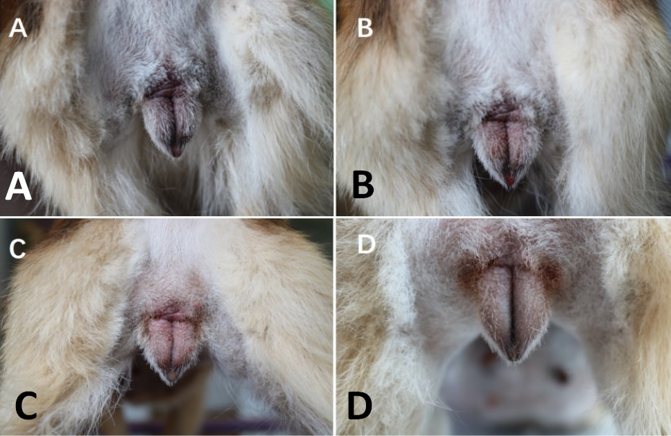
Changes in dog’s vulva. A, anestrus (day 1 to 5); B, proestrus (day 6 to 14); C, estrus (day15 to 21); D, metestrus (day 22).
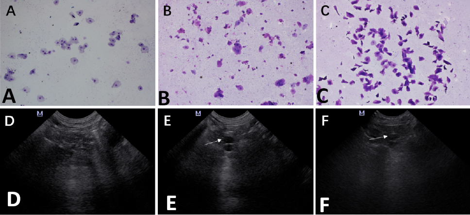
Vaginal smear (A to C) and ovary ultrasound images (D to F) of a bitch in different estrus stages. A, anestrus; B, proestrus; C, estrus; D, left ovary (day 1); E, follicle of the left ovary (day 10); F, corpus luteal of the left ovary (day 18).
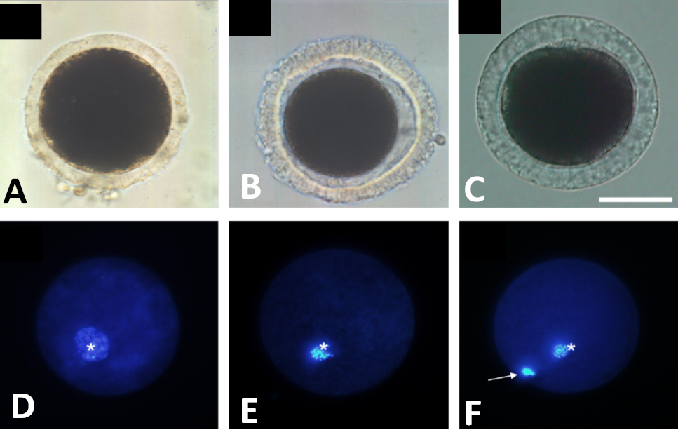
Classification of in vivo matured canine oocytes collected by flushing off allopian tubes by laparotomy (nuclear status). Three different nuclear photograph taken under fluorescence microscope (A-C) and Hoechst 33342 staining of oocytes at different stages under ultraviolet light (D-F). A, immature; B, aging; C, mature; D, GV (germinal vesicle); E, MI (metaphase I); F, MII (metaphase II); arrow indicates the first polar body, * indicates the nucleus. scale bar = 50μm.
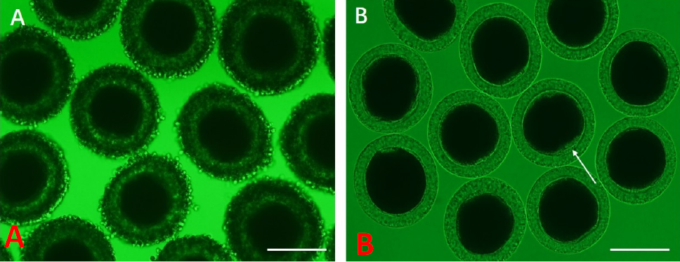
Oocytes obtained by superovulation. A, cumulus-oocyte complexes (COCs) obtained by flushing the fallopian tubes (A and B). B, Oocytes denuded cumulus cells treated by 0.1% hyaluronidase. The arrow indicates the polar body. Magnification (× 200). Scale bar = 100μm.
References
- Allen, W., 2008. Attempted oestrus induction in four bitches using pregnant mare serum gonadotrophin. J. Small Anim. Pract., 23: 223-231. https://doi.org/10.1111/j.1748-5827.1982.tb01661.x[CrossRef]
- Angel, M.A., Gil, M.A., Cuello, C., Sanchez-Osorio, J., Gomis, J., Parrilla, I., Vila, J., Colina, I., Diaz, M., Reixach, J., Vazquez, J.L., Vazquez, J.M., Roca, J., Martinez, E.A., 2014. The effects of superovulation of donor sows on ovarian response and embryo development after nonsurgical deep-uterine embryo transfer. Theriogenology, 81: 832-839. https://doi.org/10.1016/j.theriogenology.2013.12.017[CrossRef]
- Antonov, A. and Georgiev, P., 2016. Induction of estrus in bitches with the dopamine agonist bromocriptine. Int. J. Adv. Res., 3: 962-965.
- Bó, G.A. and Mapletoft, R.J., 2014. Historical perspectives and recent research on superovulation in cattle. Theriogenology, 81: 38-48. https://doi.org/10.1016/j.theriogenology.2013.09.020[CrossRef]
- Boyd, J.S., Renton, J.P., Harvey, M.J., Nickson, D.A., Eckersall, P.D. and Ferguson, J.M., 1993. Problems associated with ultrasonography of the canine ovary around the time of ovulation. J. Reprod. Fertil., Suppl., 47: 101-105.
- Chastant-Maillard, S., Viaris de Lesegno, C., Chebrout, M., Thoumire, S., Meylheuc, T., Fontbonne, A., Chodkiewicz, M., Saint-Dizier, M. and Reynaud, K., 2011. The canine oocyte: Uncommon features of in vivo and in vitro maturation. Reprod. Fertil. Dev., 23: 391-402. https://doi.org/10.1071/RD10064[CrossRef]
- Chastant-Maillard, S., Chebrout, M., Thoumire, S., Saint-Dizier, M., Chodkiewicz, M. and Reynaud, K., 2010. Embryo biotechnology in the dog: A review. Reprod. Fertil. Dev., 22: 1049-1056. https://doi.org/10.1071/RD09270[CrossRef]
- Concannon, P., Hansel, W. and McEntee, K., 1977. Changes in LH, progesterone and sexual behavior associated with preovulatory luteinization in the bitch. Biol. Reprod., 17: 604-613. https://doi.org/10.1095/biolreprod17.4.604[CrossRef]
- de Avila Rodrigues, B. and Rodrigues, J.L., 2003. Influence of reproductive status on in vitro oocyte maturation in dogs. Theriogenology, 60: 59-66. https://doi.org/10.1016/S0093-691X(02)01301-8[CrossRef]
- de Gier, J., Kooistra, H.S., Djajadiningrat-Laanen, S.C., Dieleman, S.J. and Okkens, A.C., 2006. Temporal relations between plasma concentrations of luteinizing hormone, follicle-stimulating hormone, estradiol-17beta, progesterone, prolactin, and alpha-melanocyte-stimulating hormone during the follicular, ovulatory, and early luteal phase in the bitch. Theriogenology, 65: 1346-1359. https://doi.org/10.1016/j.theriogenology.2005.08.010[CrossRef]
- England, G.C. and Yeager, A.E., 1993. Ultrasonographic appearance of the ovary and uterus of the bitch during oestrus, ovulation and early pregnancy. J. Reprod. Fertil. Suppl., 47: 107-117.
- Evans, M.J., Gastal, E.L., Silva, L.A., Gastal, M.O., Kitson, N.E., Alexander, S.L. and Irvine, C., 2006. Plasma LH concentrations after administration of human chorionic gonadotropin to estrous mares. Anim. Reprod. Sci., 94: 191-194. https://doi.org/10.1016/j.anireprosci.2006.03.029[CrossRef]
- Figueira, L.M., Alves, N.G., Maia, A.L.R.E.S., Souza-Fabjan, J.M.G.d., Batista, R.I.T.P., Morais, M.C.C., Lima, R.R., Oliveira, M.E.F. and Fonseca, J.F., 2020. Embryo yield and quality are associated with progestogen treatment during superovulation protocol in lactating Lacaune ewes. Theriogenology, 155: 132-138. https://doi.org/10.1016/j.theriogenology.2020.06.004[CrossRef]
- Fuller, J.L., 2001. Photoperiodic control of estrus in the basenji. J. Hered., 4: 1-8.
- Hirata, M., Tanihara, F., Taniguchi, M., Takagi, M., Terazono, T. and Otoi, T., 2018. Follicular development of canine ovaries stimulated by a combination treatment of eCG and hCG. Vet. Med. Sci., 4: 333-340. https://doi.org/10.1002/vms3.121[CrossRef]
- Holst, P.A. and Phemister, R.D., 1971. The prenatal development of the dog: Preimplantation events. Biol. Reprod., 5: 194-206. https://doi.org/10.1093/biolreprod/5.2.194[CrossRef]
- Jurczak, A., Domosławska, A., Bukowska, B. and Janowski, T., 2016. Equine chorionic gonadotropin and human chorionic gonadotropin stimulation increase the number of luteinized follicles and the progesterone level compared with cabergoline stimulation in anoestrus bitches. Reprod. Domest. Anim., 51: 562-568. https://doi.org/10.1111/rda.12720[CrossRef]
- Kumar, T.R., 2018. Extragonadal actions of FSH: A critical need for novel genetic models. Endocrinology, 159: 2-8. https://doi.org/10.1210/en.2017-03118[CrossRef]
- Kutzler, M.A., 2010. Effect of estrus induction on pregnancy rates in domestic bitches and queens. Clin. Theriogenol., 2: 191-207.
- Kutzler, M.A., 2018. Estrous cycle manipulation in dogs. Vet. Clin. N. Am. Small Anim. Pract., 48: 581-594. https://doi.org/10.1016/j.cvsm.2018.02.006[CrossRef]
- Lee, B.C., Kim, M.K., Jang, G., Oh, H.J., Yuda, F., Kim, H.J., Kang, S.K., Schatten, G. and Hwang, W.S., 2005. Dogs cloned from adult somatic cells. Nature, 436: 641. https://doi.org/10.1038/436641a[CrossRef]
- Lee, S.H., Oh, H.J., Kim, M.J., Kim, G.A., Choi, Y.B., Jo, Y.K., Setyawan, E.M.N. and Lee, B.C., 2017. Oocyte maturation-related gene expression in the canine oviduct, cumulus cells, and oocytes and effect of co-culture with oviduct cells on in vitro maturation of oocytes. J. Assist. Reprod. Genet., 34: 1-10. https://doi.org/10.1007/s10815-017-0910-x[CrossRef]
- Nakao, T., Aoto, Y., Fukushima, S., Moriyoshi, M. and Kawata, K., 1985. Induction of estrus in bitches with exogenous gonadotropins, and pregnancy rate and blood progesterone profiles. Nihon Juigaku Zasshi, 47: 17-24. https://doi.org/10.1292/jvms1939.47.17[CrossRef]
- Phemister, R.D., Holst, P.A., Spano, J.S. and Hopwood, M.L., 1973. Time of ovulation in the beagle bitch. Biol. Reprod., 8: 74-82. https://doi.org/10.1093/biolreprod/8.1.74[CrossRef]
- Rensis, F.D., López-Gatius, F., García-Ispierto, I. and Techakumpu, M.J.T., 2010. Clinical use of human chorionic gonadotropin in dairy cows: An update. Theriogenology, 73: 1001-1008. https://doi.org/10.1016/j.theriogenology.2009.11.027[CrossRef]
- Reynaud, K., Fontbonne, A., Saint-Dizier, M., Thoumire, S., Marnier, C., Tahir, M.Z., Meylheuc, T. and Chastant-Maillard, S., 2012. Folliculogenesis, ovulation and endocrine control of oocytes and embryos in the dog. Reprod. Domest. Anim., 47 (Suppl 6): 66-69. https://doi.org/10.1111/rda.12055[CrossRef]
- Reynaud, K., Fontbonne, A., Marseloo, N., Viaris de Lesegno, C., Saint-Dizier, M. and Chastant-Maillard, S., 2006. In vivo canine oocyte maturation, fertilization and early embryogenesis: A review. Theriogenology, 66: 1685-1693. https://doi.org/10.1016/j.theriogenology.2006.01.049[CrossRef]
- Reynaud, K., Fontbonne, A., Marseloo, N., Thoumire, S., Chebrout, M., de Lesegno, C.V. and Chastant-Maillard, S., 2005. In vivo meiotic resumption, fertilization and early embryonic development in the bitch. Reproduction (Cambridge, England) 130: 193-201. https://doi.org/10.1530/rep.1.00500[CrossRef]
- Rodrigues, B.A. and Rodrigues, J.L., 2010. In vitro maturation of canine oocytes: A unique conundrum. Anim. Reprod., 7: 1-13
- Stornelli, M.C., Mitacek, M.C.G., Giménez, F., Bonaura, M.C., Dorna, I.V., de la Sota, R.L. and Stornelli, M.A., 2012. Pharmacokinetics of eCG and induction of fertile estrus in bitches using eCG followed by hCG. Theriogenology, 78: 1056-1064. https://doi.org/10.1016/j.theriogenology.2012.04.012[CrossRef]
- Takeo, T., Mukunoki, A. and Nakagata, N., 2019. Ovulation of juvenile, mature, and aged female C57BL/6 mice following coadministration of inhibin antiserum and equine chorionic gonadotropin. Theriogenology, 135: 1-6. https://doi.org/10.1016/j.theriogenology.2019.05.028[CrossRef]
- Tsutsui, T., 1975. Studies on the reproduction in the dog: VI. Ovulation rate and transuterine migration of the fertilized ova. Jpn. J. Anim. Reprod., 21. https://doi.org/10.1262/jrd1955.21.98[CrossRef]
- Wildt, D.E., Chakraborty, P.K., Panko, W.B. and Seager, S.W., 1978. Relationship of reproductive behavior, serum luteinizing hormone and time of ovulation in the bitch. Biol. Reprod., 18: 561-570. https://doi.org/10.1095/biolreprod18.4.561[CrossRef]
- Wright, P.J., 1980. The induction of oestrus and ovulation in the bitch using pregnant mare serum gonadotrophin and human chorionic gonadotrophin. Austral. Vet. J., 56: 137-140. https://doi.org/10.1111/j.1751-0813.1980.tb05650.x[CrossRef]
Services
Article usage statistics
Loading…