
Preeclampsia (PE) is a pregnancy associated complication mainly demonstrated by the onset hypertension, proteinuria, foetal growth restriction, and maternal organ dysfunction including renal, cerebral and hepatic diseases before or after 34 weeks of gestation. As a cause of prenatal and maternal morbidity and mortality, it remains a major health concern to females worldwide. In this context, we aimed to provide an accumulative account of recent developments about the risk factors, pathophysiology, consequences and management of PE. Several genetic, acquired and environmental risk factors have been associated with the progression of PE. Pre-existing hypertension, chronic renal disease, family history, obesity, multi-foetal pregnancies and history of miscarriage are main risk factors. The disease is initiated by abnormal placentation, incomplete remodelling of spiral artery, utero-placental hypoperfusion leading to placental hypoxia and ischemia. Many molecules of foetal origin are released in to circulation and result in the dysfunction of maternal endothelium which is the hallmark of PE. The disease also has postpartum impacts on maternal and foetal health, the main consequences include cardiovascular complications, hypertension, diabetes, neurodevelopmental issues and dementia to the mother and offsprings. In the recent past, the revelation of antiangiogenic molecules and pathways has provided critical information about the pathogenesis of PE. Several molecules such as sFLT1 (soluble fms-like tyrosine kinase 1), Eng (soluble endoglin), angiotensin II, PIGF (placental growth factor), and VEGF (vascular endothelial growth factor) are regulated differently during the pathogenesis of PE. These molecules not only offer the candidate biomarkers for early disease detection but also provide therapeutic targets. Many surveillance and management strategies reported in the recent literature have also been included in the present review. Studies have emphasized that PE has a long-term threat to the health of mother and child. Therefore, the development of interventional strategies for the prevention and management of postpartum cardiovascular complications poses a challenge.
Review · Volume 1, Issue 1, 22 January 2026 · Open Access
Epidemiology-Pathogenesis-Management-of-Preeclam
· · · · · · ·
Affiliations & Notes ⌄
- 1Department of Biochemistry, Faculty of Science, King Abdulaziz University Jeddah 21589, Saudi Arabia. 2School of Biological Sciences, University of the Punjab, Lahore,cPakistan. 3Department of Zoology, Abbottabad University of Science and Technology, Abbottabad 22310, Pakistan. 4Institute of Molecular Biology and Biotechnology, University of Lahore, Lahore, Pakistan. 5Department of Gynaecology, Ayub Medical College, Abbottabad, Pakistan 6Department of Pharmacy, COMSATS University, Abbottabad, Pa
Article Info ⌄
- Journal:
- BIOMEDICUS: Journal of Biological and Biomedical Frontiers
- Published:
- 22 January 2026

From the article
- Preeclampsia, a pregnancy associated complication has serious health threat to the mother and child worldwide.
- There are several genetic and environmental risk factors associated with the disease.
- Disturbance in the balance between proangiogenic and antiangiogenic factors is the hallmark of PE and these factors provide biomarkers for the early detection of disease.
- The disease is not restricted to the pregnancy periods, it has severe postpartum consequences including CVDs, hypertension, neurological and developmental problems for the mother and child.
Selected from the article's own text.
Abstract
Introduction
Pregnancy-induced hypertension (PIH) is characterized as systolic and diastolic blood pressures after 20 weeks of gestation that includes gestational hypertension (GH) and preeclampsia (PE)1,2. According to estimates, more than 10% of pregnant women are affected by hypertension, it may be pre-existing chronic hypertension or pregnancy induced gestational hypertension. The chronic and gestational hypertension may be differentiated on the basis of their timings i.e. the chronic hypertension is a systolic and diastolic blood pressure above 140 mmHg and 90 mmHg before pregnancy or before 20 weeks and persists after pregnancy, GH is the hypertension that starts due to physiological changes after 20 weeks of pregnancy3-5. In some cases, hypertension is accompanied by increased levels of proteins in the urine known as Preeclampsia (PE). According to the definition by the International Society for the Study of Hypertension in Pregnancy (ISSHP), preeclampsia a condition characterized by a combination of gestational hypertension with organ damage and proteinuria at 20 weeks of gestation6-8.
Preeclampsia affects up to 5% pregnancies worldwide9,10 resulting in the death of 0.5 million infants and 76,000 women during pregnancy, causalities are many times higher in the developing countries9. More than 300 mg of total urine proteins per 24 h and a random urine protein to creatinine ratio above 0.30 are used to diagnose preeclampsia11. In 2013, proteinuria was excluded by the American College of Obstetricians and Gynaecologists as an essential criterion for the diagnosis of PE, now the GH with end-organ dysfunction is considered sufficient in diagnosis12. At present, the aetiology of PE is not properly understood, several risk factors have been associated with the onset of condition. The serum levels of uric acid in pregnant women also indicates the chances of preeclampsia13. Anaemia during pregnancy, history of GH, and previous second trimester abortion are associated with increased incidence of preeclampsia14. Preeclampsia is induced by many maternal, placental, foetal factors, genetic, and environmental factors15-18. The hypertension associated complications increase with the number of foetuses, singletons were found 6.5%, twins 12.7%, and triplets were found to have 20% chances of complications19, females with twins have shown 14% and those with triplets have 30% chances of preeclampsia20,21. Mothers with stage-1 hypertension have an increased chances of GH and PE22. Many mother associated risk factors include insulin resistance, abnormal lipid metabolism, obesity, family history, and late age during pregnancy23,24.
Pathogenicity of PE has been reported as a two stage process. The blastocyst implantation into the endometrium leads to its growth into an outer trophectoderm an inner cell mass that develop outer villi and embryo respectively. Placenta is developed from the outer villi and gets attached to the mother’s uterus resulting in the remodelling of spiral artery. Problems in the invasion of trophoblast to the spiral arteries result in the uteroplacental malperfusion and vascular dysfunction (stage 1)25. Ischaemia-reperfusion in the placental tissue initiates the production and of reactive oxygen species (ROS), antiangiogenic factors, and cytokines and their subsequent release into the mother’s circulatory system resulting in the systemic inflammation and endothelial dysfunction (stage 2)26,27. All the above hypertensive disorders enhance the risk of cardiovascular and renal complications during life after pregnancy28,29. GH and PE also promote the insulin resistance by increase in the ROS production via NF-kB-mediated pathway30. The low birth weight, premature delivery or mortality, and cardiac complication in infants are also associated with GH and PE31,32. Administration of aspirin can significantly (35% to 66%) reduce the chances of PE development in women with high risk of disease. Therefore, aspirin is recommended internationally to reduce the chances of PE33-35. Once the disease is diagnosed, delivery remains the ultimate solution for PE36,37. The recent developments in aetiology, health impacts and management of GH and PE are the areas of concern for the gynaecologists in particular and for the physicians in general. In this context, the present review article was aimed to accumulative account of recent developments in GH and PE.
RISK FACTORS AND EPIDEMIOLOGY
Concept of GH and PE as two different manifestations of the same disease or separate entities with similar symptoms is not very clear. In many PE patients, hypertension occurs before proteinuria on the other hand, GH does not always progress to PE38. Studies on the risk factors of GH and PE can improve the understanding of aetiological mechanisms and treatment options of these diseases39-41. According to the hypothesis considering PE as a syndromic condition, GH and PE being the progressive stages of the same disorder, the common risk factors can suggest common aspects of aetiology42. Many risk factors of PE are based on the environment, drug applications, and genetics of mother and foetus. Genetics of PE is complicated, more than 178 potentially associated genes have been described. However, the mechanism of its inheritance is not clearly understood43. Some of the prominent risk factors have been tabulated (Tables I, II).
Some other risk factors include maternal smoking habits, age at gestation, number of pregnancies, pre-gestation obesity, diabetes, hypertension, chronic kidney disease, multifetal pregnancies, trisomy 13, family history, and foetal genetics44,45. A great deal of heterogeneity is found in the epidemiology, representative clinical characteristics, and morbidity of preeclampsia. The condition can be established after 20 weeks or after 34 weeks of gestation46,47. Restriction of intrauterine growth and maternal obesity lead to early and late onset of disease. Clinically, the both types of preeclampsia are represented differently but the pattern of gene expression is similar in both cases indicating a common mechanism of maternalvascular damage83. Some of the above-mentioned proteins including PAPP-A, PlGF, sFlt, PP13, and sEng are used as diagnostic markers for PE.
Table I. Gestational hypertension and PE associated genetic risk factors (genes, genetic polymorphism).
| Description of risk factors | References | |
| Candidate gene (Gene product) | Physiological role | |
| Stox1 (Storkhead box 1) | Coagulation, trophoblast development, hypoxia, inflammation | 48 |
| Foxd1 (Forkhead box D1) | Development, inflammation, immune system | 49,50 |
| Nlrp (Nod-like receptor protein family) | Immune system, reproductive system | 51 |
| Trim28 (Tripartite motif containing 28) | Transcription inhibition, histone modification | 52,53 |
| Epas1 (Hypoxia-inducible factor 2-alpha (hif-2α)) | Regulation of physiology at different oxygen levels | 54 |
| Nos3 (Nitric oxide synthase 3) | Production of nitric oxide, blood circulation control | 55 |
| Oxgr1 (2-oxoglutarate receptor 1) | Transcription regulation | 56 |
| Igfbp1 (Insulin like growth factor binding protein 1) | Binds both insulin-like growth factors to enhance its half life | 57 |
| Mthfr (Methylen etetrahydrofolate reductase) | Re-methylation of Hcy to methionine | 58-60 |
| Agt1/agt2 (Angiotensinogen) | Regulation of blood pressure | 61, 62 |
| Agtr1 (Angiotensin II receptor type 1) | Regulation of blood pressure, salts and fluids | 63, 64 |
| TNF-α (Tumour necrosis factor-alpha) | Multifunctional proinflammatory cytokine | 65,66 |
| Rgs2 (Regulator of G protein signaling 2) | Regulates G protein-coupled receptor ignalling cascades | 67,68 |
| Vdr (Vitamin D receptor) | Cellular response to vitamin D | 69,70 |
| Ctla-4 (Cytotoxic t-lymphocyte antigen-4) | Immune response | 71 |
| Tgfb1 (Transforming growth factor beta 1) | Immunoregulation | 72 |
| Lep (Leptin) | Hormone | 73,74 |
| Papp-a (Pregnancy-associated plasma protein A) | Regulates the insulin growth factor in pregnancy | 75-78 |
| Plgf (Placental growth factor) | Plays important role in vasculogenesis and angiogenesis | 79,80 |
| sflt (Fms-like tyrosine kinase 1) | Circulating antiangiogenic protein prepared by placenta | 81,82 |
| Pp13 (Placental protein 13 produced by trophoblasts) | Vasodilation and vascularization during placenta formation | 83-85 |
| seng (Soluble endoglin) | Angiogenesis | 86-87 |
Pathogenesis
Although a considerable progress has been made during the last two decades yet the pathophysiology of PE is not clearly understood. The structural components and biochemical substances and processes contribute to the pathogenesis of PE. As for example, structure and physiology of placenta play a vital role in the pathogenesis of PE84. Placenta a link between the circulatory systems of mother and foetus is a vascular organ consisting of capillaries with about 550 km in length and 15 m2 surface area90. The extensive vasculogenesis process required for the normal physiology of placenta is influenced by proangiogenic and antiangiogenic factors. The concentration of circulating placental growth factor (PIGF) increases that interacts with a membrane anchored receptor known as fms-like tyrosine kinase 1 resulting in the activation of endothelial cells and initiation of angiogenesis. During the 30 weeks of pregnancy, highest levels of circulatory PIGF are found that helps in the maturation of vessels between uterus and placenta91. On the other hand, PIGF also interacts with a soluble fms-like tyrosine kinase 1 (sFLT1) has antiangiogenic properties. It reduces the availability of PIGF to the anchored receptor, reduces nitric oxide levels, promotes the effects of proinflammatory cytokines with endothelial cells consequently resulting in the vasoconstriction92,93. Hence, a balances of proangiogenic and antiangiogenic factors is necessary for the normal placental development and physiology. According to some reports the women suffering from PE have low concentrations of PIGF and higher concentrations of soluble fms-like tyrosine kinase 1 that is contrary to those with normal pregnancies94,95.
Table II. Mother and foetus associated risk factors.
| Risk factor | Function | References |
| Use of selective serotonin reuptake inhibitors (ssris) (Used as antidepressants, mood control, depression control) | Directly interact the transporter protein and inhibit the reuptake of serotonin / Reportedly, promote GH and PE | 96,97 |
| Assisted reproductive technology (ART) modern technology applied to treat infertility including IVF (in vitro fertilization), ICSI (intracytoplasmic sperm injection) etc. | Increased the chances of GH and PE | 98 |
| Elevated plasma uric acid levels (produced by the breakdown of purine nucleotides. Meat, dried beans are rich sources of purines) | Promotes GH and PE | 13 |
| Threatened miscarriage (Signs and symptoms like lower abdominal pain, little vaginal bleeding or spotting) | Enhanced the chances of GH and PE | 99 |
| High BMI (Body mass index) of mother (Higher BMI is also known as obesity) | High BMI at gestation time increases the chances of GH and PE | 100 |
| High consumption of soft drinks (carbonated drinks) | Excessive consumption of soft drinks during pregnancy increase the chances of GH and PE | 101 |
| Oocyte donated pregnancies (In case of IVF, if the female have weak egg, it can be donated by some healthy woman) | Pregnancies based on donated eggs have been associated with an increased chances of GH and PE | 102 |
| Obstructive sleep apnea (Blockage of air pathways by the relaxation of throat muscles and disturbance in breathing that results in snoring or obstruction in the sleep). | Reportedly increases GH and PE | 103 |
| Dysbiosis of gut microbiota (The disturbance in the normal (healthy) ecology of gut microbes) | Enhance the chances of GH and PE | 104,105 |
| Oxidative stress (Effect of reactive oxygen species (ROS)) | Promotes the onset of GH and PE | 106 |
| Pregnancies with twins (The pregnancies with two or more foetuses) | Such pregnancies have 2 to 3 times increased risk of GH and PE development | 107 |
| COVID-19 infection (COVID-19 infection during pregnancy increases the levels of pro-inflammatory cytokines) | Chances of GH and Pe are enhanced in if pregnant women suffer from COVID-19 | 108 |
Structurally, placenta consists of many different types of cells that constitute its internal and external architecture. The inner cells that differentiate in to embryo include pericytes, endothelial cells, and smooth muscle cells. The syncytial outer layer consists of trophoblasts with an ability to invade the wall of uterine and differentiate in to the outer layer of foetus and placenta. These cells forming a thick layer are known as syncytiotrophoblasts. The surface of villi are covered by the syncytiotrophoblasts which brings these cells in direct contact with the maternal circulation 109,110. Preeclampsia is a human specific disease, not found in other mammalian species. This due to the high brain to body ratio of human foetus. During the third trimester of pregnancy human foetus requires up to 60% nutritional exchange from mother’s circulation whereas in other mammalian species the nutritional exchange demand is about 20%111. Therefore, the normal development and physiology of placenta is necessary to meet the nutritional requirements of foetus112. Pathogenesis of PE is a multi-step process mainly initiated by the abnormal placentation. Uterine arteries divide into several branches that provide blood to decidua (blastocyst attachment area), myometrium or open as spiral arteries in the intravillous space. Invasion of defective trophoblasts into the spiral arteries cause injuries to the walls of blood vessels and result in the incomplete subsequent remodelling. This invasion may occur long before the detection of pregnancy and long before the symptoms of the disease are apparent 115-117. Under the normal conditions the decidualization process is prolonged to the most of the areas of endometrium and increase the chances of blood supply to the foetus by remodelling of spiral arteries. However, the molecular mechanism involved in the remodelling of spiral arteries is not clearly known,. A series of experiments involving the analysis of placental biopsy samples has shown that the spiral arteries from preeclamptic samples were significantly narrow (200 µm) as compared to those from the normal samples (500 µm). This may result in the high velocity blood flow and uterine hypoperfusion in preeclampsia-.
The abnormal placentation and failure to the remodelling of spiral arteries are central to the PE pathogenesis. Studies on the analyses of placental metabolic profiles during the first trimester have revealed that energy needs remain unaffected. Even under hypoxia, the trophoblast manage the routine proliferation by increasing the levels of HIF1α (hypoxia-inducible factor 1α), a transcription factor that also regulates the expression of nitric oxide (NO) synthase and vascular endothelial growth factor (VEGF)123,124. Normally, the level of HIFα increases at early weeks of pregnancy and then decreases. A continuously elevated level of HIFα indicates the chances of PE development125. The two stage model for the establishment of PE is supported by preclinical and clinical studies126,127. First stage is represented by incomplete remodelling of spiral artery resulting in ischemic placenta and hypoxic conditions, in the second stage ischemic placenta releases antiangiogenic factors into the maternal circulation with subsequent endothelial dysfunction (Fig. 1).
According to some studies, during spiral artery remodelling, the syncytiotrophoblast cells undergo apoptotic conditions and release a large number of syncytiotrophoblast-derived extracellular vesicles (SDEVs) into the blood circulation of mother. The level of SDEVs increase in the normal mother and becomes maximum during the third trimester. Its highest level can be found much earlier in case of PE due to hypoxic and ischemic placenta-e. A big (10% to 250%) difference of plasma SDEVs levels can be found in normal pregnancies and during PE. The difference in biological activities of these vesicles in case of normal and PE cases has been suggested128,129. In fact, the SDEVs produced during PE are strong promoters of inflammation, coagulation, endothelial damage, anti-angiogenic and vesoconstritive conditions as compared to those produced during normal pregnancies130-132. These processes not only enhance the injuries to the endothelial cells but also damage the placental vasculature and vessels of vital organs133.
MOTHER AND FOETUS ASSOCIATED POSTPARTUM OUTCOMES OF PREECLAMPSIA
According to the growing evidence, preeclampsia is not only a pregnancy disease but the patients suffering from PE have higher risk of negative health outcomes. According to the American Heart Association the mothers with PE have a long term risk of cardiovascular diseases (CVDs), and it should be considered during CVD risk evaluation134-136. According to meta-analysis the mothers with PE history have up to 4.2 times, 2.5 times, 1.8 times, and 6.7 times increased risk of heart failure, coronary artery disease, stroke, and hypertension respectively. According to similar reports such women have a double risk for thromboembolism and 4 times risk of diabetes mellitus in future137,138. The mothers with a history of PE have 3.8 times higher risk of periphery artery disease (PAD)139, more than 30% of them suffer from asymptomatic atherosclerosis140. The studies have reported increased risk of left ventricular hypertrophy or dysfunction among the mothers with PE141-142. However, the risk of all the above conditions is variable among the women from different ethnic and geographic groups143. The individuals born after PE influenced pregnancy remain at high risk of stroke and hypertension at their adult age144,. A study conducted on 45000 individuals born to preeclampsic pregnancy has demonstrated 2.39 mmHg and 1.35 mmHg increase systolic and diastolic blood pressures, respectively. These findings suggest 8% increased risk of ischemic heart disease and 12% increased chances of stroke. The cases when the mothers had early onset of preeclampsia, the children had up to 6 mmHg increase in the overall systolic blood pressure, these individuals have a specific hypertensive vascular structure. Offsprings born after GH and PE pregnancy have problems in the cardiac structure and function. The vascular remodelling demonstrates an increased risk of neuronal damage, especially the white matter and dementia-. An inverse relationship between the severity of GH, PE and development of foetus has been reported.
The women with preeclampsia have high risk of diabetes postpartum. The chances of diabetes onset increases up to 2 times in the presence or absence of GE153,154. Such women are prone to microalbuminuria and potential kidney disease155, the risk of end stage renal disease (ESRD) was reportedly increased up to 15.5% in case of three PE pregnancies156. Histopathological studies have shown focal segmental glomerulosclerosis (FSGS) associated with PE157-159. Preeclampsia associated oxidative stress, dysregulation of immune system, and growth factor has been reported to increase the risk of neurodevelopmental problems160. Studies have been conducted to associate the risk of migraine in case of PE161.
ASSESSMENT AND MANAGEMENT OF PREECLAMPSIA
The efficient management of PE depends on three main factors, disease prevention measures, availability of methods for early diagnosis and targeted procedures for the treatment. Gynaecologists can evaluate women for the risk factors, the high-risk individuals with pre-existing hypertension, antiphospholipid syndrome or renal disease should be advised according to established guidelines162,163. Women with suspected disease onset may be subjected to placental growth factor, sFlt-1/PlGF ratio, and proteinuria evaluation164-167. Plasma levels of sEng and sFlt1are significantly high among the women with PE as compared to those with normal pregnancy, the elevated levels can be used to determine the severity of PE168-172. Starting before gestation to 36 weeks post gestation a moderately high dose of calcium (above 1g/day) can reduce the risk of PE173-175. Once PE is diagnosed, regular and continuous surveillance is recommended by urine, blood pressure test, accurate anomaly and dating scans along with the assessment of foetal growth at every 4 weeks intervals. In case of severe indications of disease plasma electrolytes, liver, kidney function tests, umbilical artery Doppler test, regular measurement of respiration rate, heart rate and blood pressure are recommended after short intervals. Medicine can be prescribed or therapeutic strategies may be adopted by the physicians after the consideration of symptoms and intensity.
In case of mild PE, the experts are recommended to prescribe fluorinated steroids before 34 weeks of gestation to promote the maturation of foetus. In case of severe PE before 34 weeks of gestation termination of pregnancy is recommended after 34 weeks of gestation immediate delivery remains the only solution176. According to studies, the delayed delivery in severe PE cases can result in pulmonary edema, severe hypertension, HELLP syndrome, and liver damage to the mother. It can also cause abnormal heart rate, growth retardation, prenatal death, reverse end-diastolic flow in the umbilical artery that can be determined by Doppler ultrasound, and abruptio placentae to the foetus177. Several therapeutic strategies have also been recommended to prolong safe pregnancy in case of PE, some of these strategies are being discussed here:
Antihypertensive medication is aimed to prevent hypertensive encephalopathy, intracranial haemorrhage, and pulmonary edema. There is a universal consensus that there should not be an abrupt decrease in the blood pressure to maintain utero-placental blood flow. A 10 to 20 mm of Hg decrease in 10 to 20 min is recommended by some studies178,179. The medicines can be prescribed by a qualified physician, there is no clear advantage of one medicine on the other. According to recommendations labetalol and calcium channel blockers can be used as a first line drug against hypertension. According to some other reports the use of ketanserin, nitroprusside, chlorpromazine, and diazoxide should be avoided. MgSO4 has shown no significant effect on severe hypertension180,181. The use of antihypertensive medicines during pregnancy with PE can reduce the risk of stroke but have shown hazardous impacts to the mother182,183. According to a study involving 2200 individuals, the chances of PE are reduced up to 50% if a low dose aspirin is used from 12-16 weeks before gestation to 36 weeks of gestation173,184-186. In another clinical study 1776 pregnant females diagnosed with low PIGF levels were enrolled, a treatment with 150 mg of aspirin per day reduced the onset of PE up to 62% as compared to placebo. On the basis of these results, low dose of aspirin is recommended to the females at high risk of PE187-189. Non-specific antioxidants have not shown significant effect in prevention of PE. The studies on the sources of oxidative stress are required to define the therapeutic targets190-192.
Statins have been widely used for the activation of vascular function by stimulating the expression of haem oxygenase (HO), nitric oxide synthase (NO) and reducing the decrease in sFLT1 by placenta193,194. These medicines have shown promising results to prevent PE in pre-clinical studies195,196. Similar results have been reported in clinical studies by using pravastatin197-199. Pre-clinical studies have been conducted by using the ligands of sFLT1 such as recombinant VEGF121for the therapy against PE in rats. The results have shown an inhibition of hypertension and amelioration against renal disease without any adverse effects to the foetus200,201. Preclinical studies conducted on rodents and primates, recombinant PIGF another ligand of sFLT1 exhibited promising results against PE and hypertension. PIGF is highly specific for sFLT1 and dose not bind with sFLT2 and has no adverse effects202-204. Pre-clinical studies have shown that a pregnancy associated protein known as relaxin has potential therapeutic effects against PE induced hypertension and improves utero-placental perfusion in rats205. Apheresis is a procedure used in medical sciences in which the blood from a person is passed through an equipment to separate a specific ingredient and the remaining blood is returned to the person’s circulation. It provides an attractive option to remove antiangiogenic proteins from the blood of pregnant mothers who are at high risk of PE206. Studies have been conducted to remove LDL and sFLT1from the blood of patients that give significant positive effects207,208. The studies on the application of small molecules in the PE therapy have also been reported. Placental hypoxia plays a vital role in the pathogenesis of PE, the production of hypoxia induced factors i.e. HIF1, HIF2, and sFLT1 are inhibited by ouabain and digoxin resulting in the reduction of hypertension in rats with ischemic placenta209. Metformin is a drug that improves the sensitivity to insulin and used to treat diabetes type 2, it has been beneficial in the management of PE210-212. Small interfering RNA (siRNA) that are used to inhibit the synthesis of specific cellular proteins. The specific siRNA has been used to target sFLT1 in cell lines213.
Conclusions
Preeclampsia, a pregnancy associated complication has serious health threat to the mother and child worldwide. The characteristic signs and symptoms of disease include onset of hypertension at before or after 34 weeks of pregnancy, release of proteins in the urine, and subsequent damage to the kidneys, liver and cerebral part of brain. There are several genetic and environmental risk factors associated with the disease. The pathophysiology of disease is not completely understood. However, a two-stage mechanism of pathogenesis is well established. According to this mechanism PE is triggered by structural and functional problems in the placenta, production of defective trophoblasts, failure in proper remodelling of spirl artery, problems in the blood flow between mother and the foetus. These conditions result in the ischemic placenta, hypoxic conditions, regulation of expression of several genes including those coding for proangiogenic and antiangiogenic factors. The consequence of all these changes from normal pregnancy conditions is the onset of PE. Disturbance in the balance between proangiogenic and antiangiogenic factors is the hallmark of PE and these factors provide biomarkers for the early detection of disease. The disease is not restricted to the pregnancy periods, it has severe postpartum consequences including CVDs, hypertension, neurological and developmental problems for the mother and child. In case of severe PE, delivery is the only treatment. However, under minor to mild conditions aspirin, antihypertensive medicines and other biochemical therapies can be adopted. Restoration of angiogenic balance during the future pregnancies by administration of proangiogenic factors, removal of antiangiogenic factors can provide a useful measure to prevent PE. However, the development of strategies against postpartum outcomes for the child and mother need extensive research.
Declarations
Authors’ contribution
Conceptualization: MSN, BNM, IK. Writing original draft preparation: MSN, IU. Writing review and editing, critical revision: MNB, SJG, MMG, SIA, AU, SA.
All authors have read and agreed to the published version of the manuscript.
Funding
The study received no external funding.
Statement of conflict of interest
The authors have declared no conflict of interest.
Generative AI and AI-assisted technology statement
The authors declare that they have not used generative AI or AI-assisted technologies in this manuscript.
Publisher’s note
All claims expressed in this article are solely those of the authors and do not necessarily represent those of their affiliated organizations, and those of publishers, the editor and the reviewers. Any product that may be evaluated in this article, or claim that may be made by this manufacturer, is not gaurenteed or endorsed by the publisher.
Figures
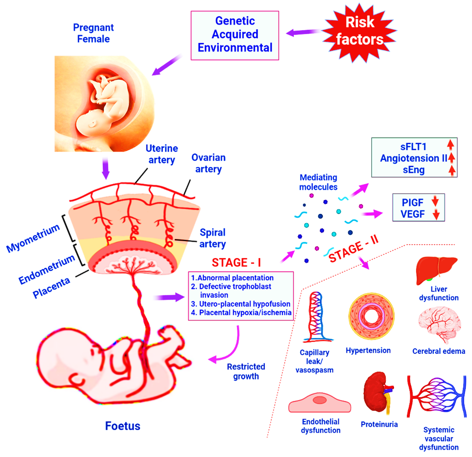
Schematic representation of stage I and stage II of preeclampsia pathogenesis.
References
- 1. Gestational Hypertension and Preeclampsia, 2020. ACOG practice bulletin summary, number 222. Obs. Gynecol. 135: 1492–1495. https://doi.org/10.1097/AOG.0000000000003892[CrossRef]
- 2. Poon, L.C., Shennan, A., Hyett, J.A., Kapur, A., Hadar, E., Divakar, H., McAuliffe, F., da Silva Costa, F., von Dadelszen, P., McIntyre, H.D., Kihara, A.B., Di Renzo, G.D. Romero, R., D’Alton, M., Berghella, V., Nicolaides, K.H. and Hod, M., 2019. The international federation of gynecology and obstetrics (FIGO) initiative on pre-eclampsia: A pragmatic guide for first-trimester screening and prevention. Int. J. Gynaecol. Obs. 145(Suppl. 1): 1–33. https://doi.org/10.1002/ijgo.12802[CrossRef]
- 3. Wiles, K., Damodaram, M. and Frise, C., 2021. Severe hypertension in pregnancy. Clin. Med., 21: e451. https://doi.org/10.7861/clinmed.2021-0508[CrossRef]
- 4. Fikadu, K., G/Meskel, F., Getahun, F., Chufamo, N. and Misiker, D., 2020. Family history of chronic illness, preterm gestational age and smoking exposure before pregnancy increases the probability of preeclampsia in Omo district in Southern Ethiopia: A case-control study. Clin. Hypert., 26: 16. https://doi.org/10.1186/s40885-020-00149-9[CrossRef]
- 5. Benschop, L., Duvekot, J.J. and Roeters van Lennep, J.E., 2019. Future risk of cardiovascular disease risk factors and events in women after a hypertensive disorder of pregnancy. Heart, 105: 1273–1278. https://doi.org/10.1136/heartjnl-2018-313453[CrossRef]
- 6. Tranquilli, A.L., Brown, M.A., Zeeman, G.G., Dekker, G. and Sibai, B.M., 2013. The definition of severe and early-onset preeclampsia statements from the International Society for the Study of Hypertension in Pregnancy (ISSHP). Pregnancy Hypertens., 3: 44-47. https://doi.org/10.1016/j.preghy.2012.11.001[CrossRef]
- 7. Brown, M.A., Lindheimer, M.D., de Swiet, M., Assche, A.V., Moutquin, J.M., 2001. The classification and diagnosis of the hypertensive disorders of pregnancy: Statement from the International Society for the Study of Hypertension in Pregnancy (ISSHP). Hypertension Pregnancy, 20: ix-xiv. https://doi.org/10.3109/10641950109152635[CrossRef]
- 8. Brown, M.A., Magee, L.A., Kenny, L.C., Karumanchi e, S.A., McCarthyF.P., Saito, S.,Hall, D.R., Warren, C.E. Adoyi, G. and Ishaku, S., 2018. International society for the study of hypertension in pregnancy (ISSHP). The hypertensive disorders of pregnancy: ISSHP classification, diagnosis and management recommendations for international practice. Pregnancy Hypertens, 13: 291-310. https://doi.org/10.1016/j.preghy.2018.05.004[CrossRef]
- 9. Malik, A., Jee, B. and Gupta, S.K., 2019. Preeclampsia: Disease biology and burden, its management strategies with reference to India. Pregnancy Hypertens., 15: 23–31. https://doi.org/10.1016/j.preghy.2018.10.011[CrossRef]
- 10. Yang, Y., Le Ray, I., Zhu, J., Zhang, J., Hua, J. and Reilly, M., 2021. Preeclampsia prevalence, risk factors, and pregnancy outcomes in Sweden and China. JAMA Network Open, 4: e218401. https://doi.org/10.1001/jamanetworkopen.2021.8401[CrossRef]
- 11. Airoldi, J. and Weinstein, L., 2007. Clinical significance of proteinuria in pregnancy. Obstet. Gynecol. Surv., 62: 117–124. https://doi.org/10.1097/01.ogx.0000253301.55009.ac[CrossRef]
- 12. August, P. and Sibai, B.M., 2017. Preeclampsia: Clinical features and diagnosis. Post TW, up to date. Waltham, MA: UpToDate. 2017 Dec.
- 13. Zhao, X., Frempong, S.T. and Duan, T., 2021. Uric acid levels in gestational hypertensive women predict preeclampsia and outcome of small-for-gestational-age infants. J. Matern. Fetal Neonat. Med., 34: 2825-2831. https://doi.org/10.1080/14767058.2019.1671339[CrossRef]
- 14. Yemane, A., Teka, H., Ahmed, S., Temesgen, H. and Langen, E., 2021. Gestational hypertension and progression towards preeclampsia in Northern Ethiopia: prospective coho https://doi.org/10.1186/s12884-021-03712-w rt study. BMC Pregnancy Childb., 21: 1-8.[CrossRef]
- 15. Garrido-Gomez, T., Dominguez, F., Quiñonero, A., Diaz-Gimeno, P., Kapidzic, M. and Gormley, M., 2017. Defective decidualization during and after severe preeclampsia reveals a possible maternal contribution to the etiology. Proc. natl. Acad. Sci., 114: E8468–E77. https://doi.org/10.1073/pnas.1706546114[CrossRef]
- 16. Rabaglino, M.B. and Conrad, K.P., 2019. Evidence for shared molecular pathways of dysregulated decidualization in preeclampsia and endometrial disorders revealed by microarray data integration. FASEB J., 33: 11682–11695. https://doi.org/10.1096/fj.201900662R[CrossRef]
- 17. Giannakou, K., Evangelou, E. and Papatheodorou, S.I., 2018. Genetic and non-genetic risk factors for pre-eclampsia: Umbrella review of systematic reviews and meta-analyses of observational studies. Ultrasound Obstet. Gynecol., 51: 720–730. https://doi.org/10.1002/uog.18959[CrossRef]
- 18. Kay, V.R., Wedel, N. and Smith, G.N., 2021. Family history of hypertension, cardiovascular disease, or diabetes and risk of developing preeclampsia: A systematic review. J. Obs. Gynaecol. Can., 43: 227–236.e19. https://doi.org/10.1016/j.jogc.2020.08.010[CrossRef]
- 19. Day, M.C., Barton, J.R., O’Brien, J.M., Istwan, N.B. and Sibai, B.M., 2005. The effect of fetal number on the development of hypertensive conditions of pregnancy. Obstet. Gynecol., 106: 927–931. https://doi.org/10.1097/01.AOG.0000182578.82926.9c[CrossRef]
- 20. Smith-Levitin, M., Kowalik, A., Birnholz, J., Skupski, D.W., Hutson, J.M., Chervenak, F.A. and Rosenwaks, Z., 1996. Selective reduction of multifetal pregnancies to twins improves outcome over nonreduced triplet gestations. Am. J. Obstet. Gynecol., 175: 878–882. https://doi.org/10.1016/S0002-9378(96)80017-2[CrossRef]
- 21. Narang, K. and Szymanski, L.M., 2021. Multiple gestations and hypertensive disorders of pregnancy: What do we know? Curr. Hypert. Rep., 23: 1-4. https://doi.org/10.1007/s11906-020-01107-4[CrossRef]
- 22. Liu, Y. and Meng, Q., 2022. Risk of gestational hypertension and preeclampsia in pregnant women with new onset blood pressure of 120–129/≤ 89 mmHg: a meta-analysis of prospective studies. Hypertens Pregnancy, 20:1-8. https://doi.org/10.1080/10641955.2021.1994588[CrossRef]
- 23. Bartsch, E., Medcalf, K.E., Park, A.L. and Ray, J.G., 2016. Clinical risk factors for pre-eclampsia determined in early pregnancy: Systematic review and meta-analysis of large cohort studies. Br. med. J., 19: 353. https://doi.org/10.1136/bmj.i1753[CrossRef]
- 24. Dawson, L.M., Parfrey, P.S., Hefferton, D., Dicks, E.L., Cooper, M.J., Young, D. and Marsden, P.A., 2002. Familial risk of preeclampsia in Newfoundland: A population-based study. J. Am. Soc. Nephrol., 13: 1901-1906. https://doi.org/10.1097/01.ASN.0000017224.24670.82[CrossRef]
- 25. Li, J., LaMarca, B. and Reckelhoff, J.F., 2012. A model of preeclampsia in rats: The reduced uterine perfusion pressure (RUPP) model. Am. J. Physiol. Heart Circ. Physiol., 303: H1–H8. https://doi.org/10.1152/ajpheart.00117.2012[CrossRef]
- 26. Staff, A.C., 2019. The two-stage placental model of preeclampsia: An Update. J. Reprod. Immunol., 134–135: 1–10. https://doi.org/10.1016/j.jri.2019.07.004[CrossRef]
- 27. Guerby, P., Vidal, F., Garoby-Salom, S., Vayssiere, C., Salvayre, R., Parant, O., Negre-Salvayre, A., 2015. Oxidative stress and preeclampsia: A review. Gynecol. Obst. Fertil., 43: 751-756. https://doi.org/10.1016/j.gyobfe.2015.09.011[CrossRef]
- 28. Tooher, J., Thornton, C., Makris, A., Ogle, R., Korda, A. and Hennessy, A., 2017. All hypertensive disorders of pregnancy increase the risk of future cardiovascular disease. Hypertension, 70: 798-803. https://doi.org/10.1161/HYPERTENSIONAHA.117.09246[CrossRef]
- 29. Mistry, H.D., Kurlak, L.O., Gardner, D.S., Torffvit, O., Hansen, A., Broughton, P.F. and Strevens, H., 2019. Evidence of augmented intrarenal angiotensinogen associated with glomerular swelling in gestational hypertension and preeclampsia: Clinical implications. J. Am. Heart Assoc., 8: e012611. https://doi.org/10.1161/JAHA.119.012611[CrossRef]
- 30. Guo, Y., Liu, Z. and Wang, M., 2021. NFKB1-mediated downregulation of microRNA-106a promotes oxidative stress injury and insulin resistance in mice with gestational hypertension. Cytotechnology, 73: 115-126. https://doi.org/10.1007/s10616-020-00448-x[CrossRef]
- 31. Wang, Y.X., Arvizu, M., Rich-Edwards, J.W., Wang, L., Rosner, B., Stuart, J.J., Rexrode, K.M., Chavarro, J.E., 2021. Hypertensive disorders of pregnancy and subsequent risk of premature mortality. J. Am. Coll. Cardiol., 77: 1302-1312. https://doi.org/10.1016/j.jacc.2021.01.018[CrossRef]
- 32. Liu, Y., Li, N., An, H., Li, Z., Zhang, L., Li, H., Zhang, Y. and Ye, R., 2021. Impact of gestational hypertension and preeclampsia on low birthweight and small-for-gestational-age infants in China: A large prospective cohort study. J. clin. Hypert., 23(4): 835-842. https://doi.org/10.1111/jch.14176[CrossRef]
- 33. Duley, L., Meher, S., Hunter, K.E., Seidler, A.L. and Askie, L.M., 2019. Antiplatelet agents for preventing pre-eclampsia and its complications. Cochrane Datab. Syst. Rev., 2019. https://doi.org/10.1002/14651858.CD004659.pub3[CrossRef]
- 34. Lausman, A., Kingdom, J. and Gagnon, R., 2013. Maternal Fetal Medicine Committee. Intrauterine growth restriction: screening, diagnosis, and management. J. Obstet. Gynaecol. Can., 35: 741-748. https://doi.org/10.1016/S1701-2163(15)30865-3[CrossRef]
- 35. Henderson, J.T., Whitlock, E.P., O’Connor, E., Senger, C.A., Thompson, J.H. and Rowland, M.G., 2014. Low-dose aspirin for prevention of morbidity and mortality from preeclampsia: A systematic evidence review for the U.S. Preventive Services Task Force. Annls. Intern. Med., 160: 695-703. https://doi.org/10.7326/M13-2844[CrossRef]
- 36. ACOG Committee on Obstetric Practice, 2022. ACOG practice bulletin. Diagnosis and management of preeclampsia and eclampsia. No. 33, January 2002. American College of Obstetricians and Gynecologists. Obstet. Gynecol., 99:159–167. https://doi.org/10.1016/S0029-7844(01)01747-1[CrossRef]
- 37. Wagner, L.K., 2004. Diagnosis and management of preeclampsia. Am. Family Phys., 70: 2317-2324.
- 38. Saudan, P., Brown, M.A., Buddle, M.L. and Jones, M., 1998. Does gestational hypertension become pre-eclampsia? BJOG Int. J. Obst. Gynaecol., 105: 1177-1184. https://doi.org/10.1111/j.1471-0528.1998.tb09971.x[CrossRef]
- 39. Boghossian, N.S., Yeung, E., Mendola, P., Hinkle, S.N., Laughon, S.K., Zhang, C. and Albert, P.S., 2014. Risk factors differ between recurrent and incident preeclampsia: A hospital-based cohort study. Annls Epidemiol., 24: 871–877e873. https://doi.org/10.1016/j.annepidem.2014.10.003[CrossRef]
- 40. McDonald, S.D., Best, C. and Lam, K., 2009. The recurrence risk of severe de novo pre-eclampsia in singleton pregnancies: A population-based cohort. Br. J. Obstet. Gynaecol., 116:1578–1584. https://doi.org/10.1111/j.1471-0528.2009.02317.x[CrossRef]
- 41. Poon, L.C., Nicolaides, K.H., Early prediction of preeclampsia. Obstet. Gynecol. Int., 2014: 297397. https://doi.org/10.1155/2014/297397.[CrossRef]
- 42. Shen, M., Smith, G.N., Rodger, M., White, R.R., Walker, M.C. and Wen, S.W., 2017. Comparison of risk factors and outcomes of gestational hypertension and pre-eclampsia. PLoS One, 12: e0175914. https://doi.org/10.1371/journal.pone.0175914[CrossRef]
- 43. Jebbink, J., Wolters, A., Fernando, F., Afink, G., van der Post, J. and Ris-Stalpers, C., 2012. Molecular genetics of preeclampsia and HELLP syndrome. A review. Biochim. Biophys. Acta, Mol. Basis Dis., 1822: 1960–1969. https://doi.org/10.1016/j.bbadis.2012.08.004[CrossRef]
- 44. Hutcheon, J.A., Lisonkova, S. and Joseph, K.S., 2011. Epidemiology of pre-eclampsia and the other hypertensive disorders of pregnancy. Best Pract. Res. Clin. Obst. Gynaecol., 25: 391–403. https://doi.org/10.1016/j.bpobgyn.2011.01.006[CrossRef]
- 45. Bartsch, E., Medcalf, K.E., Park, A.L. and Ray, J.G., 2016. Clinical risk factors for pre-eclampsia determined in early pregnancy: Systematic review and meta-analysis of large cohort studies. Br. med. J., 353: i1753. https://doi.org/10.1136/bmj.i1753[CrossRef]
- 46. Robillard, P.Y., Dekker, G., Iacobelli, S. and Chaouat, G., 2016. An essay of reflection: Why does preeclampsia exist in humans, and why are there such huge geographical differences in epidemiology? J. Reprod. Immunol., 114: 44–47. https://doi.org/10.1016/j.jri.2015.07.001[CrossRef]
- 47. Lisonkova, S. and Joseph, K.S., 2013. Incidence of preeclampsia: Risk factors and outcomes associated with early- versus late-onset disease. Am. J. Obst. Gynecol., 209: 544.e1–544.e12. https://doi.org/10.1016/j.ajog.2013.08.019[CrossRef]
- 48. Vaiman, D. and Miralles, F., 2016. Targeting STOX1 in the therapy of preeclampsia. Expert. Opin. Therapeut. Targets, 20: 1433–1443. https://doi.org/10.1080/14728222.2016.1253682[CrossRef]
- 49. Quintero-Ronderos, P. and Laissue, P., 2018. The multisystemic functions of FOXD1 in development and disease. J. mol. Med., 96: 725–739. https://doi.org/10.1007/s00109-018-1665-2[CrossRef]
- 50. Quintero-Ronderos, P., Jimenez, K.M., Esteban-Perez, C., Ojeda, D.A., Bello, S., Fonseca, D.J., Coronel, M.A., Moreno-Ortiz, H., Sierra-Díaz, D.C., Lucena, E., Barbaux, S., Vaiman, D. and Laissue, P., 2019. FOXD1 mutations are related to repeated implantation failure, intra-uterine growth restriction and preeclampsia. Mol. Med., 25: 37. https://doi.org/10.1186/s10020-019-0104-3[CrossRef]
- 51. Soellner, L., Kopp, K.M., Mutze, S., Meyer, R., Begemann, M., Rudnik, S., Rath, W., Eggermann, T. and Zerres, K., 2018. NLRP genes and their role in preeclampsia and multi-locus imprinting disorders. J. Perinat. Med., 46: 169–173. https://doi.org/10.1515/jpm-2016-0405[CrossRef]
- 52. Geis, F.K. and Goff, S.P., 2020. Silencing and transcriptional regulation of endogenous retroviruses: An overview. Viruses, 12: 884. https://doi.org/10.3390/v12080884[CrossRef]
- 53. Zhang, X. and Muglia, L.J., 2021. Baby’s best Foe-riend: Endogenous retroviruses and the evolution of eutherian reproduction. Placenta, 103: (online ahead of print, Feb 25, 2021). https://doi.org/10.1016/j.placenta.2021.02.011[CrossRef]
- 54. Ahmed, S.I., Ibrahim, M.E. and Khalil, E.A., 2017. High altitude and pre-eclampsia: Adaptation or protection. Med. Hypoth., 104: 128–132. https://doi.org/10.1016/j.mehy.2017.05.007[CrossRef]
- 55. Drews, K., Kra-nik, W., Kurzawi-ska, G., Barlik, M., Perlik, M. and Seremak-Mrozikiewicz, A., 2012. Genetic variants of endothelial nitric synthase in gestational hypertension and preeclampsia. Ginekol. Polska, 83.
- 56. Huppertz, B., Ghosh, D. and Sengupta, J., 2014. An integrative view on the physiology of human early placental villi. Prog. Biophys. Mol. Biol., 114: 33–48. https://doi.org/10.1016/j.pbiomolbio.2013.11.007[CrossRef]
- 57. Liao, S., Vickers, M.H., Taylor, R.S., Jones, B., Fraser, M., McCowan, L.M., Baker, P.N. and Perry, J.K., 2017. Maternal serum IGF-1, IGFBP-1 and 3, and placental growth hormone at 20 weeks’ gestation in pregnancies complicated by preeclampsia. Pregnancy Hypertens., 10: 149–154. https://doi.org/10.1016/j.preghy.2017.07.148[CrossRef]
- 58. Ge, J., Wang, J., Zhang, F., Diao, B., Song, Z.F., Shan, L.L., Wang, W., Cao, H.J. and Li, X.Q., 2015. Correlation between MTHFR gene methylation and pre-eclampsia, and its clinical significance. Genet. mol. Res., 14: 8021–8028. https://doi.org/10.4238/2015.July.17.10[CrossRef]
- 59. Wang, X.M., Wu, H.Y. and Qiu, X.J., 2013. Methylenetetrahydrofolate reductase (MTHFR) gene C677T polymorphism and risk of preeclampsia: An updated meta-analysis based on 51 studies. Arch. med. Res., 44: 159–168. https://doi.org/10.1016/j.arcmed.2013.01.011[CrossRef]
- 60. Xu, R., Dai, Y., Xu, X., Cong, D., Mu, R., Zhang, L., Tao, J. and Li, Z., 2018. C677T gene polymorphism of MTHFR is a risk factor for impaired renal function in pregnant women with preeclampsia in the Chinese Han population. J. Hypertens., 36: e122 https://doi.org/10.1097/01.hjh.0000548488.34059.84.[CrossRef]
- 61. Li, X., Tan, H., Zhou, S., Hu, S., Zhang, T., Li, Y., Dou, Q., Lai, Z. and Chen, F., 2016. Renin–angiotensin–aldosterone system gene polymorphisms in gestational hypertension and preeclampsia: A case–control gene-association study. Sci. Rep., 6: 1–8. https://doi.org/10.1038/srep38030[CrossRef]
- 62. Song, C., Xie, S., Wang, J., Lian, J., Diao, B. and Tang, Y., 2013. Association of angiotensinogen gene polymorphisms and angiogenic factors with preeclampsia in Chinese women. Gynecol. Obst. Invest., 76: 64–68. https://doi.org/10.1159/000352070[CrossRef]
- 63. Zhao, L., DeWan, A.T. and Bracken, M.B., 2012. Association of maternal AGTR1 polymorphisms and preeclampsia: A systematic review and meta-analysis. J. Maternal-Fetal Neonat. Med., 25: 2676–2680. https://doi.org/10.3109/14767058.2012.708370[CrossRef]
- 64. Li, C., Peng, W., Zhang, H. and Yan, W., 2018. Association of angiotensin receptor 2 gene polymorphisms with pregnancy-induced hypertension risk. Hypertens. Pregnancy, 37: 87–92 https://doi.org/10.1080/10641955.2018.1460666.[CrossRef]
- 65. Peraçoli, J.C., Rudge, M.V. and Peraçoli, M.T., 2007. Tumor necrosis factor-alpha in gestation and puerperium of women with gestational hypertension and pre-eclampsia. Am. J. Reprod. Immunol., 57: 177–185. https://doi.org/10.1111/j.1600-0897.2006.00455.x[CrossRef]
- 66. Xie, C., Yao, M.Z., Liu, J.B. and Xiong, L.K., 2011. A meta-analysis of tumor necrosis factor-alpha, interleukin-6, and interleukin-10 in preeclampsia. Cytokine, 56: 550–559. https://doi.org/10.1016/j.cyto.2011.09.021[CrossRef]
- 67. Karppanen, T., Kaartokallio, T., Klemetti, M.M., Heinonen, S., Kajantie, E., Kere, J., Kivinen, K., Pouta, A., Staff, A.C. and Laivuori, H., 2016. An RGS2 3′ UTR polymorphism is associated with preeclampsia in overweight women. BMC Genet., 17: 1–7. https://doi.org/10.1186/s12863-016-0428-8[CrossRef]
- 68. Perschbacher, K., 2016. Regulator of G protein signaling 2 (RGS2) in preeclampsia: Association, consequence, and cause. Doctoral dissertation, The University of Iowa.
- 69. Caccamo, D., Cannata, A., Ricca, S., Catalano, L.M., Montalto, A.F., Alibrandi, A., Ercoli, A. and Granese, R., 2020. Role of vitamin-D receptor (VDR) single nucleotide polymorphisms in gestational hypertension development: A case-control study. PLoS One, 15: e0239407. https://doi.org/10.1371/journal.pone.0239407[CrossRef]
- 70. Magiełda-Stola, J., Kurzawi-ska, G., O-arowski, M., Karpi-ski, T.M., Drews, K. and Seremak-Mrozikiewicz, A., 2021. The significance of VDR genetic polymorphisms in the etiology of preeclampsia in pregnant Polish women. Diagnostics, 11: 1698. https://doi.org/10.3390/diagnostics11091698[CrossRef]
- 71. Best, L., Davis, K., Bercier, S. and Anderson, C., 2010. P51 Genes associated with immune response and risk of pre-eclampsia in an American Indian population. Pregnancy Hypertens., 1: S56. https://doi.org/10.1016/S2210-7789(10)60217-6[CrossRef]
- 72. Hortolani, A.C.C., Tanaka, S.C.S.V., Paschoini, M.C. and Balarin, M.A.S., 2018. Investigation of rs1800469 and rs1800468 polymorphisms of the TGF-α1 gene in women with pre-eclampsia. Rev. Brasil. Saúde Mater. Infant., 18: 179–185. https://doi.org/10.1590/1806-93042018000100009[CrossRef]
- 73. Wiedemann, A., Vocke, F., Fitzgerald, J.S., Markert, U.R., Jeschke, U., Lohse, P. and Toth, B., 2010. Leptin gene (TTTC)n microsatellite polymorphism as well as leptin receptor R223Q and PPAR-2 P12A substitutions are not associated with hypertensive disorders in pregnancy. Am. J. Reprod. Immunol., 63: 310–317. https://doi.org/10.1111/j.1600-0897.2009.00799.x[CrossRef]
- 74. Liu, Y., Wang, Z. and Zhao, L., 2021. A potential three-gene-based diagnostic signature for hypertension in pregnancy. Int. J. Gen. Med., 14: 6847. https://doi.org/10.2147/IJGM.S331573[CrossRef]
- 75. Bonno, M., Oxvig, C., Kephart, G.M., Wagner, J.M., Kristensen, T., Sottrup-Jensen, L. and Gleich, G.J., 1994. Localization of pregnancy-associated plasma protein-A and colonization of pregnancy-associated plasma protein-A messenger ribonucleic acid and eosinophil granule major basic protein messenger ribonucleic acid in placenta. Lab. Invest., 71: 560–566.
- 76. Lawrence, J.B., Oxvig, C., Overgaard, M.T., Sottrup-Jensen, L., Gleich, G.J., Hays, L.G., Yates, J.R. and Conover, C.A., 1999. The insulin-like growth actor (IGF)-dependent IGF binding protein-4 protease secreted by human fibroblasts is pregnancy-associated plasma protein-A. Proc. natl. Acad. Sci.USA, 96: 3149–3153. https://doi.org/10.1073/pnas.96.6.3149[CrossRef]
- 77. Wright, D., Akolekar, R., Syngelaki, A., Poon, L.C. and Nicolaides, K.H., 2012. A competing risks model in early screening for preeclampsia. Fetal Diagn. Ther., 32: 171–177. https://doi.org/10.1159/000338470[CrossRef]
- 78. Akolekar, R., Syngelaki, A., Poon, L., Wright, D. and Nicolaides, K.H., 2013. Competing risks model in early screening for preeclampsia by biophysical and biochemical markers. Fetal Diagn. Ther., 33: 8–15. https://doi.org/10.1159/000341264[CrossRef]
- 79. Thadani, R., Mutter, W.P., Wolf, M., Levine, R.J., Taylor, R.N., Sukhatme, V.P., Ecker, J., Karumanchi, S.A., 2004. First trimester placental growth factor and soluble fms-like tyrosine kinase 1 and risk for preeclampsia. J. clin. Endocrinol. Metab., 89: 770–775. https://doi.org/10.1210/jc.2003-031244[CrossRef]
- 80. Verlohren, S., Herraiz, I. and Lapaire, O., 2014. New gestational phase-specific cutoff values for the use of the soluble fms-like tyrosine kinase-1/placental growth factor ratio as a diagnostic test for preeclampsia. Hypertension, 63: 346–352. https://doi.org/10.1161/HYPERTENSIONAHA.113.01787[CrossRef]
- 81. Zeisler, H., Llurba, E. and Chantraine, F., 2016. Predictive value of the sFlt-1: PlGF ratio in women with suspected preeclampsia. N. Eng. J. Med., 374: 13–22. https://doi.org/10.1056/NEJMoa1414838[CrossRef]
- 82. Hoffmann, J., Ossada, V., Weber, M. and Stepan, H., 2017. An intermediate sFlt-1/PlGF ratio indicates an increased risk for adverse pregnancy outcome. Pregnancy Hypertens, 10: 165–170. https://doi.org/10.1016/j.preghy.2017.08.003[CrossRef]
- 83. Cerdeira, A.S., Kopcow, H.D. and Karumanchi, S.A., 2012. Regulatory T cells in preeclampsia: Some answers, more questions? Am. J. Pathol., 181: 1900–1902. https://doi.org/10.1016/j.ajpath.2012.09.020[CrossRef]
- 84. Than, N.G., Romero, R., Kim, C.J., McGown, M.R., Papp, Z. and Wildman, D.E., 2012. Galectins: Guardians of eutherian pregnancy at the maternal-fetal interface. Trends Endocrinol. Metab., 23: 23–31. https://doi.org/10.1016/j.tem.2011.09.003[CrossRef]
- 85. Than, N.G., Romero, R. and Goodman, M., 2009. A primate subfamily of galectins expressed at the maternal–fetal interface that promote immune cell death. Proc. natl. Acad. Sci. USA, 106: 9731–9736. https://doi.org/10.1073/pnas.0903568106[CrossRef]
- 86. Noori, M., Donald, A.E., Angelakopoulou, A., Hingorani, A.D. and Williams, D.J., 2010. Prospective study of placental angiogenic factors and maternal vascular function before and after preeclampsia and gestational hypertension. Circulation, 122: 478–487. https://doi.org/10.1161/CIRCULATIONAHA.109.895458[CrossRef]
- 87. Romero, R., Nien, J.K., Espinoza, J., 2008. A longitudinal study of angiogenic (placental growth factor) and anti-angiogenic (soluble endoglin and soluble vascular endothelial growth factor receptor-1) factors in normal pregnancy and patients destined to develop preeclampsia and deliver a small for gestational age neonate. J. Matern. Fetal Neonatal Med., 21: 9–23. https://doi.org/10.1080/14767050701830480[CrossRef]
- 88. Chaiworapongsa, T., Romero, R., Whitten, A., Tarca, A.L., Bhatti, G., Draghici, S., Chaemsaithong, P., Miranda, J. and Hassan, S.S., 2013. Differences and similarities in the transcriptional profile of peripheral whole blood in early and late-onset preeclampsia: Insights into the molecular basis of the phenotype of preeclampsia. J. Perinat. Med., 41: 485–504. https://doi.org/10.1515/jpm-2013-0082[CrossRef]
- 89. Roberts, J.M. and Hubel, C.A., 2009. The two stage model of preeclampsia: Variations on the theme. Placenta, 30 (Suppl A): S32–S37. https://doi.org/10.1016/j.placenta.2008.11.009[CrossRef]
- 90. Burton, G.J. and Jauniaux, E., 1995. Sonographic, stereological and Doppler flow velocimetric assessments of placental maturity. Br. J. Obst. Gynaecol., 102: 818–825. https://doi.org/10.1111/j.1471-0528.1995.tb10849.x[CrossRef]
- 91. Saffer, C., Olson, G., Boggess, K.A., Beyerlein, R., Eubank, C. and Sibai, B.M., Normals Study Group, 2013. Determination of placental growth factor (PlGF) levels in healthy pregnant women without signs or symptoms of preeclampsia. Pregnancy Hypertens, 3: 124–132. https://doi.org/10.1016/j.preghy.2013.01.004[CrossRef]
- 92. Cindrova-Davies, T., Sanders, D.A., Burton, G.J. and Charnock-Jones, D.S., 2011. Soluble FLT1 sensitizes endothelial cells to inflammatory cytokines by antagonizing VEGF receptor-mediated signalling. Cardiovasc. Res., 89: 671–679.
- 93. Burton, G.J., Redman, C.W., Ro https://doi.org/10.1093/cvr/cvq346 berts, J.M. and Moffett, A., 2019. Pre-eclampsia: Pathophysiology and clinical implications. Br. med. J., 366: l2381. https://doi.org/10.1136/bmj.l2381[CrossRef]
- 94. Levine, R.J., Maynard, S.E., Qian, C., Lim, K.H., England, L.J., Yu, K.F., Schisterman, E.F., Thadhani, R., Sachs, B.P., Epstein, F.H., Sibai, B.M., Sukhatme, V.P. and Karumanchi, S.A., 2004. Circulating angiogenic factors and the risk of preeclampsia. N. Eng. J. Med., 350: 672–683. https://doi.org/10.1056/NEJMoa031884[CrossRef]
- 95. Hertig, A., Berkane, N., Lefevre, G., Toumi, K., Marti, H.P., Capeau, J., Uzan, S. and Rondeau, E., 2004. Maternal serum sFlt1 concentration is an early and reliable predictive marker of preeclampsia. Clin. Chem., 50: 1702–1703. https://doi.org/10.1373/clinchem.2004.036715[CrossRef]
- 96. Toh, S., Mitchell, A.A., Louik, C., Werler, M.M., Chambers, C.D. and Hernández-Díaz, S., 2009. Selective serotonin reuptake inhibitor use and risk of gestational hypertension. Am. J. Psych., 166: 320–328. https://doi.org/10.1176/appi.ajp.2008.08060817[CrossRef]
- 97. Kulkarni, M.T., Holzman, C., Wasilevich, E., Luo, Z., Scheid, J. and Allswede, M., 2019. Pregnancy hypertension and its associations with pre-pregnancy depression, anxiety, antidepressants, and anxiolytics. Pregnancy Hypertens, 16: 67–74. https://doi.org/10.1016/j.preghy.2019.03.003[CrossRef]
- 98. Wang, Y.A., Chughtai, A.A., Farquhar, C.M., Pollock, W., Lui, K. and Sullivan, E.A., 2016. Increased incidence of gestational hypertension and preeclampsia after assisted reproductive technology treatment. Fertil. Steril., 105: 920–926. https://doi.org/10.1016/j.fertnstert.2015.12.024[CrossRef]
- 99. Gunarathna, S.M., Nishad, A.N., Pallemulla, L., Rathnayaka, N., Rasanjana, L. and Abeysundara, P.K. 2021. The association between threatened miscarriage and development of gestational hypertension/preeclampsia. medRxiv. https://doi.org/10.1101/2021.05.07.21256696[CrossRef]
- 100. Eick, S.M., Welton, M. and Cordero, J.F. 2019. Relationship between prepregnancy overweight, obesity, and preterm birth in Puerto Rico. Matern. Child Hlth. J., 23: 925–933. https://doi.org/10.1007/s10995-018-02719-8[CrossRef]
- 101. Barbosa, J.M.A., da Silva, A.A.M., Kac, G., Simões, V.M.F., Bettiol, H., Cavalli, R.C., Barbieri, M.A. and Ribeiro, C.C.C., 2021. Is soft drink consumption associated with gestational hypertension? Results from the BRISA cohort. Braz. J. med. biol. Res., 54: e10558. https://doi.org/10.1590/1414-431x202010162[CrossRef]
- 102. Masoudian, P., Nasr, A., de Nanassy, J., Fung-Kee-Fung, K., Bainbridge, S.A. and El-Demellawy, D., 2016. Oocyte donation pregnancies and the risk of preeclampsia or gestational hypertension: A systematic review and meta-analysis. Am. J. Obst. Gynecol., 214: 328–339. https://doi.org/10.1016/j.ajog.2015.11.020[CrossRef]
- 103. Champagne, K., Schwartzman, K., Opatrny, L., Barriga, P., Morin, L., Mallozzi, A., Benjamin, A. and Kimoff, R.J., 2009. Obstructive sleep apnoea and its association with gestational hypertension. Eur. Resp. J., 33: 559–565. https://doi.org/10.1183/09031936.00122607[CrossRef]
- 104. Wang, J., Gu, X., Yang, J., Wei, Y. and Zhao, Y. 2019. Gut microbiota dysbiosis and increased plasma LPS and TMAO levels in patients with preeclampsia. Front. Cell. Infect. Microbiol., 9: 409. https://doi.org/10.3389/fcimb.2019.00409[CrossRef]
- 105. Edwards, S.M., Cunningham, S.A., Dunlop, A.L. and Corwin, E.J., 2017. The maternal gut microbiome during pregnancy. MCN: Am. J. Matern. Child Nurs., 42: 310–317. https://doi.org/10.1097/NMC.0000000000000372[CrossRef]
- 106. Guerby, P., Tasta, O., Swiader, A., Frédéric, P.O., Bujold, E., Parant, O., Vayssiere, C., Salvayre, R. and Negre-Salvayre, A., 2021. Role of oxidative stress in the dysfunction of the placental endothelial nitric oxide synthase in preeclampsia. Redox Biol., 38: 101861. https://doi.org/10.1016/j.redox.2021.101861[CrossRef]
- 107. Wang, Y., Wu, N. and Shen, H., 2021. A review of research progress of pregnancy with twins with preeclampsia. Risk Manage. Hlthc.Policy, 14: 1999–2005. https://doi.org/10.2147/RMHP.S304040[CrossRef]
- 108. Coronado-Arroyo, J.C., Concepción-Zavaleta, M.J., Zavaleta-Gutiérrez, F.E. and Concepción-Urteaga, L.A. 2021. Is COVID-19 a risk factor for severe preeclampsia? Hospital experience in a developing country. Eur. J. Obst. Gynecol. Reprod. Biol., 256: 502. https://doi.org/10.1016/j.ejogrb.2020.09.020[CrossRef]
- 109. Han, C., Han, L., Huang, P., Chen, Y., Wang, Y. and Xue, F., 2019. Syncytiotrophoblast-derived extracellular vesicles in pathophysiology of preeclampsia. Front. Physiol., 10: 1236. https://doi.org/10.3389/fphys.2019.01236[CrossRef]
- 110. Cooke, W.R., Jones, G.D., Redman, C.W. and Vatish, M., 2021. Syncytiotrophoblast derived extracellular vesicles in relation to preeclampsia. Matern. Fetal Med., 3: 151–160. https://doi.org/10.1097/FM9.0000000000000093[CrossRef]
- 111. Brett, K.E., Ferraro, Z.M., Yockell-Lelièvre, J., Gruslin, A. and Adamo, K.B., 2014. Maternal–fetal nutrient transport in pregnancy pathologies: The role of the placenta. Int. J. mol. Sci., 15: 16153–16185. https://doi.org/10.3390/ijms150916153[CrossRef]
- 112. Robillard, P.Y., Dekker, G., Iacobelli, S. and Chaouat, G., 2016. An essay of reflection: Why does preeclampsia exist in humans, and why are there such huge geographical differences in epidemiology? J. Reprod. Immunol., 114: 44–47. https://doi.org/10.1016/j.jri.2015.07.001[CrossRef]
- 113. Godfrey, K.M., 2002. The role of the placenta in fetal programming. A review. Placenta, 23(Suppl A): S20–S27. https://doi.org/10.1053/plac.2002.0773[CrossRef]
- 114. Fowden, A.L., Sibley, C., Reik, W. and Constancia, M., 2006. Imprinted genes, placental development and fetal growth. Horm. Res. Paed., 65(Suppl. 3): 50–58. https://doi.org/10.1159/000091506[CrossRef]
- 115. Sircar, M., Thadhani, R. and Karumanchi, S.A., 2015. Pathogenesis of preeclampsia. Curr. Opin. Nephrol. Hyperten., 24: 131–138. https://doi.org/10.1097/MNH.0000000000000105[CrossRef]
- 116. Brown, M.A., Magee, L.A., Kenny, L.C., 2018. The hypertensive disorders of pregnancy: ISSHP classification, diagnosis and management recommendations for international practice. Pregnancy Hypertens, 13: 291–310. https://doi.org/10.1161/HYPERTENSIONAHA.117.10803[CrossRef]
- 117. Pankiewicz, K., Szczerba, E., Maciejewski, T. and Fijałkowska, A., 2019. Non-obstetric complications in preeclampsia. Przegląd Menopauzalny, 18: 99–109. https://doi.org/10.5114/pm.2019.85785[CrossRef]
- 118. Phipps, E., Prasanna, D., Brima, W. and Jim, B., 2016. Preeclampsia: Updates in pathogenesis, definitions, and guidelines. Clin. J. Am.Soc. Nephrol., 11: 1102–1113. https://doi.org/10.2215/CJN.12081115[CrossRef]
- 119. Brosens, I.A., Robertson, W.B. and Dixon, H.G., 1972. The role of the spiral arteries in the pathogenesis of preeclampsia. Obst. Gynecol. Ann., 1: 177–191.
- 120. Zhou, Y., Damsky, C.H. and Fisher, S.J., 1997. Human cytotrophoblasts adopt a vascular phenotype as they differentiate. A strategy for successful endovascular invasion? J. clin. Invest., 99: 2139–2151. https://doi.org/10.1172/JCI119387[CrossRef]
- 121. Zhou, Y., Damsky, C.H. and Fisher, S.J., 1997. Preeclampsia is associated with failure of human cytotrophoblasts to mimic a vascular adhesion phenotype. One cause of defective endovascular invasion in this syndrome? J. clin. Invest., 99: 2152–2164. https://doi.org/10.1172/JCI119388[CrossRef]
- 122. Kingdom, J.C., 2009. Rheological and physiological consequences of conversion of the maternal spiral arteries for uteroplacental blood flow during human pregnancy. Placenta, 30: 473–482. https://doi.org/10.1016/j.placenta.2009.02.009[CrossRef]
- 123. Cindrova-Davies, T., Spasic-Boskovic, O., Jauniaux, E. and Burton, G.J., 2015. Energy status and HIF signalling in chorionic villi show no evidence of hypoxic stress during human early placental development. Mol. Human Reprod., 21: 296–308. https://doi.org/10.1093/molehr/gau105[CrossRef]
- 124. Caniggia, I., Mostachfi, H., Winter, J., Gassmann, M., Lye, S. J., Kuliszewski, M. and Post, M. 2000. Hypoxia-inducible factor-1 mediates the biological effects of oxygen on human trophoblast differentiation through TGF-β3. J. clin. Invest., 105: 577–587. https://doi.org/10.1172/JCI8316[CrossRef]
- 125. Rajakumar, A., Doty, K., Daftary, A., Harger, G. and Conrad, K.P., 2003. Impaired oxygen-dependent reduction of HIF-1α and -2α proteins in pre-eclamptic placentae. Placenta, 24: 199–208. https://doi.org/10.1053/plac.2002.0893[CrossRef]
- 126. Gilbert, J.S., Babcock, S.A. and Granger, J.P., 2007. Hypertension produced by reduced uterine perfusion in pregnant rats is associated with increased soluble fms-like tyrosine kinase-1 expression. Hypertension, 50: 1142–1147. https://doi.org/10.1161/HYPERTENSIONAHA.107.096594[CrossRef]
- 127. Makris, A., Thornton, C., Thompson, J., Thomson, S., Martin, R., Ogle, R., Waugh, R., McKenzie, P., Kirwan, P. and Hennessy, A., 2007. Uteroplacental ischemia results in proteinuric hypertension and elevated sFLT-1. Kidney Int., 71: 977–984. https://doi.org/10.1038/sj.ki.5002175[CrossRef]
- 128. Tannetta, D., Collett, G., Vatish, M., Redman, C. and Sargent, I., 2017. Syncytiotrophoblast extracellular vesicles-circulating biopsies reflecting placental health. Placenta, 52: 134–138. https://doi.org/10.1016/j.placenta.2016.11.008[CrossRef]
- 129. Desforges, M., Parsons, L., Westwood, M., Sibley, C.P. and Greenwood, S.L., 2013. Taurine transport in human placental trophoblast is important for regulation of cell differentiation and survival. Cell Death Dis., 4: e559. https://doi.org/10.1038/cddis.2013.81[CrossRef]
- 130. Bobek, G., Surmon, L., Mirabito, K. M., Makris, A. and Hennessy, A. 2015. Placental regulation of inflammation and hypoxia after TNF-alpha infusion in mice. Am. J. Reprod. Immunol., 74: 407–418. https://doi.org/10.1111/aji.12417[CrossRef]
- 131. Brown, C.E., Flynn, J., Carty, D.M., Scotland, G. and Delles, C., 2015. Lb01.05: Vascular consequences of pre-eclampsia. J. Hyperten., 33(Suppl. 1): e46. https://doi.org/10.1097/01.hjh.0000467467.39257.dd[CrossRef]
- 132. Sergeeva, O.N., Chesnokova, N.P., Ponukalina, E.V., Rogozhina, I.E. and Glukhova, T.N., 2015. Pathogenetic relationship between endothelial dysfunction and disorders of blood coagulation potential in pregnancy complicated by pre-eclampsia. Annl. Russian Arad. med. Sci., 70: 599–603. https://doi.org/10.15690/vramn.v70.i5.1448[CrossRef]
- 133. Xiao, X., Xiao, F., Zhao, M., Tong, M., Wise, M. and Stone, P., 2017. Treating normal early gestation placentae with preeclamptic sera produces extracellular micro and nano vesicles that activate endothelial cells. J. Reprod. Immunol., 120: 34–41. https://doi.org/10.1016/j.jri.2017.04.004[CrossRef]
- 134. Chen, C.W., Jaffe, I.Z. and Karumanchi, S.A., 2014. Pre-eclampsia and cardiovascular disease. Cardiovas. Res., 101: 579–586. https://doi.org/10.1093/cvr/cvu018[CrossRef]
- 135. Ahmed, R., Dunford, J., Mehran, R., Robson, S. and Kunadian, V., 2014. Pre-eclampsia and future cardiovascular risk among women: A review. J. Am. Coll. Cardiol., 63: 1815–1822. https://doi.org/10.1016/j.jacc.2014.02.529[CrossRef]
- 136. Mosca, L., 2011. Effectiveness-based guidelines for the prevention of cardiovascular disease in women—2011 update: A guideline from the American Heart Association. J. Am. Coll. Cardiol., 57: 1404–1423. https://doi.org/10.1161/CIR.0b013e31820faaf8[CrossRef]
- 137. Wu, P., Haththotuwa, R., Kwok, C.S., Babu, A., Kotronias, R.A., Rushton, C., Zaman, A., Fryer, A.A., Kadam, U., Chew-Graham, C.A. and Mamas, M.A., 2017. Preeclampsia and future cardiovascular health: A systematic review and meta-analysis. Circ. Cardiovasc. Qual. Outc., 10: e003497. https://doi.org/10.1161/CIRCOUTCOMES.116.003497[CrossRef]
- 138. Lykke, J.A., Langhoff-Roos, J., Sibai, B.M., Funai, E.F., Triche, E.W. and Paidas, M.J., 2009. Hypertensive pregnancy disorders and subsequent cardiovascular morbidity and type 2 diabetes mellitus in the mother. Hypertension, 53: 944–951. https://doi.org/10.1161/HYPERTENSIONAHA.109.130765[CrossRef]
- 139. Ray, J.G., Vermeulen, M.J., Schull, M.J. and Redelmeier, D.A., 2005. Cardiovascular health after maternal placental syndromes (CHAMPS): Population-based retrospective cohort study. Lancet, 366: 1797–1803. https://doi.org/10.1016/S0140-6736(05)67726-4[CrossRef]
- 140. Zoet, G.A., Benschop, L., Boersma, E., Budde, R.P.J., Fauser, B.C.J.M. and van der Graaf, Y., 2018. Prevalence of subclinical coronary artery disease assessed by coronary computed tomography angiography in 45- to 55-years old women with a history of preeclampsia. Circulation, 137: 877–879. https://doi.org/10.1161/CIRCULATIONAHA.117.032695[CrossRef]
- 141. Jessup, M., Abraham, W.T., Casey, D.E., Feldman, A.M., Francis, G.S. and Ganiats, T.G., 2009. 2009 focused update: ACCF/AHA guidelines for the diagnosis and management of heart failure in adults; a report of the American College of Cardiology Foundation/American Heart Association task force on practice guidelines: developed in collaboration with the International Society for Heart and Lung Transplantation. Circulation, 119: 1977–2016. https://doi.org/10.1161/CIRCULATIONAHA.109.192064[CrossRef]
- 142. Melchiorre, K., Thilaganathan, B., Giorgione, V., Ridder, A., Memmo, A. and Khalil, A., 2020. Hypertensive disorders of pregnancy and future cardiovascular health. Front. Cardiovasc. Med., 7: 59. https://doi.org/10.3389/fcvm.2020.00059[CrossRef]
- 143. Tooher, J., Thornton, C., Makris, A., Ogle, R., Korda, A. and Hennessy, A., 2017. All hypertensive disorders of pregnancy increase the risk of future cardiovascular disease. Hypertension, 70: 798–803. https://doi.org/10.1161/HYPERTENSIONAHA.117.09246.[CrossRef]
- 144. Rosenbloom, J.I., Lewkowitz, A.K., Lindley, K.J., Nelson, D.M., Macones, G.A., Cahill, A.G., Olsen, M.A. and Stout, M.J., 2020. Expectant management of hypertensive disorders of pregnancy and future cardiovascular morbidity. Obst. Gynecol., 135: 27. https://doi.org/10.1097/AOG.0000000000003567[CrossRef]
- 145. Kajantie, E., Eriksson, J.G., Osmond, C., Thornburg, K. and Barker, D.J., 2009. Pre-eclampsia is associated with increased risk of stroke in the adult offspring: The Helsinki birth cohort study. Stroke, 40: 1176–1180. https://doi.org/10.1161/STROKEAHA.108.538025[CrossRef]
- 146. Palmsten, K., Buka, S.L. and Michels, K.B., 2010. Maternal pregnancy-related hypertension and risk for hypertension in offspring later in life. Obst.Gynecol., 116: 858. https://doi.org/10.1097/AOG.0b013e3181f3a1f9[CrossRef]
- 147. Davis, E.F., Lazdam, M., Lewandowski, A.J., Worton, S.A., Kelly, B., Kenworthy, Y., Adwani, S., Wilkinson, A.R., McCormick, K. and Sargent, I., 2012. Cardiovascular risk factors in children and young adults born to preeclamptic pregnancies: A systematic review. Pediatrics, 129: e1552–e1561. https://doi.org/10.1542/peds.2011-3093[CrossRef]
- 148. Lazdam, M., De La Horra, A., Pitcher, A., Mannie, Z., Diesch, J., Trevitt, C., Kylintireas, I., Contractor, H., Singhal, A. and Lucas, A., 2010. Elevated blood pressure in offspring born premature to hypertensive pregnancy: Is endothelial dysfunction the underlying vascular mechanism? Hypertension, 56: 159–165. https://doi.org/10.1161/HYPERTENSIONAHA.110.150235[CrossRef]
- 149. Timpka, S., Macdonald-Wallis, C., Hughes, A.D., Chaturvedi, N., Franks, P.W., Lawlor, D.A. and Fraser, A., 2016. Hypertensive disorders of pregnancy and offspring cardiac structure and function in adolescence. J. Am. Heart Assoc., 5: e003906. https://doi.org/10.1161/JAHA.116.003906[CrossRef]
- 150. Barnes, J.N., Harvey, R.E., Miller, K.B., Jayachandran, M., Malterer, K.R., Lahr, B.D., Bailey, K.R., Joyner, M.J. and Miller, V.M. 2018. Cerebrovascular reactivity and vascular activation in postmenopausal women with histories of preeclampsia. Hypertension, 71: 110–117. https://doi.org/10.1161/HYPERTENSIONAHA.117.10248[CrossRef]
- 151. Siepmann, T., Boardman, H., Bilderbeck, A., Griffanti, L., Kenworthy, Y., Zwager, C., McKean, D., Francis, J., Neubauer, S., Yu, G.Z., Lewandowski, A.J., Sverrisdottir, Y.B. and Leeson, P., 2017. Long-term cerebral white and gray matter changes after preeclampsia. Neurology, 88: 1256–1264. https://doi.org/10.1212/WNL.0000000000003765[CrossRef]
- 152. Basit, S., Wohlfahrt, J. and Boyd, H.A., 2018. Pre-eclampsia and risk of dementia later in life: Nationwide cohort study. Br. med. J., 363: k4109. https://doi.org/10.1136/bmj.k4109[CrossRef]
- 153. Jiang, W., Mo, M., Si, S., Wu, J., Pu, L., Huang, M., Shao, B., Xin, X., Wang, S., Shen, Y. and Yu, Y. 2021. Association of hypertensive disorders of pregnancy with infant growth in the first 36 months of life. Eur. J. Pediat., 2021: 1–9. https://doi.org/10.1007/s00431-021-04173-1[CrossRef]
- 154. Carr, D.B., Newton, K.M., Utzschneider, K.M., Tong, J., Gerchman, F., Kahn, S.E., Easterling, T.R. and Heckbert, S.R., 2009. Preeclampsia and risk of developing subsequent diabetes. Hyperten. Pregn., 28: 435–447. https://doi.org/10.3109/10641950802629675[CrossRef]
- 155. Feig, D.S., Shah, B.R., Lipscombe, L.L., Wu, C.F., Ray, J.G., Lowe, J., Hwee, J. and Booth, G.L., 2013. Preeclampsia as a risk factor for diabetes: A population-based cohort study. PLoS Med., 10: e1001425. https://doi.org/10.1371/journal.pmed.1001425[CrossRef]
- 156. McDonald, S.D., Han, Z., Walsh, M.W., Gerstein, H.C. and Devereaux, P.J., 2010. Kidney disease after preeclampsia: A systematic review and meta-analysis. Am. J. Kidney Dis., 55: 1026–1039. https://doi.org/10.1053/j.ajkd.2009.12.036[CrossRef]
- 157. Vikse, B.E., Irgens, L.M., Leivestad, T., Skjærven, R. and Iversen, B.M., 2008. Preeclampsia and the risk of end-stage renal disease. N. Eng. J. Med., 359: 800–809. https://doi.org/10.1056/NEJMoa0706790[CrossRef]
- 158. Kwiatkowski, S., Kwiatkowska, E., Rzepka, R., Kurkiewicz, V., Mikołajek-Bedner, W. and Torbè, A., 2016. Development of a focal segmental glomerulosclerosis after pregnancy complicated by preeclampsia: Case report and review of literature. J. Matern. Fetal Neonatal Med., 29: 1566–1569. https://doi.org/10.3109/14767058.2015.1053865[CrossRef]
- 159. Kwiatkowska, E., Stefa-ska, K., Zieli-ski, M., Sakowska, J., Jankowiak, M., Trzonkowski, P., Marek-Trzonkowska, N. and Kwiatkowski, S., 2020. Podocytes, The most vulnerable renal cells in preeclampsia. Int. J. mol. Sci., 21: 5051. https://doi.org/10.3390/ijms21145051[CrossRef]
- 160. Gumusoglu, S.B., Chilukuri, A.S., Santillan, D.A., Santillan, M.K. and Stevens, H.E. 2020. Neurodevelopmental outcomes of prenatal preeclampsia exposure. Trends Neurosci., 43: 253–268. https://doi.org/10.1016/j.tins.2020.02.003[CrossRef]
- 161. Adeney, K.L. and Williams, M.A., 2006. Migraine headaches and preeclampsia: An epidemiologic review. Headache: J. Head Face Pain, 46: 794–803. https://doi.org/10.1111/j.1526-4610.2006.00432.x[CrossRef]
- 162. National Institute for Health and Care Excellence (NICE), 2010. Hypertension in pregnancy: The management of hypertensive disorders during pregnancy. National Institute for Health and Clinical Excellence, Manchester, UK. http://www.nice.org.uk/guidance/cg107/resources/guidance-hypertension-in-pregnancy-pdf. Accessed February 5, 2015.
- 163. Tsakiridis, I., Giouleka, S., Arvanitaki, A., Giannakoulas, G., Papazisis, G., Mamopoulos, A., Athanasiadis, A. and Dagklis, T., 2021. Gestational hypertension and preeclampsia: An overview of national and international guidelines. Obst. Gynecol. Surv., 76: 613–633. https://doi.org/10.1097/OGX.0000000000000942[CrossRef]
- 164. Klein, E., Schlembach, D., Ramoni, A., Langer, E., Bahlmann, F., Grill, S., Schaffenrath, H., van der Does, R., Messinger, D., Verhagen-Kamerbeek, W.D. and Reim, M., 2016. Influence of the sFlt-1/PlGF ratio on clinical decision-making in women with suspected preeclampsia. PLoS One, 11: e0156013. https://doi.org/10.1371/journal.pone.0156013[CrossRef]
- 165. Herraiz, I., Llurba, E., Verlohren, S. and Galindo, A., 2018. Update on the diagnosis and prognosis of preeclampsia with the aid of the sFlt-1/PlGF ratio in singleton pregnancies. Fetal Diagn. Ther., 43: 81–89. https://doi.org/10.1159/000477903[CrossRef]
- 166. Rana, S., Powe, C.E., Salahuddin, S., Verlohren, S., Perschel, F.H., Levine, R.J., Lim, K.H., Wenger, J.B., Thadhani, R. and Karumanchi, S.A., 2012. Angiogenic factors and the risk of adverse outcomes in women with suspected preeclampsia. Circulation, 125: 911–919. https://doi.org/10.1161/CIRCULATIONAHA.111.054361[CrossRef]
- 167. Thadhani, R., Hagmann, H., Schaarschmidt, W., Roth, B., Cingoez, T., Karumanchi, S.A., Wenger, J., Lucchesi, K.J., Tamez, H., Lindner, T. and Fridman, A., 2016. Removal of soluble fms-like tyrosine kinase-1 by dextran sulfate apheresis in preeclampsia. J. Am. Soc. Nephrol., 27: 903–913. https://doi.org/10.1681/ASN.2015020157[CrossRef]
- 168. Leaños-Miranda, A., Campos-Galicia, I., Berumen-Lechuga, M.G., Molina-Pérez, C.J., García-Paleta, Y., Isordia-Salas, I. and Ramírez-Valenzuela, K.L., 2015. Circulating angiogenic factors and the risk of preeclampsia in systemic lupus erythematosus pregnancies. J. Rheumatol., 42: 1141–1149. https://doi.org/10.3899/jrheum.141571[CrossRef]
- 169. Ramma, W., Buhimschi, I.A., Zhao, G., Dulay, A.T., Nayeri, U.A., Buhimschi, C.S. and Ahmed, A. 2012. The elevation in circulating anti-angiogenic factors is independent of markers of neutrophil activation in preeclampsia. Angiogenesis, 15: 333–340. https://doi.org/10.1007/s10456-012-9261-5[CrossRef]
- 170. Venkatesha, S., Toporsian, M., Lam, C., Hanai, J.-I., Mammoto, T., Kim, Y.M., Bdolah, Y., Lim, K.-H., Yuan, H.-T. and Libermann, T.A., 2006. Soluble endoglin contributes to the pathogenesis of preeclampsia. Nat. Med., 12: 642–649. https://doi.org/10.1038/nm1429[CrossRef]
- 171. Lim, R., Acharya, R., Delpachitra, P., Hobson, S., Sobey, C.G., Drummond, G.R. and Wallace, E.M., 2015. Activin and NADPH-oxidase in preeclampsia: Insights from in vitro and murine studies. Am. J. Obst. Gynecol., 212: 86.e1–86.e12. https://doi.org/10.1016/j.ajog.2014.07.021[CrossRef]
- 172. Rana, S., Karumanchi, S.A., Levine, R.J., Venkatesha, S., Rauh-Hain, J.A., Tamez, H. and Thadhani, R., 2007. Sequential changes in antiangiogenic factors in early pregnancy and risk of developing preeclampsia. Hypertension, 50: 137–142. https://doi.org/10.1161/HYPERTENSIONAHA.107.087700[CrossRef]
- 173. Hofmeyr, G.J., Lawrie, T.A., Atallah, A.N., Duley, L. and Torloni, M.R., 2014. Calcium supplementation during pregnancy for preventing hypertensive disorders and related problems. Cochrane Datab. Syst. Rev., 6: CD001059. https://doi.org/10.1002/14651858.CD001059.pub4[CrossRef]
- 174. Henderson, J.T., O’Connor, E. and Whitlock, E.P., 2014. Low-dose aspirin for prevention of morbidity and mortality from preeclampsia. Annls Intern. Med., 161: 613–614. https://doi.org/10.7326/L14-5020-5[CrossRef]
- 175. Xu, T.T., Zhou, F., Deng, C.Y., Huang, G.Q., Li, J.K. and Wang, X.D., 2015. Low-dose aspirin for preventing preeclampsia and its complications: A meta-analysis. J. clin. Hyperten., 17: 567–573. https://doi.org/10.1111/jch.12541[CrossRef]
- 176. Brichant, J.F. and Bonhomme, V., 2014. Preeclampsia: An update. Acta Anaesthesiol. Belg., 65: 137–149.
- 177. Publications Committee, Society for Maternal-Fetal Medicine, Sibai, B.M., 2011. Evaluation and management of severe preeclampsia before 34 weeks’ gestation. Am. J. Obstet. Gynecol., 205: 191–198. https://doi.org/10.1016/j.ajog.2011.07.017[CrossRef]
- 178. Hypertension in Pregnancy, 2010. The management of hypertensive disorders during pregnancy. Natl. Inst. Hlth. clin. Excell. clin. Guideline, pp. 107.
- 179. Dennis, A.T., 2012. Management of pre-eclampsia: issues for anaesthetists. Anaesthesia, 67: 1009–1020. https://doi.org/10.1111/j.1365-2044.2012.07195.x[CrossRef]
- 180. Duley, L., Meher, S. and Jones, L., 2013. Drugs for treatment of very high blood pressure during pregnancy. Cochrane Datab. Syst. Rev., 7: CD001449. https://doi.org/10.1002/14651858.CD001449.pub3[CrossRef]
- 181. Odigboegwu, O., Pan, L.J., Chatterjee, P., 2018. Use of antihypertensive drugs during preeclampsia. Front. Cardiovasc. Med., 5: 50. https://doi.org/10.3389/fcvm.2018.00050[CrossRef]
- 182. Engeland, A., Bjørge, T., Klungsøyr, K., Skjærven, R., Skurtveit, S., Furu, K., 2015. Preeclampsia in pregnancy and later use of antihypertensive drugs. Eur. J. Epidemiol., 30: 501–508. https://doi.org/10.1007/s10654-015-0018-5[CrossRef]
- 183. Cleary, K.L., Siddiq, Z., Ananth, C.V., Wright, J.D., Too, G., D’Alton, M.E., Friedman, A.M., 2018. Use of antihypertensive medications during delivery hospitalizations complicated by preeclampsia. Obst. Gynecol., 131: 441. https://doi.org/10.1097/AOG.0000000000002479[CrossRef]
- 184. Roberts, J.M., 2010. Vitamins C and E to prevent complications of pregnancy-associated hypertension. N. Engl. J. Med., 362: 1282–1291. https://doi.org/10.1056/NEJMoa0908056[CrossRef]
- 185. Haddad, B., 2016. Enoxaparin and aspirin compared with aspirin alone to prevent placenta-mediated pregnancy complications: a randomized controlled trial. Obstet. Gynecol., 128: 1053–1063. https://doi.org/10.1097/AOG.0000000000001673[CrossRef]
- 186. Roberge, S., Bujold, E., Nicolaides, K.H., 2018. Aspirin for the prevention of preterm and term preeclampsia: systematic review and metaanalysis. Am. J. Obstet. Gynecol., 218: 287–293. https://doi.org/10.1016/j.ajog.2017.11.561[CrossRef]
- 187. Rolnik, D.L., 2017. Aspirin versus placebo in pregnancies at high risk for preterm preeclampsia. N. Engl. J. Med., 377: 613–622. https://doi.org/10.1056/NEJMoa1704559[CrossRef]
- 188. Task Force on Hypertension in Pregnancy, 2013. Report of the American College of Obstetricians and Gynecologists’ Task Force on hypertension in pregnancy. Obstet. Gynecol., 122: 1122–1131.
- 189. LeFevre, M.L., U.S. Preventive Services Task Force, 2014. Low-dose aspirin use for the prevention of morbidity and mortality from preeclampsia: U.S. Preventive Services Task Force recommendation statement. Annls Intern. Med., 161: 819–826. https://doi.org/10.7326/M14-1884[CrossRef]
- 190. Poston, L., 2006. Vitamin C and vitamin E in pregnant women at risk for pre-eclampsia (VIP trial): randomised placebo-controlled trial. Lancet, 367: 1145–1154. https://doi.org/10.1097/01.ogx.0000234632.66314.5e[CrossRef]
- 191. Covarrubias, A.E., 2018. AP39, a modulator of mitochondrial bioenergetics, reduces anti-angiogenic response and oxidative stress in hypoxia-exposed trophoblasts: relevance for preeclampsia pathogenesis. Am. J. Pathol., 189: 104–114. https://doi.org/10.1016/j.ajpath.2018.09.007[CrossRef]
- 192. Vaka, V.R., 2018. Role of mitochondrial dysfunction and reactive oxygen species in mediating hypertension in the reduced uterine perfusion pressure rat model of preeclampsia. Hypertension, 72: 703–711. https://doi.org/10.1161/HYPERTENSIONAHA.118.11290[CrossRef]
- 193. Girardi, G., 2017. Pravastatin to treat and prevent preeclampsia. Preclinical and clinical studies. J. Reprod. Immunol., 124: 15–20. https://doi.org/10.1016/j.jri.2017.09.009[CrossRef]
- 194. Ramma, W. and Ahmed, A., 2014. Therapeutic potential of statins and the induction of heme oxygenase-1 in preeclampsia. J. Reprod. Immunol., 101–102: 153–160. https://doi.org/10.1016/j.jri.2013.12.120[CrossRef]
- 195. Kumasawa, K., 2011. Pravastatin induces placental growth factor (PGF) and ameliorates preeclampsia in a mouse model. Proc. Natl Acad. Sci. USA, 108: 1451–1455. https://doi.org/10.1073/pnas.1011293108[CrossRef]
- 196. Saad, A.F., 2016. Pravastatin effects on placental prosurvival molecular pathways in a mouse model of preeclampsia. Reprod. Sci., 23: 1593–1599. https://doi.org/10.1177/1933719116648218[CrossRef]
- 197. Brownfoot, F.C., 2016. Effects of simvastatin, rosuvastatin and pravastatin on soluble fms-like tyrosine kinase 1 (sFlt-1) and soluble endoglin (sENG) secretion from human umbilical vein endothelial cells, primary trophoblast cells and placenta. BMC Pregnancy Childb., 16: 117. https://doi.org/10.1186/s12884-016-0902-3[CrossRef]
- 198. Chaiworapongsa, T., 2017. Pravastatin for the prevention of adverse pregnancy outcome: preeclampsia and more? J. Matern. Fetal Neonatal Med., 30: 3. https://doi.org/10.3109/14767058.2015.1129779[CrossRef]
- 199. Brownfoot, F.C., 2015. Effects of pravastatin on human placenta, endothelium, and women with severe preeclampsia. Hypertension, 66: 687–697. https://doi.org/10.1161/HYPERTENSIONAHA.115.05445[CrossRef]
- 200. Li, Z., Zhang, Y., Ying Ma, J., Kapoun, A.M., Shao, Q., Kerr, I., Lam, A., O’Young, G., Sannajust, F., Stathis, P. and Schreiner, G., 2007. Recombinant vascular endothelial growth factor 121 attenuates hypertension and improves kidney damage in a rat model of preeclampsia. Hypertension, 50: 686–692. https://doi.org/10.1161/HYPERTENSIONAHA.107.092098[CrossRef]
- 201. Gilbert, J.S., Verzwyvelt, J., Colson, D., Arany, M., Karumanchi, S.A. and Granger, J.P., 2010. Recombinant vascular endothelial growth factor 121 infusion lowers blood pressure and improves renal function in rats with placental ischemia-induced hypertension. Hypertension, 55: 380–385. https://doi.org/10.1161/HYPERTENSIONAHA.109.141937[CrossRef]
- 202. Sekizawa, A., Purwosunu, Y., Farina, A., Shimizu, H., Nakamura, M., Wibowo, N., Rizzo, N. and Okai, T., 2010. Prediction of pre-eclampsia by an analysis of placenta-derived cellular mRNA in the blood of pregnant women at 15–20 weeks of gestation. BJOG Int. J. Obst. Gynaecol., 117: 557–564. https://doi.org/10.1111/j.1471-0528.2010.02491.x[CrossRef]
- 203. Makris, A., Yeung, K.R., Lim, S.M., Sunderland, N., Heffernan, S., Thompson, J.F., Iliopoulos, J., Killingsworth, M.C., Yong, J., Xu, B. and Ogle, R.F., 2016. Placental growth factor reduces blood pressure in a uteroplacental ischemia model of preeclampsia in nonhuman primates. Hypertension, 67: 1263–1272. https://doi.org/10.1161/HYPERTENSIONAHA.116.07286[CrossRef]
- 204. Spradley, F.T., Tan, A.Y., Joo, W.S., Daniels, G., Kussie, P., Karumanchi, S.A. and Granger, J.P., 2016. Placental growth factor administration abolishes placental ischemia-induced hypertension. Hypertension, 67: 740–747. https://doi.org/10.1161/HYPERTENSIONAHA.115.06783[CrossRef]
- 205. Santiago-Font, J.A., Amaral, L.M., Faulkner, J., Ibrahim, T., Vaka, V.R., Cunningham, M.W. and LaMarca, B., 2016. Serelaxin improves the pathophysiology of placental ischemia in the reduced uterine perfusion pressure rat model of preeclampsia. Am. J. Physiol. Regulat. Integr. Comp. Physiol., 311: R1158–R1163. https://doi.org/10.1152/ajpregu.00192.2016[CrossRef]
- 206. Klingel, R., Göhlen, B., Schwarting, A., Himmelsbach, F. and Straube, R., 2003. Differential indication of lipoprotein apheresis during pregnancy. Therapeut. Apher. Dial., 7: 359–364. https://doi.org/10.1046/j.1526-0968.2003.00066.x[CrossRef]
- 207. Thadhani, R., Kisner, T., Hagmann, H., Bossung, V., Noack, S., Schaarschmidt, W., Jank, A., Kribs, A., Cornely, O.A., Kreyssig, C. and Hemphill, L. 2011. Pilot study of extracorporeal removal of soluble fms-like tyrosine kinase 1 in preeclampsia. Circulation, 124: 940–950. https://doi.org/10.1161/CIRCULATIONAHA.111.034793[CrossRef]
- 208. Roberts, J.M., Myatt, L., Spong, C.Y., Thom, E.A., Hauth, J.C., Leveno, K.J., Pearson, G.D., Wapner, R.J., Varner, M.W., Thorp Jr, J.M. and Mercer, B.M., 2010. Vitamins C and E to prevent complications of pregnancy-associated hypertension. N. Eng. J. Med., 362: 1282–1291. https://doi.org/10.1056/NEJMoa0908056[CrossRef]
- 209. Rana, S., Rajakumar, A., Geahchan, C., Salahuddin, S., Cerdeira, A.S., Burke, S.D., George, E.M., Granger, J.P., Karumanchi, S.A. 2014. Ouabain inhibits placental sFlt1 production by repressing HSP27-dependent HIF-1α pathway. The FASEB J., 28(10): 4324–4334. https://doi.org/10.1096/fj.14-252684[CrossRef]
- 210. Brownfoot, F.C., Hastie, R., Hannan, N.J., Cannon, P., Tuohey, L., Parry, L.J., Senadheera, S., Illanes, S.E., Tu’uhevaha, J. and Tong, S., 2016. Metformin as a prevention and treatment for preeclampsia: Effects on soluble fms-like tyrosine kinase 1 and soluble endoglin secretion and endothelial dysfunction. Am. J. Obst. Gynecol., 214: 356.e1. https://doi.org/10.1016/j.ajog.2015.12.019[CrossRef]
- 211. Kalafat, E.R.K.A.N., Sukur, Y.E., Abdi, A., Thilaganathan, B. and Khalil, A., 2018. Metformin for prevention of hypertensive disorders of pregnancy in women with gestational diabetes or obesity: Systematic review and meta-analysis of randomized trials. Ultras. Obst. Gynecol., 52: 706–714. https://doi.org/10.1002/uog.19084[CrossRef]
- 212. Ashar-Patel, A., Kaymaz, Y., Rajakumar, A., Bailey, J.A., Karumanchi, S.A. and Moore, M.J., 2017. FLT1 and transcriptome-wide polyadenylation site (PAS) analysis in preeclampsia. Sci. Rep., 7: 1–4. https://doi.org/10.1038/s41598-017-11639-6[CrossRef]
- 213. Turanov, A.A., Lo, A., Hassler, M.R., Makris, A., Ashar-Patel, A., Alterman, J.F., Coles, A.H., Haraszti, R.A., Roux, L., Godinho, B.M. and Echeverria, D., 2018. RNAi modulation of placental sFLT1 for the treatment of preeclampsia. Nat. Biotechnol., 36: 1164–1173. https://doi.org/10.1038/nbt.4297[CrossRef]
Services
Article usage statistics
Loading…