
Thermococcus kodakaraensis is a strictly anaerobic sulfur dependent archaeon that grows optimally at 85oC by a fermentative type metabolism. An extremely thermostable bifunctional enzyme, which exhibits pyruvate ferredoxin oxidoreductase (POR) and pyruvate decarboxylase (PDC) activities, was purified to an apparent homogeneity from this archaeon. The purified enzyme exhibited optimal activities at 95oC for POR and 90oC for PDC reactions. The optimum pH for POR reaction was 8.5 and for that of PDC it was 9.5, in the absence of oxygen. The specific activities for POR and PDC reactions were 22 U/mg and 3.8 U/mg, respectively. The native enzyme had an apparent molecular weight of 240 kDa and was a dimer of heterotetramers (αβÉ£δ)2 with molecular masses of 44, 36, 20 and 12 kDa, respectively. Both of the activities were oxygen sensitive. The apparent Km values for POR reaction towards pyruvate and CoASH were 0.49 µM and 115 µM, respectively, while for PDC reaction values these values were µM 0.34 and 42 µM. To the best of our knowledge this is the first report on purification and characterization of a POR/PDC from T. kodakaraensis.
Research Article · Volume 52, Issue 3, 15 March 2020 · Open Access
Purification and Characterization of a Thermostable Pyruvate Ferredoxin Oxidoreductase/Pyruvate Decarboxylase from Thermococcus kodakaraensis
· · · · · ·
Affiliations & Notes ⌄
- 1Department of Chemistry, Biotechnology Research Laboratory, University of Balochistan, Quetta, 87300, Pakistan 2School of Biological Sciences, University of the Punjab, Quaid-e-Azam Campus, Lahore 54590, Pakistan , * Corresponding author: masoodbioeng@hotmail.com
Article Info ⌄
- Journal:
- Pakistan Journal of Zoology
- Published:
- 15 March 2020

Abstract
INTRODUCTION
Hyperthermophilic microorganisms have been isolated from different environments especially from shallow and deep sea volcanic areas. They possess a remarkable property of growing at boiling or above boiling temperature of water ; ; ; ; ; ; . The majority of hyperthermophilic archaea are sulfur dependent microorganisms that are involved in reducing elemental sulfur (So) to H2S. Most of the So-dependent hyperthermophiles are anaerobic sulfur-reducers which grow at and above 90oC and are obligatory dependent on elemental sulfur. Only a few species from Pyrococcus, Thermococcus and Hyperthermus genera can grow well without sulfur, and they do so by fermentative-type metabolism. For the oxidation of 2-keto acids, novel metabolic pathways utilized by hyperthermophiles have been discovered ; ; . Four types of extremely anaerobic fermentative oxidoreductase-type of enzymes responsible for the oxidation of 2-keto acids have been discovered in various species of hyperthermophilic archaea ; ; ; ; ; . Among these four 2-keto oxidoreductase, pyruvate ferredoxin oxidoreductase (POR) is involved in sugar metabolism, while other three including indolepyruvate ferredoxin oxidoreductase (IOR) ; ; Siddiqui et al., 1998a; ; , 2-ketoisovalarate ferredoxin oxidoreductase (VOR) ; and 2-ketoglutarate ferredoxin oxidoreductase (KGOR) are involved in amino acids metabolism. Among these four types, IOR and VOR are unique and they are neither found in bacteria nor Eukarya.
When first time native POR was purified and characterized from Pyrococcus furiosus it was reported that the enzyme was involved in the oxidative decarboxylation reaction of pyruvate to form acetyl coenzyme A and then the product of POR was finally converted by the action of enzyme acetyl coenzyme A synthetase (ACS) to generate acetate . Later on, same group reported that POR from P. furiosus was a bifunctional enzyme. It catalyzes the oxidative decarboxylation of pyruvate to produce acetyl coenzyme A (pyruvate ferredoxin oxidoreductase activity), also has the ability to catalyze the nonoxidative decarboxylation of pyruvate to form acetaldehyde exhibiting pyruvate decarboxylase activity (PDC). After the passage of more than one decade, the bifunctional POR/PDC activities were also reported from another hyperthermophilic archaeon Thermococcus guaymasensis .
Thermococcus kodakaraensis, a hyperthermophilic archaeon, was isolated from Kodakara Island located in Kagoshima prefecture, Japan . It is one of the best studied hyperthermophilic archaea. Several novel enzymes have been reported from this archaeon. Extensive work on IOR involved in amino acids metabolism has been reported from T. kodakaraensis ; Siddiqui et al., 1998a; ; but the isolation and characteristics of 2-keto acids involve in carbohydrate metabolism have not been reported from this microorganism. In order to know the characteristics of POR and confirm the bifunctionality of enzyme the protein involved in POR/PDC activity was purified from the hyperthermophilic archaeon T. kodakaraensis. The POR/PDC from this microorganism is extremely heat stable. In this report properties of this enzyme from T. kodakaraensis have been discussed.
MATERIALS AND METHODS
Chemicals and reagents
All the chemicals and reagents used in this study such as coenzyme A (CoASH), thiamine pyrophosphate (TPP), Sodium pyruvate, dichloromethane, buffers N-Cyclohexyl-2-aminoethanesulfonic acid (CHES), 3-(cyclohexylamino)-propanesulfonic acid (CAPS), lysozyme and methyl viologen (MV) were purchased from Sigma (St. Louis, Mo., USA). Bactotryptone, tryptophan and Yeast extract were purchased from Difco Laboratories Fisher Scientific. Inorganic powder was obtained from Wako Chemicals Japan. Q Sepharose FF was purchased from Pharmacia Biotech Inc. Blue Sepharose FF was obtained from GE Healthcare. Protein markers were obtained from New England Biolabs, Inc. (Nebraska, USA). Commercially available Genomic DNA Purification kit (GeneJET) was purchased from thermo scientific (Thermo Fisher Scientific Baltics UAB, Vilnius, Lithuania).
Growth conditions for T. kodakaraensis
T. kodakaraensis KOD1 was grown anaerobically according to the previously reported method ; , MA-Yt + pyr medium contained 4.8 g and 26.4 g/L of Marine Art SF agents A and B, respectively (Senju Seiyaku, Osaka, Japan), yeast extract, sodium pyruvate, and tryptone 5 g/L each in deionized water supplemented with 20 µL of polysulfide solution (20% elemental sulfur in 3 M Na2S).
Purification of POR/PDC
For the purification of enzyme, the buffer was used throughout the purification was 50 mM Tris-HCl pH 8.5 containing MgSO4 (1 mM), TPP (1 mM) and NaCl (200 mM). The protein was purified according to previously reported method used for the purification of IOR with some modifications . All purification procedures were performed at 25oC under strictly anaerobic condition and in anaerobic chamber (Coy Laboratory). Tris-HCl buffer was prepared anaerobically, the degassing was performed with reduced pressure and continuous bubbling was performed by using purified argon gas. For the purification of argon gas heated copper wire was used. Repeated degassing and bubbling was performed thrice and then to protect against trace O2 contamination 2 mM dithiotreitol (DTT) and 2 mM sodium dithionite were added to the degassed buffer.
The frozen T. kodakaraensis cells were thawed with 10 mM sodium dithionite and 50 mM Tris-base. In the resulting suspension 1 mg DNase, 1 mg RNase and MgSO4 were added and stirred overnight. The homogenate was centrifuged at 80,000 x g for half an hour to obtain 500 mL cell-free extract. The supernatant/cell free extract was used to perform ion exchange chromatography using a column (5 x 50 cm) of Q Sepharose Fast Flow equilibrated with buffer. The Q Sepharose column was washed with a (1 liter) buffer. The fractions were tested for POR activity. POR activity was eluted at 250-300 mM NaCl using a gradient (4 liters) from 200-600 mM NaCl in Tris-HCl buffer. The fractions exhibiting POR activity (350 mL) from Q Sepharose column were combined and used for hydroxyapatite chromatography. The hydroxyapatite column (5 x 30 cm) containing active POR fractions from Q Sepharose Column was first washed with (0.5 liter) Tris-HCl buffer and then with 0.5 liter same buffer with addition of 0.1 M potassium phosphate. The adsorbed protein activity was eluted at 300-400 mM potassium phosphate using a gradient (2 liters) from 100-500 mM potassium phosphate in the same buffer.
Active fractions of POR (280 mL) from the previous step were concentrated and sodium dithionite was removed, because due to its property to reduce blue Sepharose and applied to a column (2.5 × 20 cm) of blue Sepharose. 0.1 M Tris-HCl buffer (100 mL) was used to wash blue Sepharose column. POR exhibited activity between the ranges of 130-200 mM of NaCl by applying a gradient (1.5 liters) from100-600 mM NaCl in Tris-HCl buffer. The trace amount of impurities from blue Sepharose column were finally removed by using gel filtration chromatography. Sepha-acryl (gel filtration) column (2.5 x 45 cm) was loaded with combining the active fractions from the blue Sepharose. The column was washed with flow rate 1 mL/min with buffer containing 100 mM NaCl. Active fractions having brownish color were analyzed by native and SDS-gel electrophoresis. The purified active fraction were combined and concentrated by Amicon YM30 membrane and stored under anaerobic condition at -30oC until use.
POR/PDC activity assay
POR activity was measured by the reduction of methyl viologen and oxidation of pyruvate at 85oC in serum-stopped cuvettes containing rubber stopper (Aldrich, Suba seal rubber septa) filled with argon gas. In the cuvettes the standard assay mixture (1 mL) contained sodium pyruvate (5 mM), MgCl2 (1 mM), coenzyme A (0.1 mM), and methyl viologen (1.0 mM) in 50 mM TAPS buffer pH 8.5. To reduce the assay mixture slightly, traces of sodium dithionite were added. The absorbance change at 600 nm was noted using UV/Visible spectrophotometer model V-530 Jasco Nihonbunko, Japan. The enzymatic activity at or above 90ºC was determined according to the reported method . POR enzyme activity (01 unit) was defined as the oxidation of 1 µmol of pyruvate/minute. The protein concentration was calculated by the literature method using bovine serum albumin (BSA) as standard.
PDC activity was determined by measuring the acetaldehyde production according to previously reported method . The standard assay mixture (2 mL) contained sodium pyruvate (10 mM), MgCl2 (1 mM), coenzyme A (1.0 mM), and thiamine pyrophosphate TPP (0.1 mM) in 50 mM CAPS buffer pH 9.0. The reaction was initiated by using 100 µg PDC in reaction mixture. For the determination of role of ferredoxin, 5 µM ferredoxin from T. kodakaraensis was used that was purified according to previous reported method (Siddiqui et al., 1998b).
Optimum pH
The optimum pH of the enzyme POR/PDC activities were obtained by using (Sodium phosphate, Tris-HCl, CHES and CAPS) buffers for standard assay method at 85oC. The Pyruvate 5 mM was used as substrates for oxidative POR reaction by using methylviologen as electron acceptor or ferredoxin purified from T. kodakaraensis (Siddiqui et al., 1998b).
Determination of POR/PDC optimum temperature
For the determination of activity of POR/PDC at 20oC to 90oC, the reaction was started by the addition of enzyme (~0.001 unit). From 95oC to 110oC, the reaction was performed according to previous reported method , the POR/PDC enzyme was added into the assay mixture inside anaerobic chamber before capping the cuvettes. Then the capped cuvette was heated up quickly to the temperature indicated by immersing into a glycerol bath. It took one to two minutes to raise the required temperature. The heated cuvette was transferred to the cuvette holder of the spectrophotometer as quickly as possible and the absorbance increased at 600 nm was measured. After one to two minutes, which was necessary to raise the temperature of the assay mixture in the cuvette, whose temperature had been dropped maximally 5 degrees during the transportation of the cuvette, the linear reaction rate could be obtained usually after ~3 minutes after transferring the cuvette to the cuvette holder. The temperature was controlled by circulating water or glycerol to the cuvette holder or by the use of a temperature controlling unit (Nihonbunko model ETC 505).
Oxygen sensitivity
To know the effect of oxygen on both POR and PDC activities the enzyme was exposed in air at room temperature (25°C). Slowly air was passed as bubbling through enzyme solution. After 10 minutes time intervals enzyme activity was tested according to the methods described in enzyme assay portion.
N-terminal sequence determination
The N-terminal sequences of the subunits of IOR were determined using 429 Precise Protein Sequencing System (ABI) using the protocol given by the manufacturer. The subunits of the enzyme were separated by SDS-PAGE and electroblotted onto a Clear Blot Membrane-p (Atto, Tokyo, Japan) using Atto electro blotting system.
Molecular weight determination
SDS-PAGE using 12.5, 16% acrylamide was performed by the method of and Native 7.5% PAGE was performed as described previously . Samples were prepared as shown in the legend of Figure 1. The molecular weight of protein was estimated by gel filtration using Jasco Nihonbunko model 880-PU HPLC system with TSK gel G3000 SWXL column. The proteins used to calibrate the column (and their molecular weights were) ferritin, 450,000; catalase, 240,000; bovine serum albumin, 66,000; ovalbumin, 45,000; and myoglobin, 16,800. Anaerobic 50 mM Tris-HCl buffer pH 8.0 containing 0.2 M NaCl was used as the eluent.
RESULTS AND DISCUSSION
Purification of POR/PDC
Cell extracts of T. kodakaraensis from three different batches of cells were analyzed for the determination of enzymatic activities of POR/PDC. It has been noticed that activities of POR/PDC were rapidly lost under aerobic conditions, therefore, the complete purification procedure was performed under strictly anaerobic conditions. Therefore, the purification procedure was carried out under argon and all buffers contained sodium dithionite (2 mM) and DTT (2 mM) to protect against trace O2 contamination. All buffers also contained MgSO4 (1 mM) and TPP (1 mM) which seemed to stabilize POR/PDC activities during purification. Upon ultracentrifugation, no activity was detected in the particulate fraction, indicating that POR/PDC were cytoplasmic enzymes. The cell free extract (CFE) exhibited POR and PDC activities 3.5 U/mg and 0.05 U/mg respectively. By applying different protein separation chromatographic techniques such as Q Sepharose, Hydroxyapatite, Blue Sepharose and Sepha-acryl a 6.3-fold protein was purified to apparent homogeneity. Throughout purification process brown color of protein was observed due to the presence of iron sulfur cluster. About 17.0 mg of purified POR/PDC was obtained from 50 g (wet weight) of cells with a specific activity of 21.9 U/mg and 3.8 U/mg for POR and PDC respectively. The protein purification steps, %age yield and amount of POR activity has been shown in Table I.
Molecular composition
The purified enzyme gave rise to a single protein band on native gel (7%) by both the activity staining and staining by coomassie brilliant blue (data not shown). Four protein bands were observed after SDS gel electrophoresis (12.5% acrylamide) and these correspond to Mr values of 44,000 ± 3,000, 36,000 ± 2,000, 20,000 ± 1,000 and 12,000 ± 1,000 (Fig. 1). The purified enzyme was eluted from a gel filtration column (using HPLC, Jasco) as a single protein peak with an apparent Mr of 240,000 ± 20,000. The four proteins bands on SDS gel electrophoresis indicated the four subunits (αβɣδ) of hetero tetrameric protein, while gel filtration elution pattern indicated the protein was dimer of hetero tetramer (αβɣδ)2.
N-Terminal amino acid sequence determination
The N-terminal amino acid sequences (18 amino acids for α subunit, 15 amino acids for β subunit, 12 for ɣ and 12 for δ subunit) has been determined (data not shown). The sequences were 100% identical with deduced amino acids sequence as reported by the available genome sequence of T. kodakaraensis (NCBI accession # WP_011250933, Q5JIJ6, WP_011250928 and Q5JIJ8, for α, β, ɣ and δ subunits, respectively).
Catalytic properties of POR/PDC
General properties
The activity of POR was absolutely dependent upon CoA, without addition of CoA in assay mixture no activity was detected. As for the purification steps all buffers contained 1 mM TPP and 1 mM MgSO4. To check the effects of TPP and Mg2+, the buffer was changed thrice using Amicon Microconc centrifugal filter device. 16 units/mg activity was discovered without the addition of TPP and Mg2+ in the assay mixture, when only Mg2+ was added, the activity increased slightly to become 21 units/mg approximately. By the addition of TPP there was no effect on POR activity, indicating that the TPP was nonessential for enzyme activity. Similarly, the nonoxidative decarboxylation reaction catalyzed by PDC was dependent to CoA.
Table I. Purification of POR/PDC based on POR activity.
| Purification steps | Total protein (mg) | Total activity (Units) | Specific activity (Units/mg) | Yield % | Purification (fold) |
| Cell free extract | 1660 | 5880 | 3.5 | 100 | 1.0 |
| Q-Sepharose | 408 | 1920 | 4.7 | 32.7 | 1.4 |
| Hydroxyapatite | 87 | 813 | 9.3 | 13.8 | 2.7 |
| Blue Sepharose | 21 | 438 | 20.8 | 9.4 | 5.9 |
| Gel filtration | 17 | 373 | 21.9 | 6.3 | 6.3 |
Cell free mixture was prepared from 50 g (weight weight) of T. kodakaraensis cells.
Table II. Kinetic parameters of POR/PDC.
| Enzyme(Units/mg) | Pyruvate | CoA | Specific activity | ||
| Km (mM) | Vmax (U/mg) | Km (mM) | Vmax (Units/mg) | ||
| POR | 0.49 | 23.2 | 115 | 34 | 22 |
| PDC | 0.34 | 3.6 | 42 | 3.7 | 3.8 |
The calculated Km and Vmax values for pyruvate (concentration range 0.1-1.0) were 0.49 µM and 23.2 units/mg when catalyzed by POR. The Km and Vmax values of CoA (concentration range 0.01-1.3 mM) also calculated which were 115 µM and 34 units/mg respectively. T. kodakaraensis ferredoxin replaced methyl viologen as the electron acceptor for pyruvate oxidation catalyzed by POR. When the reaction was catalyzed by PDC, the calculated Km and Vmax values for pyruvate (concentration range 1.0-10.0) were 0.34 µM and 3.6 units/mg. In the case of CoA (concentration range 0.01-1.3 mM) the calculated Km and Vmax values were 42 µM and 3.7 units/mg respectively (Table II). The reaction catalyzed by PDC with pyruvate was nonoxidative that generated the product acetaldehyde with the removal of carbon dioxide and therefore, did not require ferredoxin for its activity. The results suggested that bifunctional enzyme was mainly involved in the catalysis of oxidative decarboxylation reaction of pyruvate (POR activity 22 U/mg) to produce acetyl CoA but under certain conditions it also had the limited ability (PDC activity 4 U/mg) to catalyze the nonoxidative decarboxylation of pyruvate to generate acetaldehyde. It is worth to mention that till now all available Thermococcals genome sequence do not have PDC or acetyl dehydrogenase (AcDH) gene homolog therefore, Thermococcals as well as hyperthermophilic archaea use POR/PDC enzyme for dual function.
The optimum pH (Fig. 2a) for POR activity from T. kodakaraensis was 8.5 while PDC exhibited optimal pH range 9.5 (Fig. 2b). The buffer used for different pH were Phosphate, Tris-HCl, TAPS and CAPS. Both activities could not be detected at pH 5.5 using MES buffer.
Temperature dependence
Several oxidoreductases from hyperthermophiles in general and four 2-keto acid ferredoxin oxidoreductase (indolepyruvate ferredoxin oxidoreductase IOR, 2-ketoglutarate ferredoxin oxidoreductase KGOR, pyruvate ferredoxin oxidoreductase POR and 2-ketoisovalerate ferredoxin oxidoreductase VOR) in particular have been purified and characterized from various hyperthermophiles. In these papers several temperature dependence profiles of their enzyme activities were reported. There is however, no report which describe the enzyme activity above 90oC precisely. The optimal activity for the native POR/PDC from P. furiosus has been reported above 90oC, but without mentioning any specific optimal temperature. We previously reported pullulanase activity involved in starch metabolism above 95oC ; . We measured the POR/PDC activities by two methods (a) for temperature range 30-90oC by the standard enzyme essay method, with the use of a cuvette with a rubber cap without any hole (the modified standard enzyme assay system) as described in material and method section (b) the enzymatic activity at or above 90oC was determined by instant assay method . The temperature dependence POR activity profile (Fig. 2c) shows that enzyme activity was negligible at 20 or 30oC. From 30oC to 60oC, the activity increases slightly. Above 60oC to 95oC, the activity increase became sharp. The optimum temperature for catalytic activity was found extremely high 95oC. Above 100oC, the enzyme activity dropped sharply. Similarly, the optimum activity of PDC was observed 90oC (Fig. 2d).
Though we have not measured the stability of each chemical in the enzyme assay mixture, the enzyme activity measured by the preheated assay mixture, which had been treated at 95 and 100oC for 5 minutes was the same within experimental error, as that measured by the unheated standard assay mixture. These results indicated that the chemicals in the enzyme assay mixture were not unstable at the temperatures reported for activity test. Therefore POR/PDC activities measurements at mentioned temperatures were reliable.
Oxygen sensitivity
The native POR from T. kodakaraensis loses its 50% activity (t1/2) after exposure to air within 15 minutes at room temperature (Fig. 3a). Similarly, the PDC from T. kodakaraensis exhibited half-life time 20 minutes after exposure to O2 (Fig. 3b) Both activities POR and PDC showed two phases when determined aerobically fast phase in which enzyme lost half activity within 15 to 20 minutes. The second phase is comparatively slow due to slow denaturation of enzymatic protein. The small difference between t1/2 of both activities might be due to different assay procedures.
CONCLUSION
We have purified a bi-functional enzyme from hyperthermophilic archaeon T. kodakaraensis that catalyzes the ferredoxin linked oxidative decarboxylation of pyruvate to produce acetyl coenzyme A. The enzyme also catalyzes nonoxidative decarboxylation of pyruvate to produce acetaldehyde. The reaction catalyzed by the enzyme is highly oxygen sensitive.
ACKNOWLEDGEMENT
The research was partially funded by grant from University of Balochistan Research Fund (No. UBRF-17/033).
Statement of conflict of interest
The authors declare there is no conflict of interest.
Figures
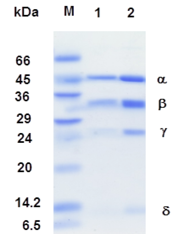
The 0.1% SDS-12.5 polyacrylamide gel electrophoresis. Sample were prepared by boiling for 3 min in a sample buffer (50 mM Tris-HCl pH 6.8, 2% SDS, 0.1% bromophenol blue, 10% glycerol. M: molecular weight markers (Albumin bovine serum, 66,000; ovalbumin, 45,000; glyceraldehydes-3-phosphate dehydrogenase, 36,000; carbonic anhydrase 29,000; trypsinogen 24,000; trypsin inhibitor, 20,000; α-lactalbumin, 14,200 and Aprotenin, 6,500).
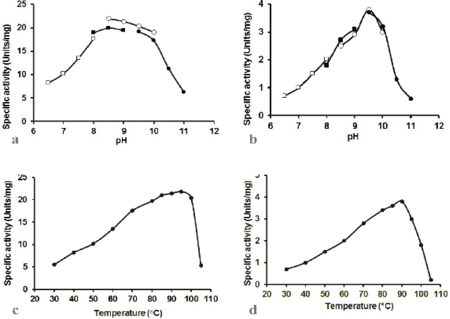
The effect of pH on (a) POR and (b) PDC specific activities of the purified enzyme from T. kodakaraensis. The open boxes indicate the pH dependent experiments with sodium phosphate buffer (pH 6.5, 7.0, 7.5 and 8.0), the closed boxes represent the experiments with Tris HCl buffer (pH 8.0, 8.5 and 9.0), open circle represent the reactions with CHES buffer (pH 8.5, 9.0, 9.5 and 10.0) and closed circles exhibit reactions with CAPS buffer (pH 9.5, 10.0, 10.5 and 11.0). Effect of temperature on (c) POR and (d) PDC specific activities of the purified enzyme from T. kodakaraensis. The assay was performed as described in Materials and method section.
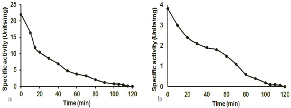
Effect of aerobic conditions on T. kodakaraensis (a) POR and (b) PDC activities. To determine the effect of oxygen on POR and PDC activities, the enzyme was incubated with shaking in the presence of air at 25°C, aliquots were taken at 5-10 min intervals and activity was determined under strict anaerobic conditions.
References
- Adams, M.W.W., and Kelly, R.M., 1992. Biocatalysis extreme temperature: Enzyme system near and above 100oC. Symp. Ser., 498, American chemical society, Washington DC.
- Adams, M.W.W., 1993. Enzyme and proteins from organisms that grow near and above 100oC. Annu. Rev. Microbiol., 47: 627-658. https://doi.org/10.1146/annurev.mi.47.100193.003211[CrossRef]
- Adams, M.W.W., 1994. Biochemical diversity among sulfur-dependent hyperthermophilic microorganism. FEMS Microbiol. Rev., 15: 267-277. https://doi.org/10.1016/0168-6445(94)90117-1[CrossRef]
- Blamey, J.M. and Adams, M.W.W., 1993. Purification and characterization of pyruvate ferredoxin oxidoreductase from the hyperthermophilic archaeon pyrococcus furiosus. Biochim. biophys. Acta, 1161: 19-27. https://doi.org/10.1016/0167-4838(93)90190-3[CrossRef]
- Blamey, J.M. and Adams, M.W.W., 1994. Characterization of an ancestral type of pyruvate ferredoxin oxidoreductase from the hyperthermophilic bacterium Thermotoga maritime. Biochemistry, 33: 1000-1007. https://doi.org/10.1021/bi00170a019[CrossRef]
- Bradford, M.M., 1976. A rapid and sensitive method for the quantitation of microgram quantities of protein utilizing the principle of protein-dye binding. Anal. Biochem., 72: 248-254. https://doi.org/10.1006/abio.1976.9999[CrossRef]
- Davis, B.J., 1964. Disc electrophoresis-II. Method and application to human serum proteins. Annls. N.Y. Acad. Sci., 121: 404-427. https://doi.org/10.1111/j.1749-6632.1964.tb14213.x[CrossRef]
- Eram, M.S., Oduaran, E. and Ma, K., 2014. The bifunctional pyruvate decarboxylase/pyruvate ferredoxin oxidoreductase from Thermococcus guaymasensis. Archaea, 2014: 349379. https://doi.org/10.1155/2014/349379[CrossRef]
- Heider, J., Mai, X. and Adams, M.W.W., 1996. Characterization of 2-ketoisovalerate ferredoxin oxidoreductase, a new and reversible coenzyme A-dependent enzyme involved in peptide fermentation by hyperthermophilic archaea. J. Bact., 178: 780-787. https://doi.org/10.1128/jb.178.3.780-787.1996[CrossRef]
- Imai, T., Yasujima, D. and Siddiqui, M.A., 2004. An instant measurement of oxidoreductase activity above 100°C by monitoring the absorbance change. J. Biosci. Bioeng., 97: 336-327. https://doi.org/10.1263/jbb.97.336[CrossRef]
- Kletzin, A. and Adams, M.W.W., 1996. Molecular and phylogenetic characterization of pyruvate and 2-ketoisovalerate ferredoxin oxidoreductase from pyrococcus furiosus and pyruvate ferredoxin oxidoreductase from Thermotoga maritime. J. Bact., 178: 248-257. https://doi.org/10.1128/jb.178.1.248-257.1996[CrossRef]
- Khalid, A., Tayyab, M., Hashmi, A.S., Yaqub, T., Awan, A.R., Wasim, M., Seed, S., Firyal, S. and Shakoori, A.R., 2019. Optimization of conditions for maximal production of recombinant thermostable cellulase from Thermotoga naphthophila using E. coli BL21-CodonPlus (DE3) as expression host. Pakistan J. Zool., 51: 1371-1377. http://dx.doi.org/10.17582/journal.pjz/2019.51.4.1371.1377[Full text][CrossRef]
- Kobayashi, T., Kwak, Y.S., Akiba, T., Kudo, T. and Horikoshi, K., 1994. Thermococcus profundus sp. Nov., A new hyperthermophilic archaeon isolated from a deep-sea hydrothermal vent System. Appl. Microbiol., 17: 232-236. https://doi.org/10.1016/S0723-2020(11)80013-5[CrossRef]
- Ma, K., Hutchins, A., Sung, S.J.S. and Adams, M.W.W., 1997. Pyruvate ferredoxin oxidoreductase from the hyperthermophilic archaeon, Pyrococcus furiosus, functions as a CoA-dependent pyruvate decarboxylase. Proc. natl. Acad. Sci. USA., 94: 9608–9613. https://doi.org/10.1073/pnas.94.18.9608[CrossRef]
- Mai, X. and Adams, M.W.W., 1994. Indolepyruvate ferredoxin oxidoreductase from a hyperthermophilic archaeon pyrococcus furiosus. J. biol. Chem. 269: 16726-16732.
- Mai, X. and Adams, M.W.W., 1996. Characterization of a four type of 2-keto acid-oxidizing enzyme from a hyperthermophilic archaeon: 2-ketoglutarate ferredoxin oxidoreductase from Thermococcus litoralis. J. Bact., 178: 5890-5896. https://doi.org/10.1128/jb.178.20.5890-5896.1996[CrossRef]
- Morikawa, M., Izawa, Y., Rashid, N., Hoaki, T., and Imanaka, T., 1994. Purification and characterization of thermostable thiol protease from a newly isolated hyperthermophilic Pyrococcus sp. Appl. environ. Microbiol. 60: 4559-4566.
- Mukund, and Adams, M.W.W., 1993. Characterization of a novel tungsten-containing formaldehyde ferredoxin oxidoreductase from the hyperthermophilic archaeon Thermococcus litoralis, a role for tungsten in peptide catabolism. J. biol. Chem. 268: 13592-13600.
- Ozawa, Y., Siddiqui, M.A., Takahashi, Y., Urushiyama, A., Ohmori, D., Yamakura, F., Arisaka, F., and Imai, T., 2012. Indolepyruvate ferredoxin oxidoreductase: An oxygen-sensitive ironesulfur enzyme from the hyperthermophilic archaeon Thermococcus profundus. J. Biosci. Bioeng., 114: 23-27. https://doi.org/10.1016/j.jbiosc.2012.02.014[CrossRef]
- Rehman, H., Siddiqui, M.A., Qayyum, A., Bano, A. and Rashid, N., 2018. Gene expression in Escherichia coli and purification of recombinant type II pullulanase from a hyperthermophilic archaeon, Pyrobaculum calidifontis. Pakistan J. Zool. 46: 1381-1386. https://doi.org/10.17582/journal.pjz/2018.50.4.1381.1386[Full text][CrossRef]
- Schafer, T., Selig, M. and Schonheit. 1993. Acetyl-CoA synthetase (ADP forming) in archaea, a novel enzyme involved in acetate formation and ATP synthesis. Arch. Microbiol., 159: 72-83. https://doi.org/10.1007/BF00244267[CrossRef]
- Siddiqui, M.A., Fujiwara, S. and Imanaka, T., 1997. Indolepyruvate ferredoxin oxidoreductase from Pyrococcus sp. KOD1 possesses a mosaic structure showing features of various oxidoreductases. Mol. Gen. Genet., 254: 433-439. https://doi.org/10.1007/PL00008607[CrossRef]
- Siddiqui, M.A., Fujiwara, S., Takagi, M. and Imanaka, T., 1998a. In Vitro heat effect on heterooligomeric subunit assembly of thermostable indolepyruvate ferredoxin oxidoreductase. FEBS. Lett., 434: 372-376. https://doi.org/10.1016/S0014-5793(98)00998-3[CrossRef]
- Siddiqui, M.A., Fujiwara, S., Takagi, M. and Imanaka, T., 1998b. Phylogenetic analysis and effect of Heat on conformational change of ferredoxin from hyperthermophilic archaeon Pyrococcus sp. KOD1. J. Ferment. Bioengn., 85: 271-277. https://doi.org/10.1016/S0922-338X(97)85674-7[CrossRef]
- Siddiqui, M.A. and Imai, T., 2007. Purification of an anaerobic oxidoreductase from anaerobic archaeon by affinity chromatography. J. Chem. Soc. Pak., 29: 390-393.
- Siddiqui, M.A., Rehman, H. and Rashid, N., 2014. Gene cloning and characterization of a type II pullulanase hydrolase from a hyperthermophilic archaeon Pyrobaculum calidifontis. Pakistan J. Zool., 46: 1077-1084.
- Stetter, K.O., 1982. Ultra thin mycelia forming organisms from submarine volcano areas having an optimum growth temperature of 105°C. Nature, 300: 258-260. https://doi.org/10.1038/300258a0[CrossRef]
- Stetter, K.O., Fiala, G., Huber, R. and Segerer, A., 1990. Hyperthermophilic microorganisms FEMS Microbiol. Rev., 75: 117-125. https://doi.org/10.1111/j.1574-6968.1990.tb04089.x[CrossRef]
- Tanaka, T., Fukui, T., Fujiwara, S., Atomi, H. and Imanaka, T., 2004. Concerted action of diacetylchitobiose deacetylase and exo-ß-D-Glucosaminidase in a novel chitinolytic pathway in the hyperthermophilic archaeon Thermococcus kodakaraensis KOD1. J. biol. Chem., 279: 30021–30027. https://doi.org/10.1074/jbc.M314187200[CrossRef]
- Weber, K. and Osborn, M., 1969. The reliability of molecular weight determination by dodecylsulfate-polyacrylamide gel electrophoresis. J. biol. Chem., 244: 4406-4412.
Services
Article usage statistics
Loading…