
TK0522 gene, encoding a carbohydrate esterase, from Thermococcus kodakarensis was cloned with its native signal sequence and expressed in Escherichia coli. Heterologous gene expression resulted in production of recombinant protein in the cytoplasm which secreted gradually to the extracellular culture medium. Determination of the N-terminal amino acid sequence of the recombinant protein, in the extracellular medium, revealed that the 19 amino acid signal peptide was cleaved between Ala19 and Gly20. It seems probable that the signal peptide of TK0522 can be used for secretion of other recombinant proteins produced in E. coli.
Short Communication · Volume 52, Issue 2, 14 February 2020 · Open Access
Escherichia coli Signal Peptidases Cleave the Signal Sequence of TK0522, a Carbohydrate Esterase from Hyperthermophilic Archaeon Thermococcus kodakarensis
· · · ·
Affiliations & Notes ⌄
- 1School of Biological Sciences, University of the Punjab, Quaid-e-Azam Campus, Lahore 54590, Pakistan , * Corresponding author: naeem.ff.sbs @pu.edu.pk
- 2naeemrashid37@hotmail.com
Article Info ⌄
- Journal:
- Pakistan Journal of Zoology
- Published:
- 14 February 2020

From the article
- In order to confirm this, we have cloned and heterologously expressed TK0522 gene, containing the native signal peptide, in E. coli.
- When the production of recombinant TK0522 was analyzed by denaturing polyacrylamide gel electrophoresis at 4 and 20 h post-induction, it was found that the amount of the protein gradually decreased in the intracellular fraction and increased in extracellular fraction (Fig. 2).
- The five amino acid residues determined at the N-terminal were X R V G Q. These results indicated that E. coli signal peptidases recognized the signal sequence of TK0522 and cleaved it between Ala19 and Gly20.
- The use of this signal peptide can potentially be helpful for the secretion of other recombinant proteins produced in E. coli.
Selected from the article's own text.
Abstract
Materials and methods
The hyperthermophilic archaeal strain T. kodakarensis KOD1 was used as a source of carbohydrate esterase gene, TK0522. E. coli DH5α was used as host for gene cloning and BL21-CodonPlus (DE3)-RIL (Stratagene) for expression of the gene. Plasmids pTZ57R/T (Thermo Fisher Scientific) and pET-21a(+) (Stratagene) were used in cloning and expression experiments, respectively. Restriction endonucleases, T4 DNA ligase and DNA polymerase were purchased from Thermo-Fisher Scientific. Nucleotide primers for amplification of the gene were commercially synthesized (Macrogen). Substrates for assay were purchased from Sigma-Aldrich.
For cloning of gene, TK0522 gene (Accession No. BAD84711) containing the N-terminal signal sequence was amplified by polymerase chain reaction using the forward (5`-CATATGAGCATGAAAGTTGCAGGCTTCC) and reverse (3`-TCACCCCTCAAAGAGCCACAGGACG) primers. The NdeI site (shown in bold underlined) was introduced in the forward primer. The amplified gene was inserted in pTZ57R/T cloning vector and the recombinant plasmid was named pTZ-TK0522. The cloned gene was liberated from pTZ-TK0522 using NdeI and HindIII restriction enzymes and inserted in pET-21a(+) expression vector. The recombinant plasmid was named pET-TK0522.
Nucleotide sequence of TK0522 gene in pET-TK0522 recombinant plasmid was determined using T7 promoter and terminator primers by the dideoxy-chain termination method. Sequencing was performed using Sequencing Ready Reaction Kit (Applied Biosystems) following the instructions of the manufacturer.
For gene expression E. coli BL21-CodonPlus (DE3)-RIL cells were transformed using pET-TK0522 recombinant plasmid. Host cells carrying pET-TK0522 vector were grown in LB medium and heterologous gene expression was induced with the addition of 0.2 mM isopropyl-β-D-1-thiogalactopyranoside. The control experiment was performed using host cells containing pET-21a(+) vector only.
For separation of intracellular and extracellular proteins cells were harvested by centrifugation at 8,000 × g for 20 min after 4, 8 and 20 h of post induction. Cell pellets were resuspended in Tris-HCl buffer (pH 8) and disrupted by sonication. TK0522, in the intracellular fraction, was partially purified by heat treatment at 85 °C for 25 min and separated by centrifugation at 14,000 × g for 15 min at 4 °C.
For purification of TK0522 from extracellular fraction, the culture medium, after harvesting the cells, was filtered through MF-Millipore Type HAWP 0.45 μm filters. The filtrate was saturated with 80% ammonium sulfate at 4 °C and incubated overnight for the precipitation of extracellular proteins. The precipitated proteins were separated by centrifugation at 14,000 × g for 30 min at 4 °C. The pellet was dissolved in 50 mM Tris-HCl (pH 8) and applied to HiTrap Phenyl HP column using AKTA purifier (GE Healthcare). The proteins bound to the column were eluted with a linear gradient of 1–0 M ammonium sulfate. The fractions containing TK0522 were pooled and dialyzed against 50 mM Tris-HCl (pH 8). The dialyzed sample was applied to Resource Q column (GE Healthcare), which was equilibrated with 50 mM Tris-HCl (pH 8.0). The proteins bound to the column were eluted with a linear gradient of 0–1 M sodium chloride.
Hydrolysis of o-nitrophenyl-β-galactoside was measured by monitoring the absorbance at 420 nm for β-galactosidase activity assay. The reaction mixture, containing 100 μL protein sample and 500 μL substrate solution (40 mM NaH2PO4, 60 mM Na2HPO4, 1.5 μL β-mercaptoethanol and 0.5 mg o-nitrophenyl-β-galactoside), was incubated at 30 °C for 90 min. The reaction was stopped by the addition of 500 μL 1 M Na2CO3.
Esterase activity was measured using p-nitrophenyl acetate as substrate. The reaction mixture containing 50 mM sodium phosphate buffer (pH 7), 40 µM p-nitrophenyl acetate and 10 µL of protein sample, in a total final volume of 1 mL, was incubated at 60 °C for 5 min. The increase in absorbance due to the production of p-nitrophenol was measured spectrophotometrically at 405 nm.
The recombinant TK0522, in the extracellular medium, was sent to Alta Bioscience (UK) for determination of the N-terminal amino acids.
Results and discussion
T. kodakarensis genome sequence harbors an open reading frame, TK0522, which is annotated as carbohydrate esterase. The open reading frame consists of 449 amino acids. Analysis of TK0522 sequence using an online available software (http://www.cbs.dtu.dk/services/signalp/) for prediction of signal peptide for Gram negative bacteria and eukaryotes displayed an N-terminal signal peptide comprising 19 amino acid residues. However, for Gram-positive bacteria, the software predicted a signal peptide of 29 amino acid residues with a cleavage site between Ala29 and Gln30. A comparison demonstrated that signal peptides of TK0522 and E. coli PhoE protein were highly similar, exhibiting an identity of more than 41% (Fig. 1). Based on this similarity, we expected that E. coli signal peptidases might be able to recognize and cleave this signal peptide. In order to confirm this, we have cloned and heterologously expressed TK0522 gene, containing the native signal peptide, in E. coli. Cloning of TK0522 gene was confirmed by digestion using NdeI and HindIII restriction enzymes, followed by DNA sequencing which confirmed the insertion of TK0522 gene with N-terminal signal sequence and no unwanted mutation.
Expression of TK0522 gene in E. coli resulted in production of recombinant protein in a significant amount. When the production of recombinant TK0522 was analyzed by denaturing polyacrylamide gel electrophoresis at 4 and 20 h post-induction, it was found that the amount of the protein gradually decreased in the intracellular fraction and increased in extracellular fraction (Fig. 2). This was further confirmed by examining the esterase activities of these fractions. At 4 h post-induction, the intracellular activity was ~98% while extracellular activity was only 2%. At 20 h post-induction, the intracellular activity decreased to ~22% while extracellular activity increased to ~77%. These results confirmed the secretion of TK0522 in the extracellular medium. We have noticed that the total esterase activity at 20 h post-induction was relatively less than the activity at 4 h post-induction. This could be due to degradation of TK 0522 by proteases of the host.
In order to check whether the observed extracellular activity is a result of the translocation of the protein through the outer membrane of the cells or lysis of the cells, we measured β-galactosidase activity. In contrast to the esterase activity, no significant amount of β-galactosidase activity was detected in extracellular fraction, indicating that TK0522 was secreted through the cell membranes and not by lysis of the cells.
Recombinant TK0522 from the extracellular medium was partially purified by hydrophobic and ion exchange column chromatographies. When the partially purified extracellular and intracellular TK0522 proteins were analyzed by denaturing polyacrylamide gel electrophoresis, there was a difference in the migration of these proteins. TK0522 from the extracellular medium migrated with a relatively faster rate compared to the intracellular TK0522 (Fig. 3).
As the extracellular TK0522 was relatively smaller in size, we therefore determined the N-terminal amino acid residues of this protein. The five amino acid residues determined at the N-terminal were X R V G Q. These results indicated that E. coli signal peptidases recognized the signal sequence of TK0522 and cleaved it between Ala19 and Gly20.
Conclusion
E. coli signal peptidases recognized and cleaved the signal peptide of TK0522. The use of this signal peptide can potentially be helpful for the secretion of other recombinant proteins produced in E. coli.
Statement of conflict of interest
The authors declare there is no conflict of interest.
Figures

Alignment of signal sequences of TK0522 from T. kodakarensis and PhoE from E. coli. Colon represent similar and asterisk show identical residues. The numbers at the right termini represent the length of the signal peptide.
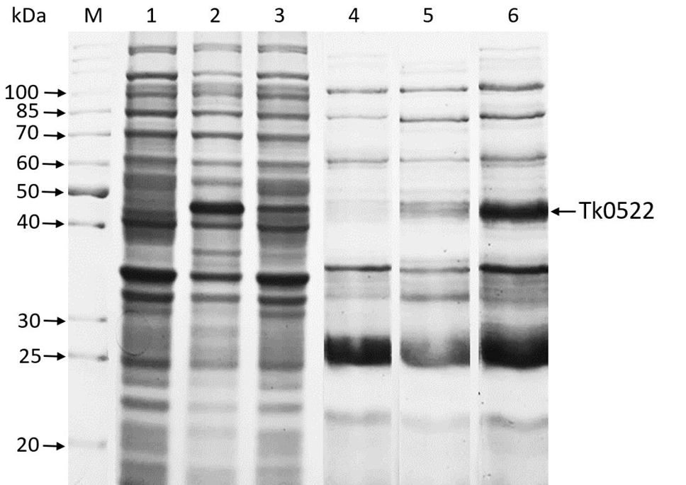
Coomassie brilliant blue stained SDS-PAGE showing recombinant TK0522 in the intracellular (lanes 1–3) and extracellular (4–6) fractions. Lane M, molecular weight marker; lane 1 and 4, cells containing pET-21a(+) at 20 h post-induction; lane 2 and 5, cells containing pET-TK0522 at 4 h post-induction; lane 3 and 6, cells containing pET-TK0522 at 20 h post-induction.
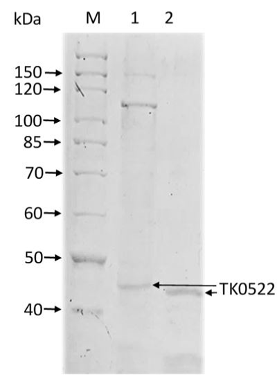
Comparison of migration of intracellular and extracellular TK0522 on SDS-PAGE. Lane M, molecular weight marker; Lane 1, partially purified TK0522 in the intracellular fraction; lane 2, partially purified TK0522 in the extracellular fraction.
References
- Albers, S.V., Szabo, Z. and Driessen, A.J.M., 2006. Nat. Rev. Microbiol., 4: 537-547. https://doi.org/10.1038/nrmicro1440[CrossRef]
- Baneyx, F., 1999. Curr. Opin. Biotechnol., 10: 411-421. https://doi.org/10.1016/S0958-1669(99)00003-8[CrossRef]
- Bosch, D., Boer, P., Bitter, W. and Tommassen, J., 1988. Biochim. biophys. Acta, 979: 69-76. https://doi.org/10.1016/0005-2736(89)90524-5[CrossRef]
- Ikemura, H., Takagi, H. and Inouye, M., 1987. J. biol. Chem., 262: 7859-7864.
- Jalal, A., Rashid, N., Ahmed, N., Iftikhar, S. and Akhtar, M. 2011. Biochemistry, 76: 347-349. https://doi.org/10.1134/S0006297911030084[CrossRef]
- Malik, B., Rashid, N., Ahmad, N. and Akhtar, M., 2013. Biochemistry, 78: 958-962. https://doi.org/10.1134/S0006297913080142[CrossRef]
- Muhammad, M.A., Falak, S., Rashid, N., Gardner, Q.A., Ahmad, N., Imanaka, T. and Akhtar, M., 2017. Biochemistry, 82: 821-825. https://doi.org/10.1134/S0006297917070070[CrossRef]
- Pugsley, A.P., 1993. Microbiol. Rev., 57: 50-108.
- Rosano, G.L. and Ceccarelli, E.A., 2014. Front. Microbiol., 5: 172. https://doi.org/10.3389/fmicb.2014.00172[CrossRef]
- Sandy, Y.M.N., Chaban, B., VanDyke, D.J. and Jarrell, K.F., 2007. Microbiology, 153: 305–314. https://doi.org/10.1099/mic.0.2006/003087-0[CrossRef]
- Schatz, G. and Dobberstein, B., 1996. Science, 271: 1519-1526. https://doi.org/10.1126/science.271.5255.1519[CrossRef]
- Shahhoseini, M., Ziaee, A.A. and Ghaemi, N., 2003. J. appl. Microbiol., 95: 1250-1254. https://doi.org/10.1046/j.1365-2672.2003.02082.x[CrossRef]
- Tariq, A., Gul, A., Muhammad, M.A., Rashid, N. and Siddiqui, M.A., 2019. Biologia, 74: 899-904. https://doi.org/10.2478/s11756-019-00243-w[CrossRef]
- Tuteja, R., 2005. Arch. biochem. Biophys., 441: 107-111. https://doi.org/10.1016/j.abb.2005.07.013[CrossRef]
- Von Heijne, G., 1983. Eur. J. Biochem., 133: 17-21. https://doi.org/10.1111/j.1432-1033.1983.tb07424.x[CrossRef]
- Yamabhai, M., Emrat, S., Sukasem, S., Pesatcha, P., Jaruseranee, N. and Buranabanyat, B., 2008. J. Biotechnol., 133: 50-57. https://doi.org/10.1016/j.jbiotec.2007.09.005[CrossRef]
- Zhang, Q., Yan, X., Zhang, L. and Tang, W., 2006. Mol. Biol., 40: 368-374. https://doi.org/10.1134/S0026893306030034[CrossRef]
Services
Article usage statistics
Loading…