
Neonatal sepsis remains one of the leading causes of newborn morbidity and mortality, requiring accurate identification of pathogens for effective treatment. Among the Gram-negative bacteria, Acinetobacter baumannii and Klebsiella pneumoniae account for high number of sepsis cases, predominantly in developing countries. The present study aims to conduct the molecular identification of multidrug resistant A. baumannii and K. pneumoniae strains isolated from neonatal sepsis patients in district Sialkot, Pakistan. A total of 10 blood samples were collected from the neonates (0-28 days) that had Gram-negative bacteria detected in their blood admitted to Allama Iqbal Memorial Hospital, Sialkot from June 2023 to November 2023. Identification of bacterial isolates was carried out using automated blood culture method and biochemical tests. Antibiotic sensitivity of isolates was assessed by Kirby-Bauer Disk diffusion method. Two highly resistant pathogens (Acinetobacter spp. and Klebsiella spp.) were subjected to molecular identification through 16S rRNA sequencing followed by the phylogenetic analysis up to the species level. The findings confirmed the isolated strains as A. baumannii and K. pneumoniae as sequences showed more than 99% similarity with reference sequences via BLAST analysis. Their phylogenetic study demonstrated a close relationship with other clinical strains reported from other regions of South Asia. The two virulent and multidrug resistant strains were submitted in Genbank with accession numbers as A. baumannii (KS7862: PP897405) and K. pneumoniae (KS7861: PP897380). This study highlights the reliability of 16S rRNA sequencing for the identification of pathogens responsible for neonatal sepsis.
Research Article · 7 October 2026 · Open Access
Molecular Identification of Acinetobacter baumannii and Klebsiella pneumoniae Isolated from Neonatal Sepsis Cases in District Sialkot, Pakistan
· · · ·
Affiliations & Notes ⌄
- 1Department of Zoology, Faculty of Science, University of Sialkot, Sialkot, Punjab, Pakistan 2Department of Microbiology, Government Khawaja Muhammad Safdar Medical College, Sialkot, Punjab, Pakistan 3Department of Biotechnology, Lahore College for Women University, Lahore, Pakistan * Corresponding author: rooma.adalat@gmail.com
Article Info ⌄
- Journal:
- South Asian Journal of Zoology
- Published:
- 7 October 2026

From the article
- Our study assessed the identification of two resistant pathogens Acineobacter spp. and Klebsiella spp. using the molecular approach through 16S rRNA sequencing which was followed by their phylogenetic analysis up to the species level.
- Likewise, a previous study reported the use of 16S rRNA gene based PCR as a diagnostic tool of neonatal sepsis.
- Despite these constraints, our findings remain significant as they provide an insight to the role of molecular identification in diagnosis of neonatal sepsis.
- This research highlights the use of 16S rRNA sequencing as a diagnostic tool in early detection of diverse pathogens causing neonatal sepsis, requiring the need to broader the surveillance at molecular level to effectively improve treatment strategies and outcomes.
Selected from the article's own text.
Abstract
Introduction
Neonatal sepsis, a critical systemic infection, occurs within the first 28 days of a newborn’s life . It is known as the third leading contributor to mortality and morbidity in infants accounting for 203,000 deaths annually, worldwide ; . However, in countries which are poor especially in terms of development, neonatal sepsis poses a considerable burden on healthcare systems due to factors such as inadequate healthcare infrastructure, limited access to medical facilities, and suboptimal hygiene practices ; . In Pakistan, sepsis is a significant contributor to the casualties in newborns, comprising 17.2% of the total deaths .
Neonatal sepsis can be classified into early-onset (EOS) or late-onset (LOS) based on time onset ; . EOS manifests within the initial three days of birth. On the other hand, LOS is diagnosed after 72 h of birth . Both LOS and EOS show distinct clinical signs, epidemiological profile, and prevalence of pathogens .
Several studies indicate that Gram-negative organisms, particularly in developing countries, are known as the causative agents of neonatal sepsis inducing morbidity and mortality . Gram-negative bacteria due to their pathogenicity can lead to severe illnesses in humans, especially those with weakened immune systems. Moreover, there is a growing rate of multidrug resistance in Gram negative bacteria which is of significant concern as they have different mechanisms to increased resistance to antimicrobials. These include reduced permeability, efflux pumps, and enzymatic degradation of antimicrobial agents . Among them, Acinetobacter spp. and Klebsiella spp. are notorious for causing neonatal sepsis and several nosocomial infections, prominently in middle and low income countries ; .
Blood culture method has various limitations as culture results take prolonged duration to be assessed and relatively has low sensitivity, even though it has been considered a gold standard. Hence, empirical treatment has to be started even in suspected sepsis as there is a great risk for neonates getting seriously affected by the infection . The reliance on empirical therapy is frequently considered inappropriate as it involves the use of unnecessary broad-spectrum antibiotics which is coupled with an extended treatment timeframe and contributes to proliferating number of antibiotic resistant bacteria; making the treatment of the infection challenging ; . Bacteria have conserved regions like 16S rRNA genes that are targeted and amplified via PCR (Polymerase chain reaction), which offers sensitive and precise diagnosis of various infectious caused by bacteria. This approach can accurately identify bacteria at both genus and species level in about 90% of the cases .
There has been very limited research conducted in Sialkot regarding the molecular identification through 16S rRNA sequencing, specifically of Klebsiella pneumoniae and Acinetobacter baumannii, isolated from neonatal sepsis patients. Therefore, this study was aimed to assess identification of the resistant strains at molecular level.
Materials and Methods
Collection of blood sample and isolation of bacteria
Blood samples (3-5 ml of blood) were collected from neonates suspected of sepsis by employing standard methods . The BacT/ALERT pediatric (yellow-30ml, BacT/ALERT PF) bottles were then inoculated with the blood sample and incubated in auotmated BACT/ALERT 3D (bioMerieoux, France) system. When positive signal was noted, culture bottles were picked out of machine and sub-cultured on Blood agar and MacConkey agar, which was followed by incubation of the inoculated plates at 37°C for 24-48 h ; .
Identification of bacteria
The plates with bacterial growth were subjected to identification methods such as Gram staining technique, colony characteristics and standard biochemical tests . The identification of some isolates was further confirmed by API 10S strips (bioMérieux, France).
Antibiotic sensitivity testing
The antibiotic sensitivity pattern of the selected isolates (n = 10) was evaluated, against a panel of antibiotics (Oxoid, UK), employing the Kirby-Bauer disk diffusion method as per the Clinical and Laboratory Standards Institute (CLSI) protocol. The isolates were inoculated onto Mueller-Hinton agar (MHA) plates. The plates were later incubated for 16 to 18 h at 37 °C. Following this, the zones of inhibition around the antibiotic discs were eventually measured using a scale and recorded in accordance with CLSI guidelines ; .
Molecular identification of Acinetobacter spp. and Klebsiella spp.
The isolates of Acinetobacter spp. and Klebsiella spp. were grown overnight in Trypticase Soy Broth to obtain pure culture for isolation of DNA . DNA was extracted from two strains of Klebsiella spp. and Acinetobacter spp. using QIAamp DNA mini extraction kits (Qiagen, Cat. No.:51306) in accordance with the instructions from manufacturer.
Amplification of 16S rRNA gene and DNA sequencing
The fragments of 16S rRNA gene were amplified using a PCR thermocycler (Applied biosystems Gene Amp PCR system 2700) with universal primers. The component mixture comprised of 6 µL DNA template, each with forward and reverse primer of 2 µL, PCR master mix from Thermoscientific (Cat. No.:K1081) of 25 µL, and sterile ddH2O of 15 µL was added to make up total volume of 50 µL. The primers added were 27F: 5 ′- AGAGTTTGATCCTGGCTCAG-3′ and 1492R: 5′- GGTTACCTTGTTACGACTT-3′ . The process of amplification was started by performing initial degradation at 94° C for 5 mins followed by denaturation with further 35 cycles for 30 secs at 94° C. The annealing temperature was 52.7°C for 30 secs, and for extension 72°C for 50 secs. Amplification was concluded at 72°C for 5 mins (final extension). A volume of around 6 µL of the amplified product was then analyzed by carrying out electrophoresis in 1% agarose gel. The gel was examined under UV light and photographed. It was followed by purification of the obtained products using Gene Jet gel extraction kit (Cat. No.: K0691) as per instructions of manufacturer and, then preserved the purified DNA at -20 °C. Finally, the purified PCR products were forwarded to Apical scientific Sdn. Bhd., Malaysia for sequencing of DNA.
Sequencing analysis
The PCR amplicons were sequenced using Sanger sequencing. The length of 16S rRNA gene amplification products was approximately 1500 bp. Molecular Evolutionary Genetics Analysis MEGA 11 software was used for analyzing the data from this sequencing. The 16S rRNA gene nucleotide sequences from pathogenic Klebsiella spp. and Acinetobacter spp. available at the GenBank were used as reference sequences. The obtained sequences of 16S rRNA gene from bacterial strains KS7861 and KS7862 were then aligned by the Clustal W program. BLAST tool was further utilized to compare these sequences with reference sequences available at the GenBank (http://www.ncbi.nlm.nih.gov/). Later, the neighbor joining algorithm method was employed to analyze the phylogenetic relationship for further characterization ; .
The obtained partial 16S rRNA sequences of both strains KS7861 and KS7862 presented in the current study under accession numbers (KS7861: PP897380.1; KS7862: PP897405.1) have been registered in GenBank (http://www.ncbi.nlm.nih.gov/).
Results
Pathogens isolated from neonatal sepsis patients
A total of 10 patients, registered in the laboratory with culture-confirmed sepsis, were selected. Blood culture method was used to identify bacterial growth. Gram negative bacterial isolates, mainly Acinetobacter spp. and Klebsiella spp. were found to be the causative agents of neonatal sepsis patients (n = 10).
Antibiotic sensitivity patterns of isolates
Acinetobacter spp. demonstrated the highest level of resistance to penicillins (96%), ceftriaxone (96%), cefoperazone (87%), gentamicin (83%), ciprofloxacin (83%). Moreover, the isolates showed increased resistance to doxycycline (78%), Sulfamethoxazole-trimethoprim (70%) and carbapenems was also observed. However, some of the isolates showed comparatively better sensitivity to cefoperazone-sulbactam (83%) and piperacillin-tazobactam (70%). In case of Klebsiella spp., the isolates showed high resistance to penicillins (100%) followed by cefoperazone (85%), ceftriaxone (85%) and doxycycline (69%). Fortunately, carbapenems were found to be effective as the isolates exhibited high sensitivity to them.
Molecular identification of isolates
Two strains were selected for molecular identification based on their high level of resistance profile. DNA was extracted from the isolated bacterial strains of Acinetobacter spp. and Klebsiella spp. 16S rRNA regions of both strains were amplified as depicted in Figure 1 and sequenced by using the universal primers. A total of around 1500 bp of 16S rRNA gene were sequenced for the identification of
the tested bacterial strains. The obtained sequences have been registered in the GenBank with accession numbers PP897380.1 and PP897405.1 for both bacterial strains KS7861 and KS7862, respectively. BLAST tool was used to compare these tested sequences against references sequences available at the Genbank. The BLAST result for strain KS7861 showed that the 16S rRNA sequence of isolated strain KS7861 shared 99.46% sequence similarity with reference K. pneumoniae subsp. rhinoscleromatis strain R-70 (NR_037084.1), K. pneumoniae strain DSM 30104 (NR_117683.1). While, BLAST result of 16S rRNA sequence of strain KS7862 showed that the isolate exhibited 99.67% sequence similarity with A. baumannii strain DSM 30007 (NR_117677.1), and A. baumannii strain ATCC 19606 (NR_117620.1).
Subsequently, two phylogenetic trees based on 16S rRNA gene sequence were generated, which shows the relatedness between the isolates KS7861 and KS7862 and their selected representative genera K. pnuemoniae and A. baumannii, respectively. Phylogenetic assessment of 16S rRNA sequences evidently showed that the isolate KS7861 belongs to genera K. pnuemoniae while KS7862 represents the genera A. baumannii, a shown in Figure 2.
Discussion
In the current study, among the isolated Gram-negative bacteria, Acinetobacter spp. and Klebsiella spp. were found to be the predominant pathogens causing sepsis in neonates, consistent with the findings from the previous studies conducted in developing countries ; . Both Klebsiella spp. and Acinetobacter spp. are frequently linked with prolonged hospitalization, increased morbidity, and higher mortality rate in infants .
This study revealed that Acinetobacter spp., exhibited significant resistance to penicillins, cephalosporins and aminoglycosides. Higher rates of resistance to carbapenems, tetracyclines and ciprofloxacin were demonstrated. Klebsiella spp., the other prominent pathogen, also exhibited high resistance to penicillins, cephalosporins and tetracyclines. In both cases, the isolates were found to be resistant against first-line and second-line antibiotics in addition to other classes of antibiotics. Previous studies have also reported the similar results ; . These results highlight the rise of multidrug resistant bacteria, which is of serious concern.
Our study assessed the identification of two resistant pathogens Acineobacter spp. and Klebsiella spp. using the molecular approach through 16S rRNA sequencing which was followed by their phylogenetic analysis up to the species level. Similar to the current study, El-Amir et al. (2019), the 16S rRNA based PCR was employed to detect bacterial isolates from neonatal sepsis patients. Likewise, a previous study reported the use of 16S rRNA gene based PCR as a diagnostic tool of neonatal sepsis. It was compared with the conventional culture method. 16S rRNA gene based PCR was found to be better in terms of rapid testing; fast detection of pathogens and increased sensitivity leading to short antibiotics courses . Several previous studies have also reported the use of molecular approach to isolate the multidrug resistant bacterial isolates in different infections ; . Additionally, the isolated strains were subjected to the phylogenetic analysis for species level identification, which is in agreement with the current study . However, culture method still cannot be replaced due to requirement of pure isolates for antibiotic sensitivity testing and better treatment outcomes .
It is vital to note that this study has several limitations as it was conducted at a local tertiary care hospital. The sample size is small, and only two strains were selected for sequencing due to limited resources. Moreover, it doesn’t include advanced molecular tools such as detecting virulent genes or whole genome sequencing. Despite these constraints, our findings remain significant as they provide an insight to the role of molecular identification in diagnosis of neonatal sepsis. Future studies are therefore recommended, particularly in developing regions, to incorporate the use of advanced molecular techniques to have deeper understanding of the epidemiologic profiling of the virulent pathogens causing infections like neonatal sepsis.
Conclusion
This research highlights the use of 16S rRNA sequencing as a diagnostic tool in early detection of diverse pathogens causing neonatal sepsis, requiring the need to broader the surveillance at molecular level to effectively improve treatment strategies and outcomes.
Declarations
Acknowledgments
The authors would like to extend their heartfelt gratitude to the Department of Pathology, Khawaja Muhammad Safdar Medical College, and Dr. Abdul Sattar’s Laboratory, Commissioner Road, Sialkot, Punjab, for their invaluable assistance in sample collection, identification, and analysis of antibiotic resistance patterns of Gram-negative bacterial isolates in neonatal sepsis patients.
Funding
This study received no financial support.
Ethical approval
The study was approved by the Ethical committee of the University of Sialkot.
Generative AI and AI assisted technology statement
The authors declare that no generative AI and AI assisted technology was used in the creation of this manuscript.
Statement of conflict of interest
The authors have declared no conflict of interest.
Figures
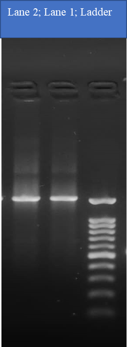
Agarose gel electrophoresis of 16S rRNA gene PCR amplification; Lane 1: PCR product of Klebsiella spp. Lane 2: PCR product of Acinetobacter spp.
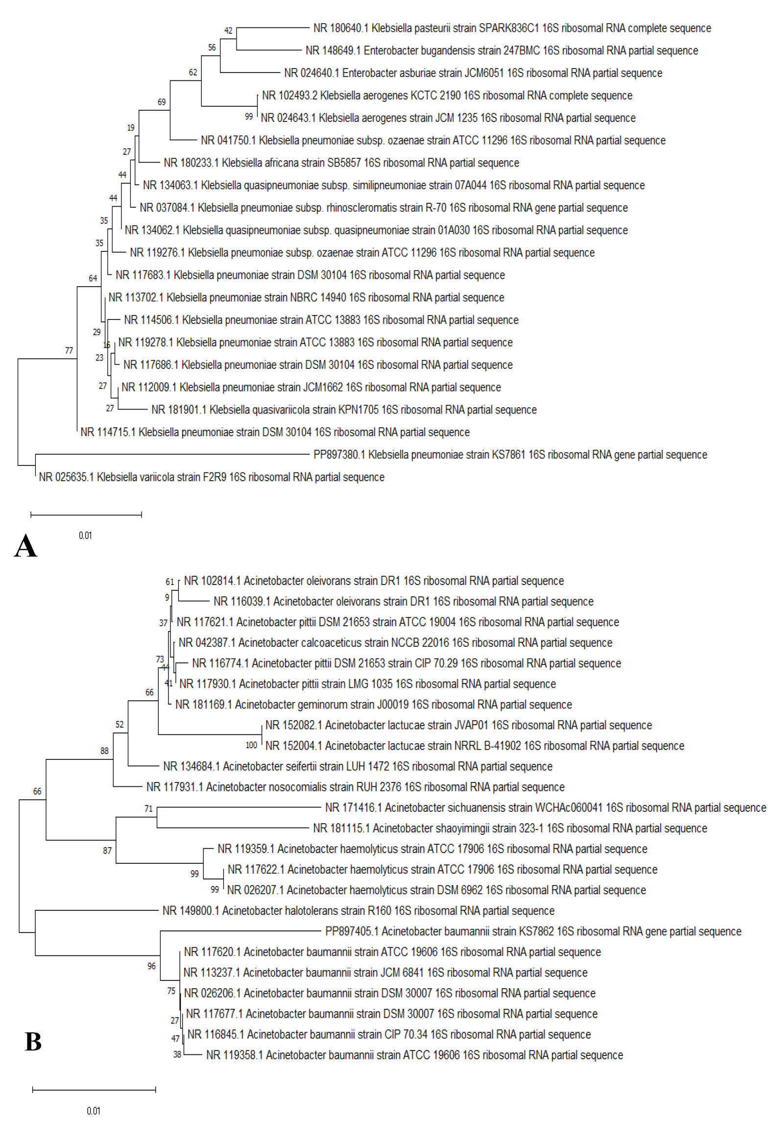
Phylogenetic trees of (A) Klebsiella pneumonaie KS7861 (PP897380.1) and (B) Acinetobacter baumannii KS7862 (PP897405.1) reveal their evolutionary relationships with regards to the closest bacteria on the basis of 16S rRNA nucleotide sequences retrieved from the GenBank database, and accession numbers of the sequences are indicated in parentheses. The phylogenetic trees were constructed employing the Neighbor-Joining tree with bootstrap values of 1000 replicates, and the bars denote Jukes-Cantor distances of 0.01 for both strains.
References
- Al-Matary, A., Almatari, F., Al-Matary, M., AlDhaefi, A., Alqahtani, M.H.S., Alhulaimi, E.A., AlOtaiby, S., Almehiny, K., John, L.S. and Alanazi, F.S., 2021. Clinical outcomes of maternal and neonate with COVID-19 infection–Multicenter study in Saudi Arabia. J. Infect. Publ. Hlth., 14: 702-708. https://doi.org/10.1016/j.jiph.2021.03.013[CrossRef]
- Almohammady, M.N., Eltahlawy, E.M. and Reda, N.M. 2020. Pattern of bacterial profile and antibiotic susceptibility among neonatal sepsis cases at Cairo University Children Hospital. J. Taibah Univ. Med. Sci., 15: 39-47. https://doi.org/10.1016/j.jtumed.2019.12.005[CrossRef]
- Atif, M., Zia, R., Malik, I., Ahmad, N. and Sarwar, S., 2021. Treatment outcomes, antibiotic use and its resistance pattern among neonatal sepsis patients attending Bahawal Victoria Hospital, Pakistan. PLoS One, 16: e0244866. https://doi.org/10.1371/journal.pone.0244866[CrossRef]
- Boscarino, G., Romano, R., Iotti, C., Tegoni, F., Perrone, S. and Esposito, S., 2024. An overview of antibiotic therapy for early-and late-onset neonatal sepsis: Current strategies and future prospects. Antibiotics, 13: 250. https://doi.org/10.3390/antibiotics13030250[CrossRef]
- Cantey, J.B. and Patel, S.J., 2014. Antimicrobial stewardship in the NICU. Infect. Dis. Clin., 28: 247-261. https://doi.org/10.1016/j.idc.2014.01.005[CrossRef]
- Chaurasia, S., Sivanandan, S., Agarwal, R., Ellis, S., Sharland, M. and Sankar, M.J., 2019. Neonatal sepsis in South Asia: Huge burden and spiralling antimicrobial resistance. Br. med. J., 364. https://doi.org/10.1136/bmj.k5314[CrossRef]
- El-Amir, M.I., El-Feky, M.A., Abo Elwafa, D.A. and Abd-Elmawgood, E.A., 2019. Rapid diagnosis of neonatal sepsis by PCR for detection of 16S rRNA gene, while blood culture and PCR results were similar in E. coli-predominant EOS cases. Infect. Drug Resist., pp. 2703-2710. https://doi.org/10.2147/IDR.S213958[CrossRef]
- Fleischmann, C., Reichert, F., Cassini, A., Horner, R., Harder, T., Markwart, R., Tröndle, M., Savova, Y., Kissoon, N. and Schlattmann, P., 2021. Global incidence and mortality of neonatal sepsis: A systematic review and meta-analysis. Arch. Dis. Childh., 106: 745-752. https://doi.org/10.1136/archdischild-2020-320217[CrossRef]
- Fleischmann-Struzek, C., Goldfarb, D.M., Schlattmann, P., Schlapbach, L.J., Reinhart, K. and Kissoon, N., 2018. The global burden of paediatric and neonatal sepsis: A systematic review. Lancet Respirat. Med., 6: 223-230. https://doi.org/10.1016/S2213-2600(18)30063-8[CrossRef]
- Ghafoor, T., Hussanain, A., Qureshi, M., Mehmood, T. and Ali, L., 2020. Antimicrobial susceptibility pattern of blood culture isolates from patients with suspected neonatal sepsis in a tertiary care hospital in Pakistan. Sri Lankan J. Infect. Dis., 10. https://doi.org/10.4038/sljid.v10i1.8244[CrossRef]
- Hassan, M.A., Abd El-Aziz, S., Elbadry, H.M., El-Aassar, S.A. and Tamer, T.M., 2022. Prevalence, antimicrobial resistance profile, and characterization of multi-drug resistant bacteria from various infected wounds in North Egypt. Saudi J. biol. Sci., 29: 2978-2988. https://doi.org/10.1016/j.sjbs.2022.01.015[CrossRef]
- Hudzicki, J., 2009. Kirby-Bauer disk diffusion susceptibility test protocol. Am. Soc. Microbiol., 15: 1-23.
- Hussein, A.H.M., Ritika, P., Sarvani, D., Tarun, S., Aayush, P., Shiny, A., Cevallos-Cueva, M., Hayes, B.K., Saranya, A. and Zain, A., 2023. Insight into neonatal sepsis: An overview. Cureus, 15. https://doi.org/10.5812/archcid.101136[CrossRef]
- Lakshmi, P., Bharadwaj, A. and Srivastava, R. K. 2020. Molecular detection and identification of bacteria in urine samples of asymptomatic and symptomatic pregnant women by 16S rRNA Gene sequencing. Arch. Clin. Infect. Dis., 15.
- Liu, C., Ai, H., Wang, W., Chen, L., Hu, H., Ye, T., Zhu, X., Wang, F., Liao, Y. and Wang, Y., 2014. Comparison of 16S rRNA gene PCR and blood culture for diagnosis of neonatal sepsis. Arch. Pédiat., 21: 162-169. https://doi.org/10.1016/j.arcped.2013.11.015[CrossRef]
- Nazir, A., 2019. Multidrug-resistant Acinetobacter septicemia in neonates: A study from a teaching hospital of Northern India. J. Lab. Phys., 11: 023-028. https://doi.org/10.4103/JLP.JLP_129_18[CrossRef]
- Nursofiah, S., Hartoyo, Y., Amalia, N., Febrianti, T., Febriyana, D., Saraswati, R., Puspandari, N., Sariadji, K., Rukminiati, Y. and Muna, F., 2021. Long-term storage of bacterial isolates by using tryptic Soy Broth with 15% glycerol in the deep freezer (-70 to -80 oC). IOP Conf. Ser. Earth Environ. Sci., IOP Publ., pp. 012070. https://doi.org/10.1088/1755-1315/913/1/012070[CrossRef]
- Oliveira, J. and Reygaert, W.C., 2023. Gram-negative bacteria. StatPearls [Internet]. StatPearls Publishing.
- Pillay, K., Ray-Chaudhuri, A., O’Brien, S., Heath, P. and Sharland, M., 2024. Acinetobacter spp. in neonatal sepsis: An urgent global threat. Front. Antibiot., 3: 1448071. https://doi.org/10.3389/frabi.2024.1448071[CrossRef]
- Salah, A., Al-Subol, I., Hudna, A., Alhaj, A., Alqubaty, A.R., Farie, W., Sulieman, D., Alnadhari, O., Alwajeeh, T. and Alobathani, F., 2021. Neonatal sepsis in Sana’a city, Yemen: A predominance of Burkholderia cepacia. BMC Infect. Dis., 21: 1108. https://doi.org/10.1186/s12879-021-06808-y[CrossRef]
- Sands, K., Carvalho, M.J., Portal, E., Thomson, K., Dyer, C., Akpulu, C., Andrews, R., Ferreira, A., Gillespie, D. and Hender, T., 2021. Characterization of antimicrobial-resistant Gram-negative bacteria that cause neonatal sepsis in seven low-and middle-income countries. Nat. Microbiol., 6: 512-523. https://doi.org/10.1038/s41564-021-00870-7[CrossRef]
- Suardana, I.W., 2014. Analysis of nucleotide sequences of the 16S rRNA gene of novel Escherichia coli strains isolated from feces of human and Bali cattle. J. Nucl. Acids, 2014: 475754. https://doi.org/10.1155/2014/475754[CrossRef]
- Tessema, B., Lippmann, N., Knüpfer, M., Sack, U. and König, B., 2021. Antibiotic resistance patterns of bacterial isolates from neonatal sepsis patients at university hospital of Leipzig, Germany. Antibiotics, 10: 323. https://doi.org/10.3390/antibiotics10030323[CrossRef]
- Tessema, B., Lippmann, N., Willenberg, A., Knüpfer, M., Sack, U. and König, B., 2020. The diagnostic performance of interleukin-6 and C-reactive protein for early identification of neonatal sepsis. Diagnostics, 10: 978. https://doi.org/10.3390/diagnostics10110978[CrossRef]
- Wayne, P., 2011. Performance standards for antimicrobial susceptibility testing. Clinical and Laboratory Standards Institute.
- Wei, X., Liang, J., Zhang, H., Yan, C., Lu, X., Chen, Y. and Li, L., 2024. Clinical features and risk factors of Klebsiella pneumoniae infection in premature infants: A retrospective cohort study. BMC Infect. Dis., 24: 1311. https://doi.org/10.1186/s12879-024-10201-w[CrossRef]
- Wen, S.C., Ezure, Y., Rolley, L., Spurling, G., Lau, C.L., Riaz, S., Paterson, D.L. and Irwin, A.D., 2021. Gram-negative neonatal sepsis in low-and lower-middle-income countries and WHO empirical antibiotic recommendations: A systematic review and meta-analysis. PLoS Med., 18: e1003787. https://doi.org/10.1371/journal.pmed.1003787[CrossRef]
- Yadav, N.S., Sharma, S., Chaudhary, D.K., Panthi, P., Pokhrel, P., Shrestha, A. and Mandal, P.K., 2018. Bacteriological profile of neonatal sepsis and antibiotic susceptibility pattern of isolates admitted at Kanti Children’s Hospital, Kathmandu, Nepal. BMC Res. Notes, 11: 301. https://doi.org/10.1186/s13104-018-3394-6[CrossRef]
Services
Article usage statistics
Loading…