
Klebsiella infections in the oral cavities of both humans and dogs have been increasingly reported and are associated with various buccal infections, as well as systemic infections. These infections appear to be rising particularly among pets and their owners, suggesting a possible bidirectional transmission between humans and dogs. Therefore, this study aimed to investigate the potential link of mixed infections involving Klebsiella pneumoniae and Enterococcus spp. Buccal cavity samples were collected from humans (n = 25) and dogs (n = 25). Samples were initially enriched in tryptic soy broth and subsequently cultured on tryptic soy agar, MacConkey agar, and blood agar. All isolates were identified using the VITEK 2 system, and eight selected isolates were further analyzed by 16S rRNA gene PCR. In humans, Klebsiella spp. were detected in 24% of samples by primary isolation, 44% by VITEK 2 analysis, and 12% were confirmed by 16S rRNA PCR. In dogs, primary isolation and VITEK 2 identification both showed a prevalence of 28%, while 20% were confirmed by 16S rRNA PCR. Among human samples, isolates were detected in 10% of males and 13.3% of females, whereas in dogs, 14.3% of males and 27.3% of females were positive. The results revealed that K. pneumoniae accounted for 66.7% and Enterococcus faecalis for 33.3% of isolates from humans, while in dogs, K. pneumoniae represented 80% and E. faecalis 20% of the isolates. These findings highlight the potential significance of transmission of these bacterial species between humans and dogs.
Research Article · Volume 42, Issue 1, 5 February 2026 · Open Access
Identification and Molecular Detection of Klebsiella spp. from the Buccal Cavity of Humans and Dogs
· · ·
Affiliations & Notes ⌄
- 1Zoonosis Diseases Research Unit, College of Veterinary Medicine, University of Baghdad, Baghdad City, Iraq
- 2Department of Laboratory Technique, Alnukhba University College, Baghdad, Iraq. *Correspondence | Hassan Falah Mohammed, Zoonosis Diseases Research Unit, College of Veterinary Medicine, University of Baghdad, Baghdad City, Iraq
- 3Email: hasan.falaah1204e@covm.uobaghdad.edu.iq
Article Info ⌄
- Journal:
- Advances in Agriculture and Animal Sciences
- Published:
- 5 February 2026
From the article
- The gender distribution of K. pneumoniae isolates in humans showed that 1 out of 10 male samples (10%) and 2 out of 15 female samples (13.3%) were positive.
- Of the human isolates sent for molecular detection by 16S rRNA PCR, 2 out of 3 samples (66.7%) were confirmed as K. pneumoniae and 1 out of 3 (33.3%) as E. faecalis.
- In our study, 16S rRNA analysis strongly indicated a close genetic relationship between K. pneumoniae isolates from humans and their dogs.
- Confirmation by 16S rRNA analysis revealed clear evidence of transmission of the same bacterial strains between humans and their dogs.
Selected from the article's own text.
Abstract
Introduction
The oral health of a human or animal depends on the presence of healthy normal flora on the surface of gums, teeth and oral cavity lining. Periodontitis is the outcome of progressive and persistent inflammation of the teeth supporting tissues, leading to clinical dental loss, alveolar bone loss, and periodontal pocket formation (; . The high prevalence of bacterial isolates of the family Enterobacteriaceae is attributed to their natural presence in the oral cavities and oesophagus ; .
Knowing the pathogenic bacteria present in the oral cavity of dogs and cats helps in finding appropriate treatment for infected bite wounds. The factors such as poor hygiene and diet lead to contamination of the dog’s oral cavities, periodontal disease is one of the most commonly diagnosed oral diseases in dogs and can result from undisturbed dental plaque. Dental prophylaxis is a routinely practiced veterinary procedure, but its effects on both the plaque and oral microbiota is not fully understood (Flancman et al., 2016; ; .
Klebsiella spp. are Gram-negative, nonmotile, usually encapsulated rod-shaped bacteria, belonging to the family Enterobacteriaceae, generally facultative anaerobic. Klebsiella spp. often occur in mucoid colonies. The genus consists of 77 capsular antigens (K antigens), leading to different serogroups . Klebsiella pneumoniae is one of the prominent pathogens found in hospital-acquired samples. They resides in the gastrointestinal (GI) microbiota of humans and animals ; . These bacteria are notoriously responsible for foodborne diseases such as septicemia, pneumonia, urinary tract infections (UTIs), liver abscess, bloodstream infection, and diarrhea ; . Enterococcus faecalis (E. faecalis) is an important ubiquitous bacterium working with other microbes in the gastrointestinal tract of humans and animals alike to maintain the gut microbes ecological homeostasis, as well as, assisting with barrier function support ; ; Kaittan and Zghair, 2024). This bacterium is a significant zoonotic infectious agent, primarily causing opportunistic infections in vulnerable human populations, while in animals it mainly exists as a commensal reservoir ; . Consequently, it represents a growing global health risk affecting both humans and animals . The detection of these isolates in humans and dogs, as well as other companion animals, suggests a potential for interspecies transmission, highlighting the importance of these pathogens in both human and animal public health. Therefore, the objective of this study was to investigate the potential association of mixed infections involving K. pneumoniae and Enterococcus spp. in the buccal cavities of humans and dogs.
Materials and Methods
Bacterial isolation
A total of 50 samples were collected from private veterinary clinics in Baghdad city Iraq from buccal cavity of human (25 samples) and dogs (25 samples). All the samples were collected from humans and dogs that showed clinical signs of diseases like coughing sneezing and fever, using sterile swabs with transport media and they were labeled and placed in cooler boxes immediately before transferring it to the College of Veterinary Medicine’s Zoonotic Diseases Unit laboratory. For a period from November 2024 - February 2025. The swabs were inoculated on Tryptic Soy Broth (TSB) and culturing on agar (TSA, MacConkey agar and Blood agar), and incubated at 37 ºC for 24 to 36 h. Samples were sent to VITEK 2 system to determine the isolates.
Primers
The primers used in this study were designed according to a previously published study and shown in Table 1. The primers were supplied in lyophilized form and were initially dissolved in nuclease-free ddH₂O to obtain a stock concentration of 100 pmol/µL. The stock solutions were stored at −20 °C. Working primer solutions (10 pmol/µL) were prepared by diluting 10 µL of the stock solution with 90 µL of nuclease-free ddH₂O to obtain a final volume of 100 µL.
Statistical analysis
Data were analyzed using descriptive statistics. The prevalence of bacterial isolates was calculated and expressed as percentages for humans and dogs, as well as according to sex. No inferential statistical tests were performed, as the study was descriptive in nature and focused on frequency and percentage distribution of the isolates.
Table 1: The primers used in the study.
| Primer | Sequence | Primer sequence | Tm (oC) | GC% | Size of product (bp |
| 16s RNA | 27F | 5'- AGAGTTTGATCCTGGCTCAG- 3' | 56.92 | 50.00 | 1250bp |
| 1492R | 5'- GGTTACCTTGTTACGACTT- 3’ | 52.20 | 42.11 |
Results
A total of 25 buccal cavity samples were collected from humans. Primary isolation by culture yielded 11 positive samples (44%), of which 6 samples (24%) were confirmed using the VITEK 2 system. In dogs, 25 buccal cavity samples were analyzed, and both primary isolation and VITEK 2 identification resulted in 7 positive samples (28%). From the total isolates, eight samples were selected for 16S rRNA gene analysis, including three human isolates and five canine isolates (Table 2, Figure 1).
Table 2: Isolation and identification of Klebsiella pneumoniae from buccal cavity samples of humans and dogs.
| Isolation source | No. of samples | Primary isolation | VITEK 2 | PCR |
| Human | 25 | 11 (44%) | 6 (24%) | 3 (12%) |
| Dogs | 25 | 7 (28%) | 7 (28%) | 5 (20%) |
The gender distribution of K. pneumoniae isolates in humans showed that 1 out of 10 male samples (10%) and 2 out of 15 female samples (13.3%) were positive. In dogs, 2 out of 14 male samples (14.3%) and 3 out of 11 female samples (27.3%) were identified as positive for K. pneumoniae (Tables 3–4).
Table 3: Number of K. pneumonie isolates from human according to gender.
| Gender | No. of samples | +ve (%) |
| Male | 10 | 1 (10%) |
| Female | 15 | 2 (13.3%) |
Table 4: Number of K. pneumonie isolates from dog according to gender.
| Gender | No. of samples | +ve (%) |
| Male | 14 | 2 (14.3%) |
| Female | 11 | 3 (27.3%) |
Of the human isolates sent for molecular detection by 16S rRNA PCR, 2 out of 3 samples (66.7%) were confirmed as K. pneumoniae and 1 out of 3 (33.3%) as E. faecalis. In dogs, 4 out of 5 samples (80%) were confirmed as K. pneumoniae and 1 out of 5 (20%) as E. faecalis, out of a total of 8 samples sent for analysis (Table 5).
Table 5: Number of bacterial isolates confirmed by 16S rRNA PCR from humans and dogs.
| Host | No. of isolates sent | K. pneumoniae | E. faecalis |
| Human | 3 | 2 (66.7%) | 1 (33.3%) |
| Dogs | 5 | 4 (80%) | 1 (20%) |
Isolates of K. pneumoniae from 6 buccal cavity samples were analyzed and sequenced, with GenBank ID: ON323049.1. Sequence identities were 100% for samples 1 and 4, while the remaining samples showed 99% identity. For E. faecalis, 2 samples were sequenced and compared to GenBank ID: OP359294.1, with 100% identity for sample 7 and 99% identity for sample 8, as shown in Table 6.
Table 6: Results of sequencing and genetic analysis of 16S rRNA gene.
| No. of sample | Source | Sequence ID (Submission) | Sequence ID (Compare) | Identities | Nucleotide change | Location | Type of substitution |
| 1 | Klebsiella pneumoniae | PV290135.1 | ON323049.1 | 100% | – | – | – |
| 2 | Klebsiella pneumoniae | PV290136.1 | ON323049.1 | 99% | G→T | 790 | Transversion |
| 3 | Klebsiella pneumoniae | PV290137.1 | ON323049.1 | 99% | C→G, G→T | 673, 790 | Transversion |
| 4 | Klebsiella pneumoniae | PV290138.1 | ON323049.1 | 100% | – | – | – |
| 5 | Klebsiella pneumoniae | PV290139.1 | ON323049.1 | 99% | G→A, C→T | 187, 209 | Transition |
| 6 | Klebsiella pneumoniae | PV290140.1 | ON323049.1 | 99% | G→T | 790 | Transversion |
| 7 | Enterococcus faecalis | PV290141.1 | OP359294.1 | 100% | – | – | – |
| 8 | Enterococcus faecalis | PV290142.1 | OP359294.1 | 99% | T→A | 775 | Transversion |
Discussion
The human oral and nasal cavities can act as reservoirs for opportunistic pathogens capable of causing acute infection. These microbes asymptomatically colonize the human oral and nasal cavities which facilitates transmission within human populations via the environment . Ahlam et al. (2025) finding of culture media and biochemical tests showed that 26.77% (34/127) of nasal swabs of human were positive samples to K. pneumoniae which not match with primary isolation 44%, but agreement with VITEK2 (24%). In our study, according to the culturing and primary isolation, the percentage of humans K. pneumoniae was (44%) and dogs was (28%), while according to VITEK results the percentage of humans K. pneumoniae was (24%) and dogs was (28%).
Hessan and Mohammed (2021) were determined that the patients provided a total of 50 samples results showed that 39 (78 percent) of the samples tested had bacterial growth, 14 of them were diagnosed as K. pneumonia. Colonization of the oral cavity by potentially pathogenic bacteria constitutes threatens its transmission to different human tissues and organs. Arkan et al. (2020) provide isolation and identification 14 isolates of K. pneumoniae recovered from oral cavity infections. While in our study appeared E. faecalis from human 33% and from dogs 20%; While K. pneumoniae isolation were 66% from human and 80% from dogs. Chen et al. (2021) examined the clonality of K. pneumoniae isolates longitudinally collected from fecal samples of a healthy married couple and their pet animals. Their study revealed that the K. pneumoniae populations in the male owner and one of the dogs were clonally diverse, whereas a dominant clone persisted in the gastrointestinal tract of the female owner, who was prone to chronic diarrhea. In contrast, the findings of the present study showed higher K. pneumoniae infection rates in females compared to males: 13.3% in human females versus 10% in human males, and 27.3% in female dogs versus 14.3% in male dogs. Similarly, Ofukwu et al. (2008) reported that 75.1% of 160 dogs were positive for at least one bacterial species, including K. pneumoniae (11.7%). These findings underscore the high risk of human infections through dog bites, direct contact with saliva, or ingestion of saliva-contaminated water and food. In our study, 16S rRNA analysis strongly indicated a close genetic relationship between K. pneumoniae isolates from humans and their dogs. A similar pattern of relatedness and significance was also observed for E. faecalis, highlighting the potential for zoonotic transmission of these bacteria between pets and their owners.
Proper medical care of local dogs, maintenance of personal hygiene, and careful selection of drugs for treating dog bite wounds or related infections are strongly recommended. In this study, the isolation rate of K. pneumoniae in dogs was 28%. Zhang et al. (2022) reported a total of 35 K. pneumoniae complex isolates (2.3%; 95% confidence interval) from 1,500 randomly collected samples across veterinary hospitals in 12 regions of China. The molecular characteristics and mobile resistance elements of these isolates allow identification of genetic loci responsible for transmission, stable inheritance, and expression of virulence or resistance genes, which confer new phenotypic traits to Klebsiella spp. .
Conclusion
These results strongly highlight the significance of Klebsiella pneumoniae and Enterococcus faecalis as opportunistic zoonotic pathogens. Confirmation by 16S rRNA analysis revealed clear evidence of transmission of the same bacterial strains between humans and their dogs. Future studies should investigate other companion animals, such as cats and birds, to further assess the zoonotic potential of these bacteria.
Acknowledgement
We acknowledge the help and thank my coworkers, the kind staff helping obtain samples, the organization for guidance, the ethics and scientific team approving my project, and thank the each other for contributing fairly.
Novelty Statement
The Research aimed to establish patterns of intercrossing infections in e. Fecalis and k. Pneumonia between dogs and their owners within an area in Iraq Baghdad
Author’s Contribution
The first author contributed to formatting support and manuscript editing.
The second author contributed to supporting lab work and manuscript writing.
The third author contributed to the conception of the idea, isolation work, and laboratory work, as well as manuscript writing.
The fourth author contributed to manuscript editing.
Generative AI and AI-assisted technology statement
There were no generative AI and AI assisted used in this manuscript.
Ethical approval
The procedures utilized in this work were authorized by the Research Ethics Committee at the University of Baghdad, College of Veterinary Medicine, under ethics number P.G./S29 on 2025.
Consent to participate
All Authors consent to participate in the research.
Consent to publish declarations
All authors approved to send this research to publishing.
Funding declaration
No funding was obtained for this research.
Conflict of interest
The authors have declared no conflict of interest.
Figures
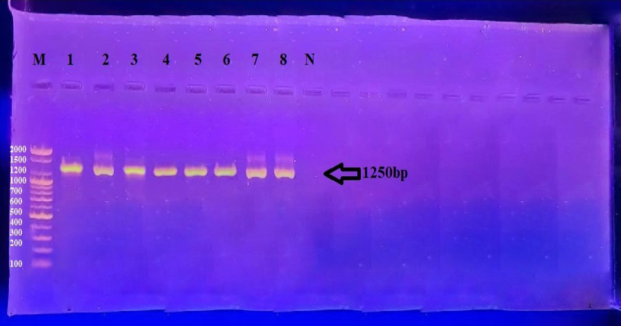
PCR amplification of Klebsiella pneumoniae showing a band size of 1250 bp. Lanes 1–3 represent human isolates, while lanes 4–8 represent canine isolates. PCR products were separated by electrophoresis on 1.5% agarose gel at 5 V/cm² using 1× TBE buffer for 1 h 30 min. Lane M: 100 bp DNA ladder.
References
- Abdullah, F.H. and Al-Gburi, N.M., 2023. Risk factors assessment and antimicrobial resistance of Salmonella isolates from apparently healthy and diarrheal dogs in Baghdad, Iraq. Iraqi J. Vet. Sci., 38(1): 155–162. https://doi.org/10.33899/ijvs.2023.139817.2984[CrossRef]
- Ahlam A. S. Al-Galebi , Mithal K. A. Al-Hassani , Hadaf Mahdi Kadhim , Hasanain A. J. Gharban.(2025). A periodical of the Network for the Veterinarians of Bangladesh (BDvetNET) ORIGINAL ARTICLE Phenotypic and molecular phylogeny of Klebsiella pneumoniae isolated from respiratory-diseased pet cats in Iraq. Journal of advanced Veterinary and animal research. 12(2): 621–628. http://doi.org/10.5455/javar.[CrossRef]
- Al-Galebi, A.A., Al-Hassani, M.K., Kadhim, H.M. and Gharban, H.A., 2025. Phenotypic and molecular phylogeny of Klebsiella pneumoniae isolated from respiratory-diseased pet cats in Iraq. J. Adv. Vet. Anim. Res., 12(2): 621. https://doi.org/10.5455/javar.2025.l926[CrossRef]
- Ali, I.A.A. and Neelakantan, P., 2022. Antibiofilm activity of phytochemicals against Enterococcus faecalis: A literature review. Phytother. Res., 36(7): 2824–2838. https://doi.org/10.1002/ptr.7488[CrossRef]
- Arkan, M., Yosif, Z. and Hussein, L.M., 2020. Phenotypic study of Klebsiella pneumoniae and Enterobacter cloacae isolated from oral cavity infections in Najaf Province, Iraq. Technol. Rep. Kansai Univ. 62(7): 7.
- Bata, S.I., Andua, O.A., Maimadu, A., Sabo, J.A., Mayowa, O. and Waziri, I.A., 2020. Oral cavities multidrug resistant bacteria colonization in apparently healthy dogs in Jos, Plateau state, Nigeria. Sci. World J., 15: no. 1: 15–20.
- Bengoechea, J.A. and Pessoa, S.A., 2019. Klebsiella pneumoniae infection biology: Living to counteract host defences. FEMS Microbiol. Rev., 43(2): 123-144. https://doi.org/10.1093/femsre/fuy043[CrossRef]
- Cattoir, V., 2021. The multifaceted lifestyle of enterococci: Genetic diversity, ecology and risks for public health. Curr. Opin. Microbiol., 65: 73–80. https://doi.org/10.1016/j.mib.2021.10.013[CrossRef]
- Chen, C.M., Tang, H.L., Chiou, C.S., Tung, K.C., Lu, M.C. and Lai, Y.C., 2021. Colonization dynamics of Klebsiella pneumoniae in the pet animals and human owners in a single-household. Vet. Microbiol., 256: 109050. https://doi.org/10.1016/j.vetmic.2021.109050[CrossRef]
- Da Silva, L., Grecellé, C.Z., Frazzon, A.P.G., Streck, A.F., Kipper, D., Fonseca, A.S.K., Ikuta, N. and Lunge, V.R., 2024. Multidrug-resistant Enterococcus faecium and Enterococcus faecalis isolated from dogs and cats in Southern Brazil. Microbiol. Res., 15(3): 1083–1090. https://doi.org/10.3390/microbiolres15030071[CrossRef]
- Dong, N., Yang, X., Chan, E. W. C., Zhang, R., & Chen, S. (2022). Klebsiella species: taxonomy, hypervirulence and multidrug resistance. EBioMedicine, 79.
- Eke, P.I., Thornton-Evans, G.O., Wei, L., Borgnakke, W.S., Dye, B.A. and Genco, R.J., 2018. Periodontitis in US adults: National health and nutrition examination survey 2009-2014. J. Am. Dental Assoc., 149(7): 576-588. https://doi.org/10.1016/j.adaj.2018.04.023[CrossRef]
- Flancman, R., Singh, A. and Weese, J.S., 2018. Evaluation of the impact of dental prophylaxis on the oral microbiota of dogs. PLoS One, 13(6): e0199676. https://doi.org/10.1371/journal.pone.0199676[CrossRef]
- Gerald, P.C., 2013. Oral microbiome homeostasis: The new frontier in oral care therapies. J. Dent. Oral Disord. Ther., 1(3). https://doi.org/10.15226/jdodt.2013.00105[CrossRef]
- Hessan, H.S., and Mohammed, R.A.K. (2021). Isolation and molecular detection of Klebsiella pneumoniae isolated from patients with oral inflammation plus pneumonia and its effect on secretory Iga levels.
- Huang, J., 2025. Multidrug-resistant Enterococcus faecalis from Yak Feces: Isolation, genomic characterization and functional insights. Vet. Sci., 12(11): 1077. https://doi.org/10.3390/vetsci12111077[CrossRef]
- Janda, J.M. and Abbott, S.L., 2006. The genera Klebsiella and Raoultella. Enterobacteria, 2: 115-129.
- Kaittan, Z.Q. and Zghair, Z.R., 2025. Enterococcus spp. from the oral cavity and wounds of slaughterhouse workers in Baghdad city. J. Med. Pharm. Chem. Res., 7: 1174-1186.
- Lateef, L.I., Mohammed, H.F., Al-Nasiry, B.S. and Kamal, I.M., 2025. The antibacterial effect of cinnamon ethanolic extract against Proteus spp. isolated from dogs. J. Anim. Health Prod., 13(s1). https://doi.org/10.17582/journal.jahp/2025/13.s1.278.284[Full text][CrossRef]
- Liu, J., Spencer, N., Utter, D.R., Grossman, A., Santos, N.C.D., Shi, W., Baker, J.L., Hasturk, H., He, X. and Bor, B., 2023. Persistent enrichment of multidrug resistant Klebsiella in oral and nasal communities during long-term starvation. bioRxiv, 12(18): 572173. https://doi.org/10.1101/2023.12.18.572173[CrossRef]
- Mohammed, H.F. and Al-Gburi, N.M., 2023. Antimicrobial resistance and biofilm formation of enterococcus spp isolated from human and pet animals. Eastern J. Agric. Biol. Sci., 3(1).
- Mohammed, H.F. and Kamal, I.M., 2025. Review of multidrug sensitivity and resistance in Enterococcus.
- Ofukwu, R.A., Akwuobu, C.A. and Oboegbulem, S., 2008. Presence and isolation pattern of zoonotic bacteria in oral cavities of dogs in peri-urban areas of Makurdi, Nigeria. J. Appl. Biosci., 11: 602-606. www.biosciences.elewa.org 602.
- Razali, K., Kaidi, R., Abdelli, A., Menoueri, M.N. and Ait-Oudhia, K., 2020. Oral flora of stray dogs and cats in Algeria: Pasteurella and other zoonotic bacteria. Vet. World, 13(12): 2806–2814. https://doi.org/10.14202/vetworld.2020.2806-2814[CrossRef]
- Siddique, A.B., Ahmed, S., Shahadat, M.N., Hasan, M., Ahmed, A. and Rahman, M.H., 2025. Uncovering antimicrobial resistance patterns in Pseudomonas aeruginosa and Enterococcus faecalis isolated from farm animals in Bangladesh. Discov. Bacteria, 2(1). https://doi.org/10.1007/s44351-025-00018-6[CrossRef]
- Srinivasan, R., Karaoz, U., Volegova, M., MacKichan, J., Kato-Maeda, M., Miller, S., Nadarajan, R., Brodie, E. L. and Lynch, S.V., 2015. Use of 16S RRNA gene for identification of a broad range of clinically relevant bacterial pathogens. PLoS One, 10(2): e0117617. https://doi.org/10.1371/journal.pone.0117617[CrossRef]
- Werner, G., Neumann, B., Weber, R.E., Kresken, M., Wendt, C., Bender, J.K., Becker, K., Borgmann, S., Diefenbach, A., Hamprecht, A., Hogardt, M., Wichelhaus, T., Kemp, V., Huebner, N., Kaasch, A., Geginat, G., Kohnen, W., Menzer, A., Krause, T. and Döring, S., 2020b. Thirty years of VRE in Germany – “expect the unexpected: The view from the National Reference Centre for Staphylococci and Enterococci. Drug Resist. Updates, 53: 100732. https://doi.org/10.1016/j.drup.2020.100732[CrossRef]
- Zhang, S., Yang, G., Ye, Q., Wu, Q., Zhang, J. and Huang, Y., 2018. Phenotypic and genotypic characterization of Klebsiella pneumoniae isolated from retail foods in China. Front. Microbiol., 9: 289. https://doi.org/10.3389/fmicb.2018.00289[CrossRef]
- Zhang, Z., Zhang, L., Dai, H., Zhang, H., Song, Y., An, Q., Wang, J. and Xia, Z., 2022. Multidrug-Resistant Klebsiella pneumoniae complex from clinical dogs and cats in China: Molecular characteristics, phylogroups, and hypervirulence-associated determinants. Front. Vet. Sci., 10(9): 816415. https://doi.org/10.3389/fvets.2022.816415[CrossRef]
Services
Article usage statistics
Loading…