
Oocytes grow and develop within several distinct ovarian follicles during the fetal and postnatal periods, and they ovulate in each estrous or menstrual cycle from ovarian follicles after puberty. Ovarian follicles start to form when oogonia are surrounded by a single layer of pregranulosa follicular cells and are called primordial follicles. Primordial follicles grow and develop into further stages upon activation. The growth and development of primordial follicles to pre-ovulatory follicles in mammals lasts several months. Oocytes acquire developmental competence during the preantral and antral ovarian follicle stages. The final maturation stage of ovarian follicles occurs during estrous or menstrual cycles. Changes occur in the cytoplasts, nuclei, and the surrounding follicular cells of oocytes for completion of meiotic maturation, fertilization, and further embryo development either in vivo or in vitro. The cytoplasmic maturation of oocytes helps to surpass the maternal zygotic block and progress for further embryo development. Hence, the cytoplasts of enucleated germinal vesicle (GV) or mature (metaphase II; MII) oocytes control development after embryonic/somatic nuclear transfer. Therefore, this review was conducted to collect and consolidate the current knowledge of cytoplast and nuclear material effects on maturation of intact and reconstructed oocytes and further developmental competence of embryos in vivo and in vitro and their importance for nucleus reprogramming and fertility.
Review · Volume 57, Issue 5, 29 August 2025 · Open Access
Cytoplasmic and Nuclear Maturation of Intact and Reconstructed Oocytes Controlling the Developmental Competence of Embryos
· · · · · ·
Affiliations & Notes ⌄
- 1Department of Animal and Fish Production, King Faisal University, P.O. Box 402, Al-Ahsa 31982, Saudi Arabia 2College of Human Medicine, Assiut University 7111, Egypt 3Department of Biology, College of Science, Taibah University, Al-Madinah Al-Munawwarah, 41321, Saudi Arabia. * Corresponding author: aamohammed@kfu.edu.sa
Article Info ⌄
- Journal:
- Pakistan Journal of Zoology
- Published:
- 29 August 2025

From the article
- Oocytes grow and develop during fetal and postnatal periods within several distinct ovarian follicles, and they ovulate in each estrous or menstrual cycle from ovarian follicles after puberty.
- The higher the follicle diameter and oocyte quality, the higher the maturation, fertilization, and developmental competence of the embryo.
- Estrous or menstrual cycle synchronization and superovulation protocols for ovarian follicles have been used in different species for embryo transfer.
- Assisted reproductive techniques (ART) are used to treat infertility, save endangered species, enhance meat and milk production through in vitro manipulations of germ cells, follicles, oocytes, and sperm.
Selected from the article's own text.
Abstract
INTRODUCTION
Oocytes grow and develop during fetal and postnatal periods within several distinct ovarian follicles, and they ovulate from the ovarian follicles after puberty in each estrous or menstrual cycle. The formation of ovarian follicle begins with primordial follicles, where oogonia are surrounded by pregranulosa cells ; . The growth and development of primordial follicles occur upon activation of pregranulosa cells to reach the preantral and antral follicle stages ; (Fig. 1).
The changes that occur during follicle development include granulosa cell proliferation, oocyte growth, and theca cell differentiation . Preantral follicle growth is gonadotropin-independent, whereas antral follicle growth is gonadotropin-dependent . Tang et al. (2012) investigated the role of follicle stimulating hormone (FSH) in combination with growth and differentiation factor-9 or basic fibroblast growth factor on the survival, activation, and growth of bovine primordial follicles, which improved after long-term culture of ovarian tissue.
Most of the information on ovarian follicle growth and development stems from studies on ruminants, swine,
rodent species, and humans ; ; ; ; . Several factors such as species ; , nutrition and feed additives ; ; , 2018; , 2019; ; ; ; ; ; , b) and males have effect on ovarian follicle growth and development. In addition, recently adapted techniques of ovarian synchronization, ovarian superovulation, and ovarian transplantation for dysfunction treatment and the maximizing of reproduction are known to change ovarian follicle growth and the development of the oocytes they contain ; (Fig. 2). Superovulation protocol via gonadotrophin stimulation might result in abnormal changes in cytoplast and nucleus structure, lower the concentration of estradiol hormone, and disrupt cumulus cell mRNA ; . Moreover, Kanitz et al. (2002) found that high doses of FSH could lead to defects in ovulation at the level of the pituitary gland and ovaries. Consequently, the transfer of GV nuclei, nucleoli, or cytoplasts of highly competent oocytes to abnormal oocytes through adapted techniques may treat abnormalities in oocytes.
Oocytes acquire developmental competence during ovarian follicle development through changes in the nucleus and cytoplast. Therefore, the recipient cytoplast and donor nucleus stages of artificial gametes affect developmental competence. Such artificial gametes may be used for different purposes, such as studying of embryonic/somatic nucleus reprogramming in recipient cytoplasts, production of cloned and transgenic animals, and treatment of infertility ; ; ; ; . The roles of cytoplasmic maturation and nuclear progression to the MII stage of intact and reconstructed oocytes in controlling developmental competence of oocytes to embryos are compiled and discussed in this review.
ASPIRATION AND DEVELOPMENTAL COMPETENCE OF CUMULUS-ENCLOSED GERMINAL VESICLE OOCYTE
Cumulus-enclosed germinal vesicle oocytes (COCs) can be aspirated or picked-up (OPU) from different animals, humans, or slaughterhouse ovaries ; ; ; ; , b) for in vitro embryo production. The OPU approach has been applied in humans to overcome abnormalities such as hidden ovaries, severe tubal diseases, or multiple adhesions with a success rate lower than 50%. Oocytes are picked up from genetically superior animals before puberty, early gestation, and early postpartum through laparoscopic and ultrasonic methods to shorten the interval between generations and increase animal production ; . COCs have been used for in vitro embryo production through oocyte maturation (IVM), fertilization or activation (IVF), and culture (IVC) of different animal species and humans. The development of COCs to blastocyst stage does not exceed 30-40% ; . Several factors, such as ovary storage time, wave of estrous cycle, follicle diameter, oocyte quality, incubation time, maturation environment, nutrition and feed additives, and male effect, affect the developmental competence of aspirated oocytes to embryos ; ; , 2018; (Table I).
The development of oocytes to embryos increases when slaughterhouse ovaries are exposed to warm conditions for a few h before oocyte aspiration. Storage of ovaries at 30 °C for 3-4 h after slaughter has led to an increase in the number of oocytes developed to embryos. It was suggested that a storage period of 4 h resulted in the formation of an environment within the 3-8 mm follicles, similar to that which occurs in preovulatory follicles. In our study, changes in follicular fluid (FF) composition at times of zero, 4, and 8 h in goat ovaries were investigated. The most notable changes in FF composition were the concentrations of glucose, cholesterol, and triglycerides, which are sources of energy for oocytes. This might partially explain the increase in the developmental competence of oocytes collected from the ovaries 4 h after slaughter but decrease thereafter.
Table I. Factors affecting developmental competence of aspirated oocytes in vitro.
| Ovaries | Treatments | Effects | References |
| Slaughterhouse ovaries | Storage the ovaries 30 °C for 3-4 h | Increase developmental competence of oocytes to embryos | Luu et al., 2011 |
| Defined and undefined media | Variable effects on oocyte maturation and embryo development | Mohammed et al., 2005; Madkour et al., 2016; Spacek and Carnevale, 2018; Abdoon et al., 2018 | |
| IGF-1 and FF | Variable effects on oocyte maturation and embryo development | Oberlender et al., 2013 | |
| Fetal calf serum | Increase developmental competence of oocytes to embryos | Mohammed et al., 2005; Moulavi and Hosseini, 2019 | |
| Follicular fluid | Variable effects on oocyte maturation and embryo development depending on follicle sizes collected and added percentage to the maturation medium | Mohammed et al., 2005; Sinclair et al., 2008 | |
| Co-culture cumulus and oviduct cells | Increase developmental competence of oocytes to embryos | Lee et al., 2018 | |
| Cumulus cells surrounding the oocytes | Increase developmental competence of oocytes to embryos | Mohammed 2006; Mohammed et al., 2005, 2008, 2010, 2019; Al-Zeidi et al., 2022 | |
| Hormone | Increase developmental competence of oocytes to embryos | Moulavi and Hosseini, 2019 | |
| Follicule size | Variable effects where the increase of follicle sizes enhance developmental competence of oocytes to embryos | Shabankareh et al., 2014; Gordon, 2003; Maylinda et al., 2018 | |
| Live organisms | Follicule size | Variable effects on oocyte maturation and embryo development where the higher follicle size the higher developmental competence of oocyte to embryos | Hasler et al., 1998; Maylinda et al., 2018; Raj et al., 2018 |
| Follicular wave | Variable effects on oocyte maturation and embryo development | Gordon et al., 2003; Cavalieri et al., 2018; Baby and Bartlewski, 2011 | |
| Follicular and luteal stages | Variable effects on oocyte maturation and embryo development | Gordon et al., 2003 | |
| Nutrition | Variable effects on oocyte maturation and embryo development | Gordon et al., 2003 | |
| Feed additives | Variable effects on oocyte maturation and embryo development | Senosy et al., 2017, 2018; Mohammed and Al-Hozab, 2018 |
The more cumulus cells surround the oocytes and homogeneity of the oocytes cytoplasts, the greater the developmental competence of oocytes to embryos . In addition, the male effect stimulation of super ovulated females during the last phase of the FSH regimen increase the number and diameter of large follicles, in addition to the number of corpora lutea and viable embryos, compared to unstimulated ewes . Therefore, these factors should be taken into consideration for enhancement of oocyte quality, oocyte maturation, fertilization, and development of embryos.
OOCYTE QUALITY AND GENE EXPRESSION
Oocyte quality is critical for maturation, fertilization, and embryonic and fetal development. The higher the oocyte quality, the higher the developmental competence of the resulting embryo and fetus. Oocytes are graded morphologically according to the cumulus cells surrounding the oocytes, diameter of the oocyte, and homogeneity of the cytoplasts ; ; . Castaneda et al. (2013) found that both active mitochondria and lipid content were correlated with oocyte diameter. In addition, brilliant cresyl blue staining (BCB) were used to select developmentally competent GV oocytes of different species ; ; . Bovine GV oocytes stained with +BCB had a relatively higher developmental competence than non-stained BCB oocytes. A previous study investigated the relationship between BCB staining, lipid content, and active mitochondria. They concluded that higher lipid content of bovine oocytes stained with BCB might provide higher developmental competence. In addition, Sinclair et al. (2008) investigated the amino and fatty acid content in the follicular fluid of bovine follicles as a predictor of embryo development. They found that two amino acids (alanine and glycine) had the highest value for predicting early embryonic development, and this might serve as a useful tool in measuring COCs quality.
In recent years, transcriptome and proteome have been substantially explored as biomarkers for oocyte quality owing to the progression of the new technologies. The approach is an invaluable tool for clinical practice in assisted reproductive technologies, obstetrics, and gynecology . The expression patterns of glucose metabolism genes (G6PDH, LDH, and PDH) were investigated in buffalo oocytes matured under different glucose concentrations . The pattern of G6PDH expression during oocyte maturation and early embryonic development is predictive of oocyte quality and developmental competence of embryos. However, Liu et al. (2021) found in good prognosis IVF patients that mitochondrial DNA copy number of cumulus cells is not linked to embryo implantation. Therefore, further studies on gene expression are required to determine the relation between oocyte quality and further development of oocytes into preimplantation and post implantation embryo stages.
NUCLEAR AND CYTOPLASMIC MATURATION OF INTACT OOCYTES
Oocytes require several months to acquire developmental competence before maturation ; . Small follicles contain inhibiting maturation substances within the follicles, which disappear in the preovulatory follicle simultaneously with the LH surge. Therefore, oocytes resume the first meiotic division to the metaphase II (MII) stage in vivo after the LH surge and in vitro when they are removed from antral follicles and cultured under favorable in vitro conditions (38.5 °C, 90% humidity, 5.0% CO2).
Oocyte maturation in vitro was described nearly 85 years ago when rabbit and human oocytes were aspirated from follicles and cultured in a favorable medium . It has been indicated that the developmental competence of in-vitro matured oocytes is lower than that of in-vivo matured oocytes ; ; due to insufficient cytoplasmic maturity . The longer the maturation time of oocytes, the lower the developmental competence of the resulting embryos, as indicated in swine and camel oocytes, compared to oocytes of other species ; ; .
The morphological maturation stages of intact GV or reconstructed GV oocytes (Figs. 3 and 4) include oocyte nuclear and cytoplasmic maturation, in addition to cumulus expansion of the surrounding cells (Fig. 4). The germinal vesicle of denuded oocytes is visible in rodent, rabbit, and human oocytes, whereas it is not visible in ruminant oocytes because of the presence of lipid droplets in the cytoplasm. Gordon (2003) described the chronology of events during bovine oocyte maturation as inductive and synthetic phases. The nuclear changes included germinal vesicle breakdown (GVBD), which occurred at 2-3 h in rodent oocytes and 6-8 h in ruminant oocytes, respectively. Rodent oocytes reached the MI stage at 11-13 h and MII stage at 15-17 h. The ruminant oocytes reached the MI stage at 15-21 h and MII stage at 24 h ; ; , 2010, 2019).
Follicle size, follicular wave during the estrous cycle, follicular and luteal stage of estrous, species and age ; , nutrition and feed additives ; , 2019; ; ; ; ; ; , and male effects were found to affect oocyte maturation. In addition, super stimulation of ovarian follicles via gonadotropin injections resulted in changes in small, medium, and large follicles according to the number and dose of gonadotropin injections and side of the ovary . FSH stimulates transcription and translation in ovarian granulosa cells, which are essential for female reproductive endocrine regulation . The timing of fully grown germinal vesicle oocytes to reach the MII stage in vitro for different species and the various approaches for improving maturation are presented in Table II ; ; . Enrichment of maturation media with cumulus cells, hormones, growth factors, and other factors improves oocyte maturation and embryo development.
CYTOPLASMIC MATURATION AND NUCLEAR REPROGRAMMING OF RECONSTRUCTED OOCYTES AND DEVELOPMENTAL COMPETENCE
Germinal vesicle nucleus, nucleolar, or cytoplasmic dysfunction of oocytes may be associated with infertility in animal and human oocytes ; ; . Oocyte GV nuclei, nucleoli, MII spindles, and cytoplast transfer techniques have been used for the treatment of oocyte dysfunction due to cytoplasmic, nuclear, or nucleolar reasons or to increase their developmental competence ; ; Benc et al., 2018). Hoseini et al. (2016) concluded that cytoplast transfer technique is not effective for cytoplasmic maturity of recipient GV oocytes; however, the presence of cumulus cells during oocyte maturation or GV nucleus or nucleolus transfer is effective in increasing the developmental competence of the resulting embryos. Chang et al. (2005) found that the developmental incompetency of denuded mouse GV oocytes matured in vitro is ooplasmic in nature and is associated with aberrant Oct-4 expression.
One of the fundamental factors for successful assisted reproductive technologies (ART) in reconstructed oocytes is the interaction between the recipient cytoplast and donor nucleus ; ; , 2010; . Oocytes at stages GV, ProMI, MI, and MII were enucleated to obtain recipient cytoplasts. The obtained cytoplasts can be used in cell biology for basic research and reprogramming of the introduced germ, embryonic, or somatic nuclei during transfer ; ; , 2010, 2019). Such approaches might solve the problem of low developmental competence of oocytes from prepubertal or advanced maternal age (Hassold and Chiu, 2003) due to developmental incompetency of ooplasmic or nuclear nature . The following questions arise: Does the transfer of nuclei from non-growing, growing, or aging GV oocytes into fully grown GV cytoplasts solve infertility problem? Does the transfer of MII spindles to competent mature MII cytoplasts rescue infertility in animals and humans? Does the transfer of competent nucleoli from donor fully grown GV oocytes to recipient GV oocytes containing abnormal nucleoli rescues infertility?
Table II. Duration of oocyte maturation in vitro from germinal vesicle to metaphase II stage using different supplements to maturation media in different animal species and human.
| Species | Duration, h | Treatments | References |
| Rodents | 17 | Cumulus cells Cumulus cells Cumulus cells Perfluorooctane CRH and ACTH | Grabarek et al., 2004 Mohammed 2006, 2008 Mohammed et al., 2006, 2008, 2010 Wei et al., 2021 Gong et al., 2021 |
| Goat | 24 | Cysteamine, leukemia inhibitory factor, and Y27632 Physiological oocyte maturation Leptin | An et al., 2018 Suresh et al., 2021 de Senna Costa et al., 2022 |
| Sheep | 24 | Resveratrol Oxygen tension C-type natriuretic peptide | Zabihi et al., 2020 Sánchez-Ajofrín et al., 2020 Zhang et al., 2018 |
| Cattle | 24 | FF and FCS Retinoic acid Lycopene | Mohammed et al., 2005 Borges et al., 2021 Residiwati et al., 2021 |
| Buffalo | 24 | - Brain-derived neurotrophic factor Retinoic acid | Marin et al., 2019 Zhao et al., 2019 Gad et al., 2018 |
| Rabbit | 24 | Gonadotropin-releasing hormone - | Yoshimura et al., 1991 Arias-Álvarez et al., 2017 |
| Human | 24 | - - Coenzyme Q10 A fertilin-derived peptide | Chian et al., 2004 Baruffi et al., 2004 Ma et al., 2020 Sallem et al., 2022 |
| Camel | 42-48 | - Macromolecule Roscovitine | Wani et al., 2018 Moulavi and Hosseini, 2019 Wani and Hong 2018; 2020 |
| Pig | 48 | Dihydroartemisinin Zearalenone CRH and ACTH Allicin | Luo et al., 2018 Wang et al., 2022 Gong et al., 2021 Li et al., 2022 |
Therefore, the techniques of enucleation of denuded GV, cumulus-enclosed GV, and enucleolation of GV oocytes were adapted for such studies ; Mohammed et al., 2008, 2010, 2019). Our unique enucleation and enucleolation techniques (Figs. 5 and 6) of denuded and cumulus-enclosed GV oocytes enabled us to confirm the role of cumulus cells, nuclear material, and nucleoli on oocyte maturation and embryo development. Complete enucleation of denuded GV oocytes removes the entire GV nucleus. Therefore, the resulting cytoplast does not contain any GV components or cumulus effects. Selective enucleation of denuded GVs involves the removal of nuclear membrane, leaving the nucleolus and nuclear sap in the cytoplast; however, selective enucleation of cumulus-enclosed GVs leaves the effect of nucleolus, nuclear sap, and cumulus cells in the cytoplast (Fig. 5). Therefore, the aforementioned techniques and enucleolation confirmed the role of cumulus cells, nuclear material, and nucleolus in oocyte maturation and developmental competence.
Our results of selective enucleation of GV oocytes yielded GV cytoplasts in similar quantity as did complete enucleation of GV oocytes ; , 2010). Therefore, the aforementioned techniques and enucleolation confirmed the role of cumulus cells, nuclear material, and nucleolus in oocyte maturation and further developmental competence of embryos. Our results indicated that GVsel cytoplasts provided proper timing of maturation after reconstruction with GV, contrary to GV cytoplasts, which accelerated timing of maturation. However, this did not improve haploidization, as measured with abnormal nuclear morphology. Abnormal acceleration of maturation cannot be rescued by any particular cell cycle stage or developmental stage of blastomeres 1/2, 1/4 or 1/8 used as nuclear donor. Highest maturation efficiency of GV cytoplasts together with their least disturbance from abnormal meiosis were provided by G2 donor blastomere nuclei ; , 2010, 2019, 2022).
The nucleolus and cumulus cells were confirmed to be dispensable for GV oocyte maturation, but the resulting embryos are blocked at the two-cell stage with an anucleolated nucleus (Fig. 7). Therefore, selective enucleation as countered by enucleolation revealed the influence of GV contents on pronuclear formation. Furthermore, complete enucleation of denuded GV oocytes followed by G2/M embryonic or fetal fibroblast nuclear transfer indicate maturation with abnormalities such as acceleration of polar body extrusion, large polar body, and misalignment of chromosomes over the MII spindle, in addition to blocked embryo development at the one-cell stage after activation or fertilization. Surprisingly, no nucleoli were observed in the developed embryos. In addition, upon fertilization, one pronucleus was formed in the cytoplast, and was expected to be somatic, whereas the male pronucleus was not formed and was found as scattered chromosomes in the cytoplast (Fig. 8). This expectation is based on the fact that the pronucleus close to the polar body is somatic and the pronucleus farthest from the polar body is spermatic. This was confirmed through maturation of GV cytoplasts for 17 h followed by fertilization. Fertilizing sperm in the cytoplasm remained in a highly condensed head form , 2010, 2019, 2022).
Upon selective enucleation of denuded GV oocytes, reconstructed with G2/M embryonic nucleus, followed by maturation and fertilization, both pronuclei were formed in addition to further developmental competence to morula/blastocyst (Fig. 9 and 10). Although activated reconstructed GVsel cytoplasts were also cleaved and few of them developed to the blastocyst stage the frequency of cleavage to the 4-cell stage and blastocyst formation was evidently lower than those of reconstructed GVCsel cytoplasts (38.5% vs 71.4% and 7.7% and 23.8%, respectively). Also, the mean cell number in the obtained blastocysts was slightly higher in blastocysts derived from activated reconstructed GVCsel cytoplasts. What is deserved to observe is the absence or lower cells of inner cell mass in the cloned developed blastocysts over somatic or embryonic nuclear transfer to GVCsel cytoplasts compared to normal developed blastocysts. The number and quality of inner cell mass is the most important character of the developed blastocysts to give birth. Zhu et al. (2022) reported that the poor morphology of inner cell mass (grade C) of the developed embryos increases birth weight and large for gestational age compared to inner cell mass of grade A. in addition, Plana-Carmona et al. (2022) confirmed that the trophectoderm acts as a niche for the inner cell mass through C/EBPα-regulated IL-6 signaling. Therefore, further studies are required for investigating the reasons of absence or poor quality inner cell mass of the developed blastocysts from GV cytoplasts after embryonic nuclear transfer.
This very interesting technique of enucleation denuded or cumulus-enclosed GV oocytes could be applied with visible GV nucleus as in human, rodent and rabbit oocytes. In ruminant GV oocytes, ultra-centrifugation to 15,000 rpm is required for visualization of GV nucleus due to lipid droplets and the cumulus is stripped off during such centrifugation. In addition, the GV nucleus could be visible approximately in two third of the centrifuged GV oocytes.
Concerning the cell cycle stage of the introduced donor nuclei, our results and others indicate that G0/G1 cells or nuclei could be transferred to metaphase II cytoplasts, whereas G2/M cells or nuclei could be transferred to germinal vesicle cytoplasts ; , 2010, 2019, 2022). Donor cells or nuclei introduced in the S phase to the germinal vesicle cytoplast result in abnormalities in oocyte maturation and block further developmental competence of embryos. To the best of our knowledge, embryonic or somatic nuclear transfer to metaphase II cytoplasts results in deliveries with low efficiency ; , whereas with embryonic or somatic nuclear transfer to GVCsel cytoplast, the embryonic development reached to the hatched blastocyst stage , 2010). Upon morula and blastocysts transferred to the uterine of surrogate mothers, no delivery was confirmed in mice (Mohammed unpublished data).
Our complete and selective enucleation of GV yielded GV cytoplasts with high efficiency ; , 2010, 2019, 2022). The sole success of the recipient germinal vesicle cytoplast is the meiotic maturation of germinal vesicle karyoplasts; germ cells; primary or secondary spermatogonia ; . Germinal vesicle recipient cytoplasts are required in basic research for investigations of cell biology and donor cell reprogramming. Further studies are still required for improvement the outcomes of manipulation germinal vesicle oocytes in different animal species and human as well.
CONCLUSIONS
Oocytes grow and develop during fetal and postnatal periods within several distinct ovarian follicles, and they ovulate in each estrous or menstrual cycle from ovarian follicles after puberty. Trials were unsuccessful when germ cells were used for in vitro embryo production during the fetal period; however, they succeeded when germ cells were used of the prepubertal and post pubertal periods for in vitro embryo production. The higher the follicle diameter and oocyte quality, the higher the maturation, fertilization, and developmental competence of the embryo. Estrous or menstrual cycle synchronization and superovulation protocols for ovarian follicles have been used in different species for embryo transfer. In addition, ovarian tissue transplantation has been adopted for the treatment of infertility and conservation of species. Furthermore, defects in oocytes can be repaired via the nucleolus, germinal vesicle, and spindle transfer to the recipient oocyte or cytoplast. Primary and secondary spermatocytes and spermatids have been successfully used to fertilize GV and MII oocytes. Furthermore, germinal vesicle, embryonic, and somatic cells at different cell cycle stages (G0, S, and G/M stages) have been transferred to different cytoplasts (GV, ProMI, MI, and MII stages) with variable results. Assisted reproductive techniques (ART) are used to treat infertility, save endangered species, enhance meat and milk production through in vitro manipulations of germ cells, follicles, oocytes, and sperm. Studies on the kinetics or progression of oocyte maturation and further developmental competence of embryos are required, in addition to ovarian follicle gene expression. Although assisted reproductive techniques are still relatively rare and expensive, especially in third-world countries, their use has doubled over the past decade and thus necessitates further development.
Declarations
Acknowledgments
The authors would like to acknowledge the Deanship of Scientific Research, Vice Presidency for Graduate Studies and Scientific Research, King Faisal University, Saudi Arabia (GrantA051).
Funding
The study was funded by Funding of Scientific Research Deanship of King Faisal University (GrantA051)
IRB approval
The approval of the study was granted by the The Ethical Research Committee of King Faisal University.
Ethical statement
The proceedures and material of the current study was approved of the Scientific Research Deanship Ethical Standards of King Faisal University.
Statement of conflict of interest
The authors have declared no conflict of interest.
Figures
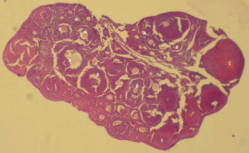
Ovarian follicle structures upon fallopian tube ligation in mice; preantral follicles (A), antral follicles (B) and corpora lutea (C) structures. N.B. Differences in sizes of oocytes and the layers of surrounding cells inside the follicles.
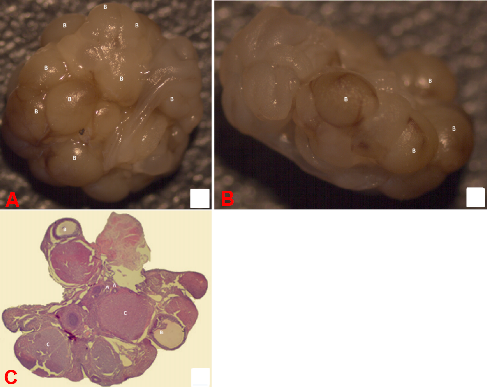
The structures of ovarian follicle in control (A) and ovarian transplanted tissues in rats (B, C). The images show preantral follicles (A), antral follicles (B), and corpora lutea (C).
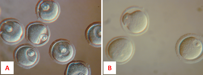
The germinal vesicle karyoplasts placed under the zona pellucida of enucleated germinal vesicle oocytes (A) and fused with cytoplasts (B).
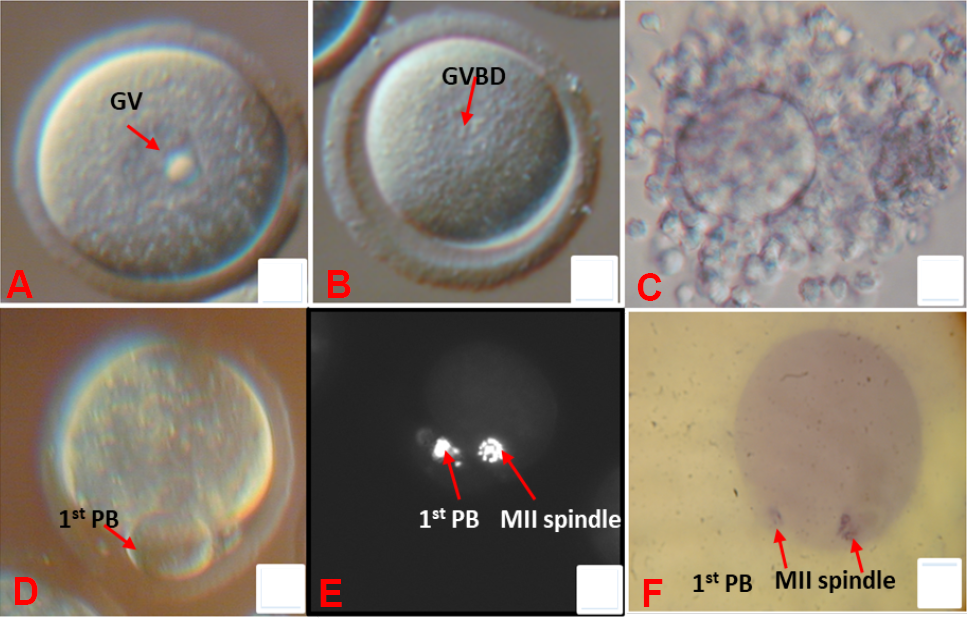
Oocyte maturation; denuded germinal vesicle oocyte (A), germinal vesicle breakdown (B), cumulus expansion (C), polar body extrusion (D), fluorescence staining of matured oocyte (E), hematoxylin stain of matured oocyte (F).
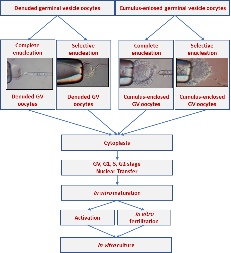
Obtaining types of recipient germinal vesicle cytoplasts, donor nuclei transfer and further meiotic maturation and developmental competence; GV germinal vesicle nucleus of fully-grown oocyte, G1 gap 1 stage, S S-phase stage, G2 gap 2 stage.
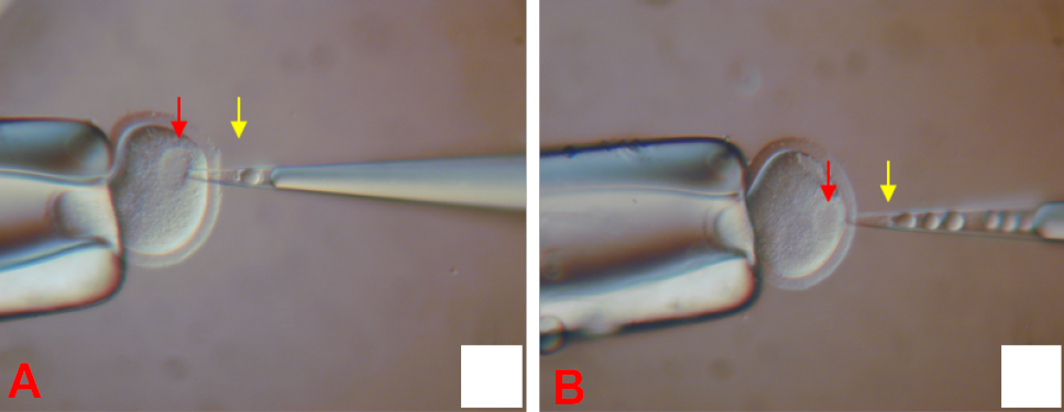
Enucleolation of fully grown germinal vesicle oocyte (A and B) the nucleolus-free germinal vesicles are visible (red arrows) in the cytoplasm. In the enucleolation pipettes, the nucleolus of GV oocyte undergoing enucleolation (A, yellow arrow) and several nucleoli of previously enucleolated GV oocytes (B yellow arrows) are visible.
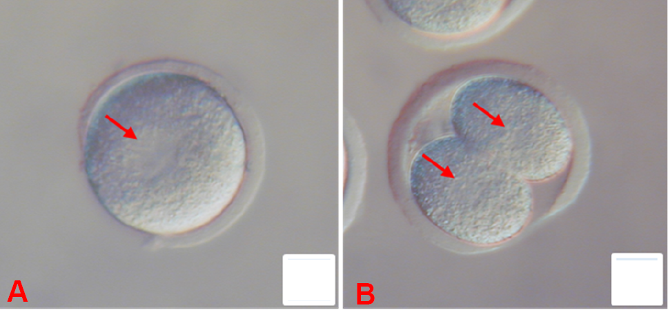
Pronuclei (arrow) in the zygote (A) and nuclei (arrow) in 2-cell embryo (B) obtained from enucleolated and fertilized fully grown germinal vesicle oocytes. Nucleoli are not visible both in pronuclei and in blastomere nuclei. In the zygote the second pronucleus is out of focus.
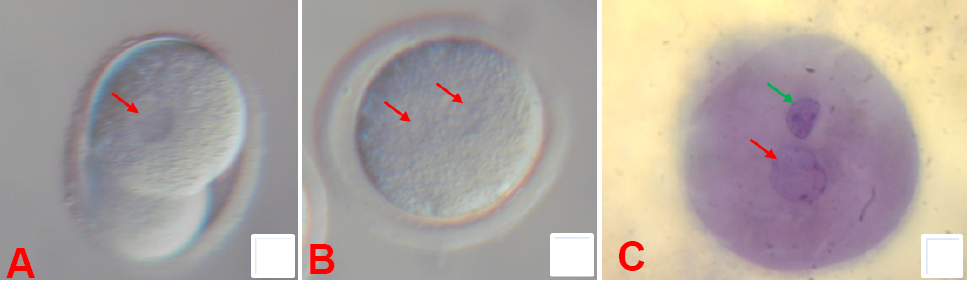
Pronuclear morphology in GV (A) and GVsel cytoplasts (B and C) reconstructed with embryonic/somatic nuclei and fertilized after in vitro maturation (light microscope, DIC). (A) One pronucleus formed of somatic origin without nucleoli (arrow) and extruded large PB; (B) Two pronuclei were formed of somatic and sperm origin and both contained nucleoli; (C) stained two pronuclei were formed of somatic (green arrow) and sperm (red arrow) origin and both contained nucleoli.
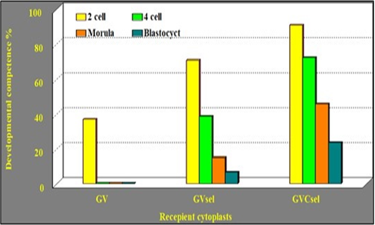
Effects of nuclear material and cumulus cells on developmental competence of cytoplasts reconstructed with ½-blastomere nuclei. GV removal the whole germinal vesicle of GV oocyte, GVsel removal nuclear material and membrane leaving nucleolus and nuclear sap in the cytoplast of denuded GV oocytes, GVCsel removal nuclear material and membrane leaving nucleolus and nuclear sap in the cytoplast of culumus enclosed GV oocytes.
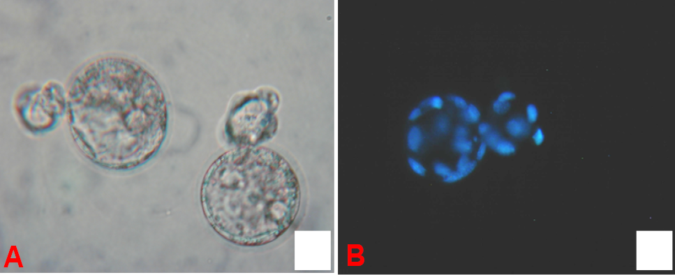
Hatching blastocysts developed from GVCsel cytoplasts reconstructed with 1/2-blastomere nuclei at G2/M phase (A) two hatching blastocysts under light microscope (DIC) and (B) one hatching blastocyst after Hoechst staining (fluorescence microscope).
References
- Abdelnaby, E.A., El-Maaty, A.M.A. and El-Badry, D.A., 2021. Evaluation of ovarian hemodynamics by color and spectral Doppler in cows stimulated with three sources of follicle-stimulating hormone. Reprod. Biol., 21: 100478. https://doi.org/10.1016/j.repbio.2020.100478[CrossRef]
- Abdoon, A.S., Fathalla, S.I., Shawky, S.M., Kandil, O.M., Kishta, A.A. and Masoud, S.R., 2018. In vitro maturation and fertilization of donkey oocytes. J. Equine Vet. Sci., 65: 118–122. https://doi.org/10.1016/j.jevs.2018.04.007[CrossRef]
- Al-Masruri, H., Al-Zeidi, R., Al-Mufarji, A., Mohammed, A.A., Al-Madani, A. and Mohammed, H., 2022. Leverage of Moringa oleifera supplementation on performances, biochemical, and milk profiles in mammals. Adv. Anim. Vet. Sci., 10: 2043-2050. https://doi.org/10.17582/journal.aavs/2022/10.9.2043.2050[Full text][CrossRef]
- Al-Masruri, H., Al-Zeidi, R., Al-Mufarji, A. and Mohammed, A.A., 2022a. Moringa oleifera leaves supplementation to anaesthetized mice associated with changes of thermo-tolerance parameters, blood and plasma indices. Fresen. Environ. Bull., 8: 8179-8187.
- Al-Mufarji, A., Mohammed, A.A., Al-Zeidi, R., Al-Masruri, H. and Mohammed, A., 2022b. Effects of Moringa oleifera on follicular development, blood and metabolic profiles of subtropical ewes during peripartum. Adv. Anim. Vet. Sci., 10: 1706-1712. https://doi.org/10.17582/journal.aavs/2022/10.8.1706.1712[Full text][CrossRef]
- Al-Zeidi, R., Al-Masruri, H., Al-Mufarji, A. and Mohammed, A.A., 2022a. Role of cumulus cells and follicular fluid on oocyte maturation and developmental competence of embryos: Intact and reconstructed oocytes. Adv. Anim. Vet. Sci., 10: 1219-1226. https://doi.org/10.17582/journal.aavs/2022/10.6.1219.1226[Full text][CrossRef]
- Al-Zeidi, R., Al-Masruri, H., Al-Mufarji, A., Mohammed, A.A. and Mohammed, H., 2022b. Changes in nucleus and cytoplast during oocyte maturation: Involvement in embryo production. Adv. Anim. Vet. Sci., 10: 2081-2089. https://doi.org/10.17582/journal.aavs/2022/10.9.2081.2089[Full text][CrossRef]
- Algriany, O.A., Vos, P., Groenendaal, H., van Gastel, A., Sirard, M.A. and Dieleman, S.J., 2007. Effects of oFSH stimulation on steroid concentrations in the fluid of preovulatory follicles during final maturation in relation to the LH surge in the cow. Expression analysis of genes implicated in meiotic resumption in vivo and developmental competence of bovine oocytes, pp. 33.
- An, L., Liu, J., Du, Y., Liu, Z., Zhang, F., Liu, Y., Zhu, X., Ling, P., Chang, S. and Hu, Y. 2018. Synergistic effect of cysteamine, leukemia inhibitory factor, and Y27632 on goat oocyte maturation and embryo development in vitro. Theriogenology, 108: 56–62. https://doi.org/10.1016/j.theriogenology.2017.11.028[CrossRef]
- Arias-Álvarez, M., García-García, R.M., López-Tello, J., Rebollar, P.G., Gutiérrez-Adán, A. and Lorenzo, P.L., 2017. In vivo and in vitro maturation of rabbit oocytes differently affects the gene expression profile, mitochondrial distribution, apoptosis and early embryo development. Reprod. Fertil. Dev., 29: 1667–1679. https://doi.org/10.1071/RD15553[CrossRef]
- Ata, B. and Telek, S.B., 2021. Assisted reproductive technology for women with endometriosis, a clinically oriented review. Curr. Opin. Obstet. Gynecol., 33: 225-231. https://doi.org/10.1097/GCO.0000000000000710[CrossRef]
- Baby, T.E. and Bartlewski, P.M., 2011. Circulating concentrations of ovarian steroids and follicle-stimulating hormone (FSH) in ewes with 3 or 4 waves of antral follicle emergence per estrous cycle. Reprod. Biol., 11: 19–36. https://doi.org/10.1016/S1642-431X(12)60061-8[CrossRef]
- Baerwald, A. and Pierson, R., 2020. Ovarian follicular waves during the menstrual cycle: Physiologic insights into novel approaches for ovarian stimulation. 2020. Fertil. Steril., 114: 443–457. https://doi.org/10.1016/j.fertnstert.2020.07.008[CrossRef]
- Bao, S., Hitoshi, U., Hirose, A., Aono, F., Ono, Y. and Kono, T., 2003. Development of bovine oocytes reconstructed with a nucleus from growing stage oocytes after fertilization in vitro. Theriogenology, 59: 1231-1239. https://doi.org/10.1016/S0093-691X(02)01174-3[CrossRef]
- Baruffi, R.L., Avelino, K.B., Petersen, C.G., Mauri, A.L., Garcia, J.M. and Franco, J.G., 2004. Nuclear and cytoplasmic maturation of human oocytes cultured in vitro. Fertil. Steril., 82: S265–S266. https://doi.org/10.1016/j.fertnstert.2004.07.709[CrossRef]
- Benc, M., Strejček, F., Morovič, M., Murín, M., Martínková, S., Jettmarová, D. and Laurinčík, J., 2019. Enucleolation and nucleolus transfer in mammalian oocytes and zygotes. Int. J. Dev. Biol., 63: 253–258. https://doi.org/10.1387/ijdb.190002mb[CrossRef]
- Blondin, P., Coenen, K., Guilbault, L.A. and Sirard, M.A., 1997. In vitro production of bovine embryos: Developmental competence is acquired before maturation. Theriogenology, 47: 1061–1075. https://doi.org/10.1016/S0093-691X(97)00063-0[CrossRef]
- Borges, M.A., Sousa, F.S.S., Paschoal, J.D., Lopes, I.A.R., Feijó, A.L.S., Neto, A.C.P.S., Silva, P.L., Seixas, F.K. and Collares, T., 2021. Effect of supplementation of medium with Bauhinia for ficata recombinant lectins on expression of oxidative stress genes during in vitro maturation of bovine oocytes. Reprod. Toxicol., 103: 64–70. https://doi.org/10.1016/j.reprotox.2021.05.012[CrossRef]
- Bunel, A., Nivet, A.L., Blondin, P., Vigneault, C., Richard, F.J. and Sirard, M.A., 2020. The effects of LH inhibition with cetrorelix on cumulus cell gene expression during the luteal phase under ovarian coasting stimulation in cattle. Domest. Anim. Endocrinol., 72: 106429. https://doi.org/10.1016/j.domaniend.2019.106429[CrossRef]
- Campbell, K.H., Loi, P., Otaegui, P.J. and Wilmut, I., 1996. Cell cycle co-ordination in embryo cloning by nuclear transfer. Rev. Reprod., 1: 40–46. https://doi.org/10.1530/ror.0.0010040[CrossRef]
- Castaneda, C.A., Kaye, P., Pantaleon, M., Phillips, N., Norman, S., Fry, R. and Michael, J.D., 2013. Lipid content, active mitochondria and brilliant cresyl blue staining in bovine oocytes. Theriogenology, 79: 417–422. https://doi.org/10.1016/j.theriogenology.2012.10.006[CrossRef]
- Cavalieri, F.L.B., Morotti, F., Seneda, M.M., Colombo, A.H.B., Andreazzi, M.A., Emanuelli, I.P. and Rigolon, L.P., 2018. Improvement of bovine in vitro embryo production by ovarian follicular wave synchronization prior to ovum pick-up. Theriogenology, 117: 57–60. https://doi.org/10.1016/j.theriogenology.2017.11.026[CrossRef]
- Chakravarthi, V.P., Ratri, A., Masumi, S., Borosha, S., Ghosh, S., Christenson, L.K., Roby, K.F., Wolfe, M. W. and Rumi, M.A.K., 2021. Granulosa cell genes that regulate ovarian follicle development beyond the antral stage: The role of estrogen receptor β. Mol. Cell. Endocrinol., 528: 111212. https://doi.org/10.1016/j.mce.2021.111212[CrossRef]
- Chang, H.C., Liu, H., Zhang, J., Grifo, J. and Krey, L.C., 2005. Developmental incompetency of denuded mouse oocytes undergoing maturation in vitro is ooplasmic in nature and is associated with aberrant Oct-4 expression. Hum. Reprod., 20: 1958–1968. https://doi.org/10.1093/humrep/dei003[CrossRef]
- Chian, R.C., Buckett, W.M. and Tan, S.L., 2004. In-vitro maturation of human oocytes. Reprod. Biomed. Online, 8: 148–166. https://doi.org/10.1016/S1472-6483(10)60511-1[CrossRef]
- Dai, X.X., Jiang, Z.Y., Wu, Y.W., Sha, Q.Q., Liu, Y., Ding, J.Y., Xi, W.D., Li, J. and Fan, H.Y. 2021. CNOT6/6L-mediated mRNA degradation in ovarian granulosa cells is a key mechanism of gonadotropin-triggered follicle development. Cell Rep., 37: 110007. https://doi.org/10.1016/j.celrep.2021.110007[CrossRef]
- de Senna Costa, J.A., Cezar, G.A., Monteiro, P.L.J., Silva, D.M.F., Silva, R.A.J.A., Bartolomeu, C.C., dos Santos Filho, A.S., Wischral, A. and Batista, A.M., 2022. Leptin improves in-vitro maturation of goat oocytes through MAPK and JAK2/STAT3 pathways and affects gene expression of cumulus cells. Reprod. Biol., 22: 100609. https://doi.org/10.1016/j.repbio.2022.100609[CrossRef]
- Demyda-Peyrás, S., Dorado, J., Hidalgo, M., Anter, J., de Luca, L., Genero, E. and Moreno-Millán, M., 2013. Effects of oocyte quality, incubation time and maturation environment on the number of chromosomal abnormalities in IVF-derived early bovine embryos. Reprod. Fertil. Dev., 25: 1077–1084. https://doi.org/10.1071/RD12140[CrossRef]
- Dolma, P., Ali, A., Sobti, R.C. and Chauhan, A., 2022. Animal cloning: Perspectives for futuristic medicine. In: Advances in animal experimentation and modeling, Elsevier. pp. 53–67. https://doi.org/10.1016/B978-0-323-90583-1.00008-8[CrossRef]
- Fernandes, C.C.L., Martins, L.T., Gaudêncio Neto, S., Tavares, K.C.S., Aguiar, L.H., Calderón, C.E.M., Silva, A.M., Alves, J.P.M., Silva, C.M.G., Rossetto, R., Bertolini, L.R., Bertolini, M. and Rondina, D., 2016. Gene expression, oocyte quality and embryo production by cloning in goats supplemented with different diets. Small Rumin. Res., 144: 255-262. https://doi.org/10.1016/j.smallrumres.2016.09.023[CrossRef]
- Fulka, H., Mrazek, M. and Fulka Jr, J., 2004. Nucleolar dysfunction may be associated with infertility in humans. Fertil. Steril., 82: 486–487. https://doi.org/10.1016/j.fertnstert.2003.12.042[CrossRef]
- Furukawa, E., Chen, Z., Ueshiba, H., Wu, Y., Chiba, H., Yanagawa, Y., Katagiri, S., Nagano, M. and Hui, S.P., 2021. Postpartum cows showed high oocyte triacylglycerols concurrently with high plasma free fatty acids. Theriogenology, 176: 174–182. https://doi.org/10.1016/j.theriogenology.2021.09.034[CrossRef]
- Gad, A., Hamed, S.A., Khalifa, M., Amin, A., El-Sayed, A., Swiefy, S.A. and El-Assal, S., 2018. Retinoic acid improves maturation rate and upregulates the expression of antioxidant-related genes in in vitro matured buffalo (Bubalus bubalis) oocytes. Int. J. Vet. Sci. Med., 6: 279–285. https://doi.org/10.1016/j.ijvsm.2018.09.003[CrossRef]
- Girsh, E., 2021. Chapter 8- ovum pickup (OPU) from section 2- assisted reproductive procedures. In: A textbook of clinical embryology. Cambridge University Press. pp. 89–94. https://doi.org/10.1017/9781108881760.009[CrossRef]
- Gong, S., Jiang, L., Cheng, H., Pan, L.Z., Xu, M.T., Zhang, M., Yuan, H.J. and Tan, J.H., 2022. Effects of CRH and ACTH exposure during in vitro maturation on competence of pig and mouse oocytes. Theriogenology, 173: 211–220. https://doi.org/10.1016/j.theriogenology.2021.06.025[CrossRef]
- Gordon, I., 2003. Establishing pregnancies with IVP Embryos. In: Laboratory production of cattle embryos. (2nd), pp. 303–321. https://doi.org/10.1079/9780851996660.0303[CrossRef]
- Grabarek, J.B., Płusa, B., Modliński, J.A. and Karasiewicz, J., 2004. Reconstruction of enucleated mouse germinal vesicle oocytes with blastomere nuclei. Zygote, 12: 163–172. https://doi.org/10.1017/S0967199404002746[CrossRef]
- Gutiérrez-Añez, J.C., Lucas-Hahn, A., Hadeler, K.G., Aldag, P. and Niemann, H., 2021. Melatonin enhances in vitro developmental competence of cumulus-oocyte complexes collected by ovum pick-up in prepubertal and adult dairy cattle. Theriogenology, 161: 285–293. https://doi.org/10.1016/j.theriogenology.2020.12.011[CrossRef]
- Hashimoto, S., Minami, N., Takakura, R. and Imai, H., 2002. Bovine immature oocytes acquire developmental competence during meiotic arrest in vitro. Biol. Reprod., 66: 1696–1701. https://doi.org/10.1095/biolreprod66.6.1696[CrossRef]
- Hasler, J.F., 1998. The current status of oocyte recovery, in vitro embryo production, and embryo transfer in domestic animals, with an emphasis on the bovine. J. Anim. Sci., 76: 52–74. https://doi.org/10.2527/1998.76suppl_352x[CrossRef]
- Hassold, T. and Chiu, D., 1985. Maternal age-specific rates of numerical chromosome abnormalities with special reference to trisomy. Hum. Genet., 70: 11–17. https://doi.org/10.1007/BF00389450[CrossRef]
- Hoseini, F.S., Salsabili, N., Akbari-Asbagh, F., Aflatoonian, R. and Aghaee-Bakhtiari, S.H., 2016. Comparison of gene expression profiles in human germinal vesicle before and after cytoplasmic transfer from mature oocytes in Iranian infertile couples. J. Family Reprod. Hlth., 10: 71.
- Jewgenow, K., Fernandez-Gonzalez, L., Jänsch, S., Viertel, D. and Zahmel, J., 2019. Brilliant cresyl blue staining allows the selection for developmentally competent immature feline oocytes. Theriogenology, 126: 320–325. https://doi.org/10.1016/j.theriogenology.2018.12.021[CrossRef]
- Jin, J.I., Ghanem, N., Kim, S.S., Choi, B.H., Ha, A.N., Lee, K.L., Sun, D.W., Lim, H.T., Lee, J.G. and Kong, I.K., 2016. Interaction of donor age, parity and repeated recovery of cumulus–oocyte complexes by ovum pick-up on in vitro embryo production and viability after transfer. Livest. Sci., 188: 43–47. https://doi.org/10.1016/j.livsci.2016.04.002[CrossRef]
- Kanitz, W., Becker, F., Schneider, F., Kanitz, E., Leiding, C., Nohner, H.P. and Pöhland, R., 2002. Superovulation in cattle: Practical aspects of gonadotropin treatment and insemination. Reprod. Nutr. Dev., 42: 587–599. https://doi.org/10.1051/rnd:2002045[CrossRef]
- Ko, Y., Kim, J.H., Lee, S.R., Kim, S.H. and Chae, H.D., 2021. Influence of pretreatment of insulin on the phosphorylation of extracellular receptor kinase by gonadotropin-releasing hormone and gonadotropins in cultured human granulosa cells. Eur. J. Obstet. Gynecol. Reprod. Biol., 262: 113–117. https://doi.org/10.1016/j.ejogrb.2021.05.016[CrossRef]
- Korzekwa, A.J. and Kotlarczyk, A.M., 2021. Artificial reproductive technology (ART) applied to female cervids adapted from domestic ruminants. Animals, 11: 2933. https://doi.org/10.3390/ani11102933[CrossRef]
- Kumar, P., Rajput, S., Verma, A., De, S. and Datta, T.K., 2013. Expression pattern of glucose metabolism genes in relation to development rate of buffalo (Bubalus bubalis) oocytes and in vitro–produced embryos. Theriogenology, 80: 914–922. https://doi.org/10.1016/j.theriogenology.2013.07.018[CrossRef]
- Lee, S.H., Oh, H.J., Kim, M.J., Kim, G.A., Choi, Y. Bin, Jo, Y.K. and Lee, B.C. 2018. Effect of co-culture canine cumulus and oviduct cells with porcine oocytes during maturation and subsequent embryo development of parthenotes in vitro. Theriogenology, 106: 108–116. https://doi.org/10.1016/j.theriogenology.2017.09.015[CrossRef]
- Leibfried-Rutledge, M.L., Critser, E.S., Eyestone, W.H., Northey, D.L. and First, N.L., 1987. Development potential of bovine oocytes matured in vitro or in vivo. Biol. Reprod., 36: 376–383. https://doi.org/10.1095/biolreprod36.2.376[CrossRef]
- Li, M., Yan, J., Zhi, X., Wang, Y., Hang, J. and Qiao, J., 2019. Gene expression during oogenesis and oocyte development. In: The Ovary. Elsevier, pp. 205–216. https://doi.org/10.1016/B978-0-12-813209-8.00013-3[CrossRef]
- Li, Q., Zhang, Y., Li, W., Yan, K., Liu, Y., Xu, H., Lu, Y., Liang, X. and Yang, X., 2022. Allicin protects porcine oocytes against LPS-induced defects during maturation in vitro. Theriogenology, 182: 138-147. https://doi.org/10.1016/j.theriogenology.2022.02.007[CrossRef]
- Liang, S., Kang, J., Jin, H., Liu, X., Li, J., Li, S., Lu, Y., Wang, W. and Yin, X.J., 2012. The influence of 9-cis-retinoic acid on nuclear and cytoplasmic maturation and gene expression in canine oocytes during in vitro maturation. Theriogenology, 77: 1198–1205. https://doi.org/10.1016/j.theriogenology.2011.10.027[CrossRef]
- Liu, W., Guo, J., Li, C., Liao, H., Qin, Y. and Huang, G., 2021. Mitochondrial DNA copy number of cumulus cells is not linked to embryo implantation in good prognosis IVF patients. Reprod. Biomed. Online, 42: 901–908. https://doi.org/10.1016/j.rbmo.2021.01.002[CrossRef]
- Lopes, G., Rocha, A. and Sousa, M., 2010. Comparative ultrastructural analysis of diestrous and anestrous canine Grade 1 cumulus-oocyte complexes. Anim. Reprod. Sci., 122: 244-252. https://doi.org/10.1016/j.anireprosci.2010.08.018[CrossRef]
- Luo, Y., Che, M.J., Liu, C., Liu, H.G., Fu, X.W. and Hou, Y.P., 2018. Toxicity and related mechanisms of dihydroartemisinin on porcine oocyte maturation in vitro. Toxicol. appl. Pharmacol., 341: 8–15. https://doi.org/10.1016/j.taap.2018.01.002[CrossRef]
- Luu, V.V., Namula, Z., Kaedei, Y., Tanihara, F. and Otoi, T., 2011. Effects of ovary storage time on the quality and meiotic competence of cat oocytes. Reprod. Fertil. Dev., 24: 205–206. https://doi.org/10.1071/RDv24n1Ab187[CrossRef]
- Ma, L., Cai, L., Hu, M., Wang, J., Xie, J., Xing, Y., Shen, J., Cui, Y., Liu, X.J. and Liu, J., 2020. Coenzyme Q10 supplementation of human oocyte in vitro maturation reduces postmeiotic aneuploidies. Fertil. Steril. 114: 331–337. https://doi.org/10.1016/j.fertnstert.2020.04.002[CrossRef]
- Madkour, A., Bouamoud, N., Kaarouch, I., Louanjli, N., Saadani, B., Assou, S., Aboulmaouahib, S., Sefrioui, O., Amzazi, S. and Copin, H., 2018. Follicular fluid and supernatant from cultured cumulus-granulosa cells improve in vitro maturation in patients with polycystic ovarian syndrome. Fertil. Steril., 110: 710–719. https://doi.org/10.1016/j.fertnstert.2018.04.038[CrossRef]
- Margalit, T., Ben-Haroush, A., Garor, R., Kotler, N., Shefer, D., Krasilnikov, N., Tzabari, M., Oron, G., Shufaro, Y. and Sapir, O., 2019. Morphokinetic characteristics of embryos derived from in-vitro matured oocytes and their in-vivo matured siblings after ovarian stimulation. Reprod. Biomed. Online, 38: 7–11. https://doi.org/10.1016/j.rbmo.2018.10.002[CrossRef]
- Marin, D.F.D., da Costa, N.N., Santana, P.P.B., de Souza, E.B. and Ohashi, O.M., 2019. Importance of lipid metabolism on oocyte maturation and early embryo development: Can we apply what we know to buffalo? Anim. Reprod. Sci., 211: 106220. https://doi.org/10.1016/j.anireprosci.2019.106220[CrossRef]
- Maylinda, S., Sarah, O.L. and Busono, W., 2018. Role of seasons on the production and reproduction performance of Kacang goats (Kambing kacang) in north middle Timor regency. Ind. J. Anim. Res., 52: 1-5. https://doi.org/10.18805/ijar.B-668[CrossRef]
- Miao, J.K., Liu, Y.H., Liu, S., Liu, X.M., Wang, P.C., Du, Z.Q. and Yang, C.X., 2019. Lysosomal dysfunction disturbs porcine oocyte maturation and developmental capacity by disorganizing chromosome/cytoskeleton and activating autophagy/apoptosis, Theriogenology, 140: 44-51. https://doi.org/10.1016/j.theriogenology.2019.08.019[CrossRef]
- Mohammed, A.A. and Al-Hozab, A.A., 2020. + (-) catechin raises body temperature, changes blood parameters, improves oocyte quality and reproductive performance of female mice. Ind. J. Anim. Res., 54: 543–548. https://doi.org/10.18805/ijar.B-981[CrossRef]
- Mohammed, A.A. and Attaai, A.H., 2011. Effects of dietary urea on timing of embryo cleavages and blood components in mice. Vet. World, 4: 360. https://doi.org/10.5455/vetworld.2011.360-363[CrossRef]
- Mohammed, A.A., Karasiewicz, J., Kubacka, J., Gręda, P. and Modliński, J.A., 2010. Enucleated GV oocytes as recipients of embryonic nuclei in the G1, S, or G2 stages of the cell cycle. Cell. Reprogram., 12: 427–435. https://doi.org/10.1089/cell.2009.0107[CrossRef]
- Mohammed, A.A., Karasiewicz, J. and Modliński, J.A., 2008. Developmental potential of selectively enucleated immature mouse oocytes upon nuclear transfer. Mol. Reprod. Dev., 75: 1269–1280. https://doi.org/10.1002/mrd.20870[CrossRef]
- Mohammed, A.A., Karasiewicz, J., Papis, K. and Modlinski, J.A., 2005. Oocyte maturation in the presence of randomly pooled follicular fluid increases bovine blastocyst yield in vitro. J. Anim. Feed Sci., 14: 501. https://doi.org/10.22358/jafs/67048/2005[CrossRef]
- Mohammed, A.A., 2006. Meiotic maturation and developmental competence of mouse oocytes reconstructed with G2/M somatic nuclei. In: Biotechnology. Scientific Redagogical Publishing, C. Budejovice, pp. 1117-1120.
- Mohammed, A.A., 2018. Development of oocytes and preimplantation embryos of mice fed diet supplemented with dunaliella salina. Adv. Anim. Vet. Sci., 6: 33-39. https://doi.org/10.17582/journal.aavs/2018/6.1.33.39[Full text][CrossRef]
- Mohammed, A.A., 2019. Nigella sativa oil improves physiological parameters, oocyte quality after ovarian transplantation, and reproductive performance of female mice. Pakistan J. Zool., 51: 2225-2231. https://doi.org/10.17582/journal.pjz/2019.51.6.2225.2231[Full text][CrossRef]
- Mohammed, A.A., Al-Mufarji, A. and Alawaid, S., 2022. Developmental potential of ovarian follicles in mammals: involvement in assisted reproductive techniques. Pakistan J. Zool., 54: 2919-2929. https://doi.org/10.17582/journal.pjz/20220128140127[Full text][CrossRef]
- Mohammed, A.A., Al-Suwaiegh, S. and Al-Shaheen, T., 2019. Do the cytoplast and nuclear material of germinal vesicle oocyte support developmental competence upon reconstruction with embryonic/somatic nucleus. Cell. Reprogram, 21: 163–170. https://doi.org/10.1089/cell.2019.0032[CrossRef]
- Moulavi, F. and Hosseini, S.M., 2019. Effect of macromolecule supplement on nuclear and cytoplasmic maturation, cryosurvival and in vitro embryo development of dromedary camel oocytes. Theriogenology, 132: 62–71. https://doi.org/10.1016/j.theriogenology.2019.03.027[CrossRef]
- O’Connell, J.M. and Pepling, M.E., 2021. Primordial follicle formation. Some assembly required. Curr. Opin. Endocr. Metab. Res., 18: 118–127. https://doi.org/10.1016/j.coemr.2021.03.005[CrossRef]
- Oberlender, G., Murgas, L.D.S., Zangeronimo, M.G., da Silva, A.C., de Alcantara Menezes, T., Pontelo, T.P., and Vieira, L.A., 2013. Role of insulin-like growth factor-I and follicular fluid from ovarian follicles with different diameters on porcine oocyte maturation and fertilization in vitro. Theriogenology, 80: 319–327. https://doi.org/10.1016/j.theriogenology.2013.04.018[CrossRef]
- Ogura, A., Suzuki O., Tanemura K., Mochida K., Kobayashi Y. and Matsuda J., 1998. Development of normal mice from metaphase I oocytes fertilized with primary spermatocytes. Proc. natl. Acad. Sci. USA, 12: 5611–5615. https://doi.org/10.1073/pnas.95.10.5611[CrossRef]
- Ongaratto, F.L., Cedeño, A.V., Rodriguez-Villamil, P., Tríbulo, A. and Bó, G.A., 2020. Effect of FSH treatment on cumulus oocyte complex recovery by ovum pick up and in vitro embryo production in beef donor cows. Anim. Reprod. Sci., 214: 106274. https://doi.org/10.1016/j.anireprosci.2020.106274[CrossRef]
- Opiela, J. and Kątska-Książkiewicz, L., 2013. The utility of Brilliant Cresyl Blue (BCB) staining of mammalian oocytes used for in vitro embryo production (IVP). Reprod. Biol., 13: 177–183. https://doi.org/10.1016/j.repbio.2013.07.004[CrossRef]
- Patton, B.K., Madadi, S. and Pangas, S.A., 2021. Control of ovarian follicle development by TGF-β family signaling. Curr. Opin. Endocr. Metab. Res., 18: 102–110. https://doi.org/10.1016/j.coemr.2021.03.001[CrossRef]
- Pincus, G. and Enzmann, E.V., 1935. The comparative behavior of mammalian eggs in vivo and in vitro: I. The activation of ovarian eggs. J. exp. Med., 62: 665–675. https://doi.org/10.1084/jem.62.5.665[CrossRef]
- Plana-Carmona, M., Stik, G., Bulteau, R., Segura-Morales, C., Alcázar, N., Wyatt, C., Klonizakis, A., Andrés-Aguayo, L., Gasnier, M., Tian, T.V., Garcia, G.T., Vila-Casadesús, M., Plachta, N., Serrano, M., Francesconi, M. and Graf, T., 2022. The trophectoderm acts as a niche for the inner cell mass through C/EBPα-regulated IL-6 signaling. Stem Cell Reports, 17: 1991-2004. https://doi.org/10.1016/j.stemcr.2022.07.009[CrossRef]
- Polanski, Z., Hoffmann, S. and Tsurumi, C., 2005. Oocyte nucleus controls progression through meiotic maturation. Dev. Biol., 281: 184–195. https://doi.org/10.1016/j.ydbio.2005.02.024[CrossRef]
- Pournaghi, M., Khodavirdilou, R., Saadatlou, M.A.E., Nasimi, F.S., Yousefi, S., Mobarak, H., Darabi, M., Shahnazi, V., Rahbarghazi, R. and Mahdipour, M., 2021. Effect of melatonin on exosomal dynamics in bovine cumulus cells. Process Biochem., 106: 78–87. https://doi.org/10.1016/j.procbio.2021.03.008[CrossRef]
- Raj, M.P., Naidu, G.V., Srinivas, M., Raghunath, M. and Rao, K.A., 2018. Effect of preovulatory follicle on fertility in Graded murrah buffaloes (Bubalus bubalis). Ind. J. Anim. Res., 52: 834–838. https://doi.org/10.18805/ijar.B-3302[CrossRef]
- Residiwati, G., Azari-Dolatabad, N., Tuska, H.S.A., Sidi, S., van Damme, P., Benedetti, C., Montoro, A.F., Luceno, N.L., Pavani, K.C. and Opsomer, G., 2021. Effect of lycopene supplementation to bovine oocytes exposed to heat shock during in vitro maturation. Theriogenology, 173: 48–55. https://doi.org/10.1016/j.theriogenology.2021.07.014[CrossRef]
- Richards, J.S. and Pangas, S.A., 2010. The ovary: Basic biology and clinical implications. J. clin. Investig., 120: 963–972. https://doi.org/10.1172/JCI41350[CrossRef]
- Saini, S., Sharma, V., Ansari, S., Kumar, A., Thakur, A., Malik, H., Kumar, S. and Malakar, D., 2022. Folate supplementation during oocyte maturation positively impacts the folate-methionine metabolism in pre-implantation embryos. Theriogenology, 182: 63-70. https://doi.org/10.1016/j.theriogenology.2022.01.024[CrossRef]
- Sakaguchi, K. and Nagano, M., 2020. Follicle priming by FSH and pre-maturation culture to improve oocyte quality in vivo and in vitro. Theriogenology, 150: 122–129. https://doi.org/10.1016/j.theriogenology.2020.01.023[CrossRef]
- Sallem, A., Denizot, A.L., Ziyyat, A., L’Hostis, A., Favier, S., Burlet, P., Lapierre, J.M., Dimby, S.F., Patrat, C. and Sifer, C., 2022. A fertilin-derived peptide improves in vitro maturation and ploidy of human oocytes. F and S Sci., 3: 21–28. https://doi.org/10.1016/j.xfss.2021.10.004[CrossRef]
- Sánchez-Ajofrín, I., Iniesta-Cuerda, M., Sánchez-Calabuig, M.J., Peris-Frau, P., Martín-Maestro, A., Ortiz, J. A., del Rocío Fernández-Santos, M., Garde, J.J., Gutiérrez-Adán, A. and Soler, A.J., 2020. Oxygen tension during in vitro oocyte maturation and fertilization affects embryo quality in sheep and deer. Anim. Reprod. Sci., 213: 106279. https://doi.org/10.1016/j.anireprosci.2020.106279.[CrossRef]
- Schwartz, J., Schneider, M.R., Rodrigues, J.L. and Reichenbach, H.D., 1998. Effect of short-term storage of bovine oocytes in different media and temperatures on the subsequent in vitro embryo development. Theriogenology, 1: 217. https://doi.org/10.1016/S0093-691X(98)90570-2[CrossRef]
- Senosy, W., Kassab, A.Y. and Mohammed, A.A., 2017. Effects of feeding green microalgae on ovarian activity, reproductive hormones and metabolic parameters of Boer goats in arid subtropics. Theriogenology, 96: 16–22. https://doi.org/10.1016/j.theriogenology.2017.03.019[CrossRef]
- Senosy, W., Kassab, A.Y., Hamdon, H.A. and Mohammed, A.A., 2018. Influence of organic phosphorus on reproductive performance and metabolic profiles of anoestrous Farafra ewes in subtropics at the end of breeding season. Reprod. Domest. Anim., 53: 904–913. https://doi.org/10.1111/rda.13183[CrossRef]
- Shabankareh, H.K., Azimi, G. and Torki, M., 2014. Developmental competence of bovine oocytes selected based on follicle size and using the brilliant cresyl blue (BCB) test. Iran. J. Reprod. Med., 12: 771.
- Sinclair, K.D., Lunn, L.A., Kwong, W.Y., Wonnacott, K., Linforth, R.S.T. and Craigon, J., 2008. Amino acid and fatty acid composition of follicular fluid as predictors of in-vitro embryo development. Reprod. Biomed. Online, 16: 859–868. https://doi.org/10.1016/S1472-6483(10)60153-8[CrossRef]
- Singh, P., Singh, P.K. and Singh, R.L., 2022. Transgenic animals. In: Emerging issues in climate smart livestock production (eds. S. Mondal, R.L. Singh), Academic Press, pp. 171-208. https://doi.org/10.1016/B978-0-12-822265-2.00001-6[CrossRef]
- Somfai, T., Inaba, Y., Watanabe, S., Geshi, M. and Nagai, T., 2012. Follicular fluid supplementation during in vitro maturation promotes sperm penetration in bovine oocytes by enhancing cumulus expansion and increasing mitochondrial activity in oocytes. Reprod. Fertil. Dev., 24: 743–752. https://doi.org/10.1071/RD11251[CrossRef]
- Spacek, S.G. and Carnevale, E.M., 2018. Impact of equine and bovine oocyte maturation in follicular fluid from young and old mares on embryo production in vitro. J. Equine Vet. Sci., 68: 94–100. https://doi.org/10.1016/j.jevs.2018.04.009[CrossRef]
- Suresh, A., Shukla, M.K., Kumar, D., Shrivastava, O.P. and Verma, N., 2021. Simulated physiological oocyte maturation (SPOM) improves developmental competence of in vitro produced goat embryos. Theriogenology, 172: 193–199. https://doi.org/10.1016/j.theriogenology.2021.06.003[CrossRef]
- Taira, A.R., Brandão, F.Z., Brair, V.L., Cosentino, I.O., Leal, F.S.C., Ribeiro, A.C.S., Balaro, M.F.A., Batista, R.I.T.P., Souza-Fabjan, J.M.G. and da Fonseca, J.F., 2022. Biostimulation with the ram effect increases the follicle recruitment, ovulatory diameter, and embryo viability rate in super ovulated ewes. Theriogenology, 181: 140-146. https://doi.org/10.1016/j.theriogenology.2022.01.021[CrossRef]
- Tang, K., Yang, W.C., Li, X., Wu, C.J., Sang, L. and Yang, L.G., 2012. GDF-9 and bFGF enhance the effect of FSH on the survival, activation, and growth of cattle primordial follicles. Anim. Reprod. Sci., 131: 129–134. https://doi.org/10.1016/j.anireprosci.2012.03.009[CrossRef]
- Tian, H., Qi, Q., Yan, F., Wang, C., Hou, F., Ren, W., Zhang, L. and Hou, J., 2021. Enhancing the developmental competence of prepubertal lamb oocytes by supplementing the in vitro maturation medium with sericin and the fibroblast growth factor 2-leukemia inhibitory factor-insulin-like growth factor 1 combination. Theriogenology, 159: 13–19. https://doi.org/10.1016/j.theriogenology.2020.10.019[CrossRef]
- Virant-Klun, I., Bauer, C., Ståhlberg, A., Kubista, M. and Skutella, T., 2018. Human oocyte maturation in vitro is improved by co-culture with cumulus cells from mature oocytes. Reprod. BioMed. Online, 36: 508-523. https://doi.org/10.1016/j.rbmo.2018.01.011[CrossRef]
- von Wolff, M., Eisenhut, M., Stute, P. and Bersinger, N.A., 2022. Gonadotrophin stimulation reduces follicular fluid hormone concentrations and disrupts their quantitative association with cumulus cell mRNA. Reprod. Biomed. Online, 44: 193–199. https://doi.org/10.1016/j.rbmo.2021.08.018[CrossRef]
- Wang, Y., Xing, C.H., Chen, S. and Sun, S.C., 2022. Zearalenone exposure impairs organelle function during porcine oocyte meiotic maturation. Theriogenology, 177: 22–28. https://doi.org/10.1016/j.theriogenology.2021.10.008[CrossRef]
- Wani, N.A. and Hong, S.B., 2018. Source, treatment and type of nuclear donor cells influences in vitro and in vivo development of embryos cloned by somatic cell nuclear transfer in camel (Camelus dromedarius). Theriogenology, 106: 186–191. https://doi.org/10.1016/j.theriogenology.2017.10.010[CrossRef]
- Wani, N.A., Hong, S. and Vettical, B.S., 2018. Cytoplast source influences development of somatic cell nuclear transfer (SCNT) embryos in vitro but not their development to term after transfer to synchronized recipients in dromedary camels (Camelus dromedarius). Theriogenology, 118: 137–143. https://doi.org/10.1016/j.theriogenology.2018.05.021[CrossRef]
- Wani, N.A. and Hong, S.B., 2020. Effect of roscovitine pretreatment on in vitro maturation of oocytes and their subsequent developmental after chemical activation in dromedary camel (Camelus dromedarius). Theriogenology, 157: 176–180. https://doi.org/10.1016/j.theriogenology.2020.07.025[CrossRef]
- Wei, K.N., Wang, X.J., Zeng, Z.C., Gu, R.T., Deng, S.Z., Jiang, J., Xu, C.L., Li, W. and Wang, H.L., 2021. Perfluorooctane sulfonate affects mouse oocyte maturation in vitro by promoting oxidative stress and apoptosis induced by mitochondrial dysfunction. Ecotoxicol. Environ. Saf., 225: 112807. https://doi.org/10.1016/j.ecoenv.2021.112807[CrossRef]
- Wilmut, I., Schnieke, A.E., McWhir, J., Kind, A.J. and Campbell, K.H.S., 1997. Viable offspring derived from fetal and adult mammalian cells. Nature, 385: 810–813. https://doi.org/10.1038/385810a0[CrossRef]
- Wu, Z., Liu, K. and and Zhang, C., 2021. Clinical applications of in vitro primordial follicle activation. Curr. Opin. Endocr. Metab. Res., 18: 194–200. https://doi.org/10.1016/j.coemr.2021.03.020[CrossRef]
- Yoshimura, Y., Nakamura, Y., Yamada, H., Nanno, T., Ubukata, Y., Ando, M. and and Suzuki, M., 1991. Gonadotropin-releasing hormone agonists induce meiotic maturation and degeneration of oocytes in the in vitro perfused rabbit ovary. Fertil. Steril., 55: 177–183. https://doi.org/10.1016/S0015-0282(16)54079-5[CrossRef]
- Zabihi, A., Shabankareh, H.K., Hajarian, H. and Foroutanifar, S., 2021. In vitro maturation medium supplementation with resveratrol improves cumulus cell expansion and developmental competence of Sanjabi sheep oocytes. Livest. Sci., 243: 104378. https://doi.org/10.1016/j.livsci.2020.104378[CrossRef]
- Zhang, H., Risal, S., Gorre, N., Busayavalasa, K., Li, X., Shen, Y., Bosbach, B., Brännström, M. and Liu, K., 2014. Somatic cells initiate primordial follicle activation and govern the development of dormant oocytes in mice. Curr. Biol., 24: 2501–2508. https://doi.org/10.1016/j.cub.2014.09.023[CrossRef]
- Zhang, J. Hui Liu, H., 2015. Cytoplasm replacement following germinal vesicle transfer restores meiotic maturation and spindle assembly in meiotically arrested oocytes. Reprod. BioMed. Online, 31: 71-78. https://doi.org/10.1016/j.rbmo.2015.03.012[CrossRef]
- Zhang, T., Fan, X., Li, R., Zhang, C. and Zhang, J., 2018. Effects of pre-incubation with C-type natriuretic peptide on nuclear maturation, mitochondrial behavior, and developmental competence of sheep oocytes. Biochem. biophys. Res. Commun., 497: 200–206. https://doi.org/10.1016/j.bbrc.2018.02.054[CrossRef]
- Zhao, X., Du, F., Liu, X., Ruan, Q., Wu, Z., Lei, C., Deng, Y., Luo, C., Jiang, J. and Shi, D., 2019. Brain-derived neurotrophic factor (BDNF) is expressed in buffalo (Bubalus bubalis) ovarian follicles and promotes oocyte maturation and early embryonic development. Theriogenology, 130: 79–88. https://doi.org/10.1016/j.theriogenology.2019.02.020[CrossRef]
- Zhu, J., Lian, Y., Zhuang, X., Lin, S., Zheng, X., Li, J., Rong, L. and Ping, L.P., 2022. Poor morphology of inner cell mass increases birth weight and large for gestational age. Reprod. BioMed. Online, 45: 890-897. https://doi.org/10.1016/j.rbmo.2022.06.025[CrossRef]
Services
Article usage statistics
Loading…