
Ovarian follicles develop through several distinct phases during fetal and postnatal periods and they release their matured ova upon puberty. A finite number of primordial follicles form in the fetal ovary from primordial germ cells (PG) during the first stage of fetal development. The primordial follicles consist of oocytes surrounded by a single layer of pregranulosa follicular cells and they remain dormant in the meiotic prophase I stage. Primordial pregranulosa follicular cells initiate activation of primordial follicle and govern the development of dormant oocytes. The primordial follicles take about 6 months to grow and develop to ovulatory graafian follicles in cattle and humans. Growth of preantral follicles is gonadotropin-independent whereas growth of antral follicles is gonadotropin-dependent. Changes occur during these stages in mammalian ovarian follicles to prepare the oocyte for successful maturation, fertilization and further embryonic development. The changes enable the zygotes to overcome maternal zygotic transition stage and follow their developmental competence to fetus. The changes were affected by in vivo and in vitro molecules and factors of the organisms and the surrounding conditions, respectively. Because of the importance of follicular changes during growth and development stages, which reflected in the developmental competence of oocytes, an attempt was made in this review to collect and combine the current knowledge on growth, development and maturation of ovarian follicles and resulting oocytes and their applications in assisted reproductive techniques.
Review · Volume 54, Issue 6, 20 September 2022 · Open Access
Developmental Potential of Ovarian Follicles in Mammals: Involvement in Assisted Reproductive Techniques
· ·
Affiliations & Notes ⌄
- 1Department of Animal and Fish Production, College of Agriculture and Food Sciences, King Faisal University, P.O. Box 400, Al-Hassa, Kingdom of Saudi Arabia * Corresponding author: aamohammed@kfu.edu.sa
Article Info ⌄
- Journal:
- Pakistan Journal of Zoology
- Published:
- 20 September 2022

From the article
- Regulating ovarian follicles activation, growth and development during fetal, before and after puberty in mammals is considered the determinant of the reproductive lifespan of mammals.
- Involvement of assisted reproductive techniques is used nowadays for treatment of infertility, enhancement of meat and milk production and saving endangered species through in vitro manipulations of follicles, oocytes and sperm.
- Percentage of infants born in the United States every year are 2.1% using assisted reproductive techniques.
Selected from the article's own text.
Abstract
INTRODUCTION
Matured oocytes (metaphase II stage) originate from primordial germ cells (PGs) by extensive processes starting since early embryo stage to cycling organism. Primordial germ cells in the early embryo migrate to the gonadal ridge to continue their development from oogonia to oocytes. Oogonia in ovaries of embryos are found in germ cell cysts, which developed to form primordial follicles through their interaction with pregranulosa cells and they arrest in meiotic prophase I ; ; .
The total number of ovarian primordial follicles is considered the determinant of the reproductive lifespan of mammals . Primordial follicle pool in humans and cattle is established around 15-22 and 13 weeks of gestation and continued to increase until just after birth. A limited number of primordial follicles are only initial recruited into the growing follicle pool and the remaining is either maintained in a quiescent state or die directly from this quiescent state. Majority of ovarian follicles degenerates through process known as follicle atresia by puberty, leaving only about 400,000 follicles available in the reproductive life in human. About 400 of these ovarian follicles will be ovulated. Primordial follicles are in quiescent state and is suggested to activate through autocrine and/or paracrine actions. About 1,000 dormant primordial follicles are activated each cycle in human (Fig. 1). Majority of activated follicles degenerates but only a few reaches pre-ovulatory follicle stage.
It has been found that primordial follicle granulosa cells control the activation of primordial follicles through signaling . Primordial follicles, upon activation, grow and develop to reach the preantral and antral follicle stages, respectively. During the ovarian stages, oocytes grow, granulosa cells proliferate and theca cells differentiate . Growth of preantral follicles is gonadotropin-independent whereas growth of antral follicles is gonadotropin dependent follicles. It has been well known that reproduction in cycling mammals requires synchronization of intraovarian and extraovarian signals from hypothalamus, pituitary, and ovary (HPO) axis.
With the advent of assisted reproductive techniques (ARTs) in mammals , b, 2018, 2019a, b; , 2021; , 2020; , regulating ovarian follicles development either in vivo or in vitro becomes necessitate ; , 2021; to combat fertility disorders and enhance reproductive performances , 2018). Improvement of assisted reproductive techniques through ovarian and oocyte manipulation, in vitro embryo production (IVEP) and cryopreservation has been intensively investigated , 2008, 2010, 2011a, b, 2019a, b; . In addition, ovarian tissue cryopreservation and transplantation are more recently implemented methods of preserving ovarian tissues due to infertility of cancer treatments or preserving endangered animals’ species. Therefore, there is an unmet need for a more detailed understanding of the regulatory mechanisms of primordial follicle recruitment and follicle growth for ARTs improvement in mammals.
INITIAL AND CYCLIC RECRUITMENT OF OVARIAN FOLLICLES
The survival, activation, and growth of ovarian follicles from the smallest primordial follicles to the largest preovulatory follicles are dependent on multiple extrafollicular and intrafollicular factors. They are stage-specific growth and hormonal factors. Tang et al. (2012) found that growth and differentiation factor-9 (GDF-9) and basic fibroblast growth factor (bFGF) also known as FGF or FGF-β enhance FSH effects on the survival, activation, and growth of cattle primordial follicles in vitro. The duration of follicles’ formation from primordial follicle stage to ovulation is 6 months or longer in cattle and human ; van den Hurk and Santos, 2009; and developmental competence of oocytes are acquired during oogenesis .
The term initial recruitment or cyclic recruitment has been used to describe two processes of follicle development . The term initial recruitment describes recruitment of dormant primordial follicles continuously into the growing follicle pool whereas cyclic recruitment describes recruitment of antral follicles each reproductive cycle. The initial recruitment begins after formation and continues throughout life. The follicles’ pathway remains dormant and their oocytes starting to grow and are not capable of undergoing germinal vesicle breakdown (GVBD). The cyclic recruitment begins after puberty onset and the follicles pathway are follicular atresia through cell apoptosis . Follicular atresia is dependent on the follicular developmental stage where majority found in the transitional stage between the preantral and early antral follicles. Different autocrine and paracrine factors control the cell death of ovarian follicles.
Multiple waves of antral ovarian follicular development during bovine estrous cycle (2, 3 and 4 waves) and human menstrual cycle (2 and 3 waves) were reported in several studies ; ; (Figs. 2 and 3). There is a pool of early antral follicles at the onset of follicular phase from which the ovulatory follicle(s) is continuously selected thereafter. An early antral follicle has been estimated to takes about 40 days to develop to the preovulatory follicle in cattle. In recent years, interest has grown in the use of aspirated oocytes from ovarian follicles during prepubertal and post pubertal periods for in vitro maturation, fertilization and embryo production , b). Furthermore, oocytes were picked-up (OPU) during first stage of pregnancy in cattle for embryo production .
Follicle stimulating hormone (FSH) is preceded emergence of follicular wave ; . Around the time of follicular selection, granulosa cells acquire luteinizing hormone (LH) receptors that are essential for further development ; ; . LH hormone receptors increase as follicle grow in both theca and granulosa cells. Preovulatory follicles are characterized with high aromatase expression in the granulosa cells in addition to high concentration of estradiol hormone in follicular fluid ; ; . Diameter of gonadotrophin dependent follicle 3-4 mm whereas diameter of follicles which their granulosa cells acquire LH receptors is 9-10 mm ; ; . Ovulation occurs in cattle within 24 h. after LH surge. Ovarian follicular sizes and their follicular fluid composition are affected by follicles development and/or nutritional level during estrous cycle ; , 2012; , 2018). This in turn affects oocytes’ quality and developmental competence of the subsequent embryo development .
OOGENESIS
There is a continual initial recruitment of small numbers of primordial follicles in the ovary to start folliculogenesis, which it takes 6 months or longer. This initial recruitment continues until exhaustion of primordial follicles around the age of fifty years. During ovarian folliculogenesis, primordial follicles and their containing oocytes start growth followed later by follicle selection and final maturation. Oogenesis of oocyte occurs simultaneously during folliculogenesis through two phases; the growth phase followed by a maturation phase. The oocyte growth is a lengthy and multi-step process where it enlarges from 35 µm to 120 µm in diameter of human or bovine oocytes, produces large amounts of stable RNA, acquires the nuclear and cytoplasmic maturity to undergo fertilization and support early embryonic development.
Oocyte growth and differentiation requires a complex bidirectional communication between germ cell and the companion granulosa cells. Oocytes and follicle growth are coordinated by paracrine factors secreted from both the germ cell and the somatic cell compartments in the juvenelle ovary . Meiotic competence is acquired progressively during oocyte growth and necessitates an accumulation of cell cycle regulatory molecules, p34cdc2 and/or cyclin B . In addition, meiotic resumption is also associated with an increase in the nuclear concentration of both cyclin B and p34cdc2 and with further translational and posttranslational modifications of mitotic kinases ; .
The accumulation of cell cycle-related kinases in oocytes at diplotene stage results in the acquisition of several characteristics typical of somatic cells at G2/M stage, i.e., growing oocytes undergo dynamic changes in microtubule and chromatin configuration ; ; . The distribution of microtubules observed in growing oocytes changes when multiple microtubule organizing centers appear in the cytoplasm of fully grown mouse oocytes upon meiotic competence acquisition ; . Furthermore, during oocyte growth, the nuclear morphology undergoes dynamic modifications and changes from a decondensed chromatin configuration (nonsurrounded nucleolus, NSN) typically found in the nucleoplasm of growing oocytes toward a progressive condensation and redistribution of chromatin around the nucleolus (surrounded nucleolus, SN. Both decondensed (NSN) and condensed (SN) chromatin configurations are found in the fully grown GV oocytes obtained from the large antral follicles . Heterogeneity in morphology of GV nuclei results in profound changes in the oocyte’s metabolic properties. Therefore, synthesis and storage of transcripts during oocyte growth are essential components in the establishment of the maternal program for maternal zygotic transition. The proportion of oocytes with the SN configuration has been found to increase with gonadotropins treatment of mature females ; .
Oocyte communication with compartment of granulosa cells is probably essential for both oocyte growth and acquisition of meiotic competence . However, both cyclin B and p34cdc2 accumulate in cumulus-enclosed and denuded GV oocytes . This suggests that meiotic competence is developmentally regulated by an oocyte-intrinsic program , and granulosa cells are important regulators of final oocyte differentiation events ; .
The maturation phase of oocyte requires relatively less time where it is 24 h. in human and ruminants in vitro (Fig. 4) Oocyte maturation is the most important stage for further embryo development ; . Follicular wave, follicle size, type of organism and nutrition, follicular and luteal stages are some of the factors affecting the quality of oocytes and maturation rate in vivo and in vitro , 2012a, 2020, 2021). The maturation media and their supplementations (hormones, FF, BSA, glutamine, amino acids etc.) and culture conditions (oxygen, CO2, humidity, light) in vitro were indicated to affect maturation, fertilization and embryo development ; ; .
CUMULUS CELLS
Cumulus cells are a cluster of cells that surround and communicate the oocyte through intermediate and gap junctions. It has been thought that gap junctions between oocytes and cumulus cells is absolutely necessary for oocytes’ growth and maturation and further embryonic development. Communication of cumulus cells with the germinal vesicle oocytes (cumulus oocytes complexes, COCs) or addition of cumulus cells to maturation media , 2008; , 2008, 2010, 2019b, 2020; during oocytes maturation in vitro effects on maturation rates and development of the resulting embryos thereafter. Cumulus cells is essential for transfer of some nutrients as amino acids to the oocytes. Amino acids (AAs) are uptake first by cumulus cells and transfer thereafter to the oocyte via gap junctions. There are several roles of AAs in cytoplasts of oocytes and the resulting embryos as energy sources and protein synthesis , osmolytes , intracellular buffers , antioxidant compounds and heavy metal chelators .
Follicular fluid (FF) is a semi-viscous and yellow liquid filled the follicular antrum and surround the oocyte. Its components are mainly synthesized from secretions of granulosa cells and from blood plasma transudate. Follicular fluid composition changes during estrous or menstrual cycle upon follicular development . The fluid is rich in hyaluronic acid or hyaluronan (HA), a polysaccharide molecule. Follicular fluid contents of amino acids were associated with morphological quality of cumulus-oocyte complexes (COC) and with post-fertilization embryo development to the blastocyst stage . It has been found that addition of specific amino acids in culture media facilitates embryo hatching in some species ; , helping to alleviate cultured-induced arrest. Therefore, cumulus cells or their secretions (FF) can improve cytoplasmic maturation of oocytes ; .
INVOLVEMENT OF ASSISTED REPRODUCTIVE TECHNIQUES FOR SUCCESSFUL REPRODUCTION
Assisted reproductive techniques (ART) includes reproductive procedures used primarily to address infertility as artificial insemination, embryo transfer, in vitro fertilization, gamete/embryo micromanipulation, semen sexing, genome resource banking, and somatic cell nuclear transfer for mammalian species , 2006, 2008, 2010, 2011a, b, 2012a, b, 2019a, b). Such techniques could be applied of fetal, prepubertal and post pubertal male and female gonads (Fig. 5).
OVARIAN TISSUE CRYOPRESERVATION AND TRANSPLANTATION
Recent interest of cryopreserved ovarian transplantation has grown in the last decade as an option for infertility treatment or cryopreservation of genetic materials in human and animals as well (Fig. 6). Laboratory animals have been used for investigating ovarian transplantation in several studies because of the limited availability of humans and primates’ ovarian tissues for such experiments ; ; . Our studies indicated that ovarian transplants restore functions in mice and rats ; . Restoration of ovarian function might be affected by site of transplantation and age. Youm et al. (2015) compared different ovarian tissue transplantation sites as subcutaneous, capsule back muscle, kidney and fat pad in mice. The obtained results indicated highest numbers of collected oocytes and their maturation were from ovaries transplanted in kidney capsule site and the lowest were from subcutaneous site. The number of patients with cancer has been increased in the recent years. Cancer treatments such as doses of radiotherapy and/or chemotherapy cause ovarian follicular structure degeneration resulting in infertility . Therefore, this trend of cryopreserved ovarian tissue transplantation has become an applied tool for restoring follicular development in cancer patients .
FOLLICLE AND OOCYTE CULTURES
Follicles and oocytes were cultured for growth and maturation ; . There is a continual decrease of ovarian primordial follicles with increasing age of females. Artificial ovary is a natural ovarian substitute used to imitate the ovarian functions. It is a polymer biomaterial in which growth factors, stromal cells and ovarian follicles are encapsulated with biomaterials to simulate the ovarian functions: Oocyte and steroid hormone release . Moreover, bovine oocytes derived from early antral follicles were cultured for in vitro growth in a gas-permeable culture device for 8 days . The study indicated that low oxygen and astaxanthin supplementation promotes blastocyst yield of oocytes after 8-day in vitro growth.
Fully grown germinal vesicle (GV) oocytes of humans and ruminants were cultured for in vitro maturation 24 h. Oocyte maturation is the most important process for embryo development. The oocytes re-enter first meiotic division and nuclear and cytoplasmic changes occur for successful fertilization and early embryo development . There are different factors effect on oocyte maturation and embryo development including species, age, follicle size in addition to the maturation conditions . Our study and others indicated that oocyte quality, follicular fluid supplementation, cumulus cells affected oocyte maturation rate and timing of embryo cleavages in addition to blastocyst rate and hatching . Furthermore, nutrition and feed additive has been indicated to influence on follicular and embryonic development b; ; , 2019, 2020).
ARTIFICIAL OOCYTES “GAMETES”
In recent years, interest has grown in the use of enucleated GV, MI and MII oocytes as recipients cells of GV, embryonic and somatic nuclei (Fig. 7) . In addition, nucleolus transfer has been applied to enucleolated oocytes . The reconstructed oocytes seem to be an interesting model for studying the mechanisms of meiotic maturation, treatment of reproductive disorders or for embryonic and somatic cloning. Therefore, the recipient cytoplasts (GV, MI and MII) and donor nuclei (GV, embryonic and somatic) at different cell cycle stages (G0/G1, G2/M and S stages) affect the maturation or fertilization efficiency in addition to the developmental competence of the resulting embryos.
Transfer of germinal vesicle nucleus to the GV ooplast derived from matured young mice could not rescue ageing-associated chromosome misalignment in meiosis of oocytes from the aged mice . Chang et al. (2005) reported that the developmental incompetency of denuded mouse oocytes undergoing maturation in vitro is ooplasmic in nature and is associated with aberrant Oct-4 expression. In addition to developmental incompetency of denuded cytoplasm, it has been suggested that nucleolus dysfunction in oocytes and embryos may be associated with infertility in humans . Thus, for better understanding the background of difficulties in co-operation between foreign nucleus and cytoplasm in GV reconstutructed oocytes, the development of new micromanipulation techniques and/or new culture systems of oocytes are required which might also help to overcome the existing problems and to increase the developmental competence of resulting embryos , 2010, 2019). Meiotic maturation of enucleated oocytes reconstructed with embryonic/somatic nuclei might enable creating the new type of oocytes carrying the complete introduced nuclear genome. Such “artificial” gametes could subsequently be fertilized by spermatozoa or artificially activated. In cases of male infertility with complete absence of the germline, the male somatic cell nuclei could be introduced into intact oocytes without previous enucleation. Male somatic cell nuclei haploidization would occur in the presence of the original female nucleus (triploid to diploid reduction), hopefully leading to the formation of a diploid embryo. So far, only a few trials concerning the meiotic maturation of enucleated GV oocytes reconstructed with embryonic/somatic nuclei were undertaken whereas in vitro fertilization of such matured oocytes has been studied in our study , 2010, 2019).
CONCLUSION
Regulating ovarian follicles activation, growth and development during fetal, before and after puberty in mammals is considered the determinant of the reproductive lifespan of mammals. Involvement of assisted reproductive techniques is used nowadays for treatment of infertility, enhancement of meat and milk production and saving endangered species through in vitro manipulations of follicles, oocytes and sperm. Although the use of assisted reproductive techniques is still relatively rare and expensive specially in the third world countries, their use were doubled over the past decade. Percentage of infants born in the United States every year are 2.1% using assisted reproductive techniques.
ACKNOWLEDGEMENTS
This work was supported through the Annual Funding track by the Deanship of Scientific Research, Vice Presidency for Graduate Studies and Scientific Research, King Faisal University, Saudi Arabia [AN000419].
Statement of conflict of interest
The authors have declared no conflict of interest.
Figures
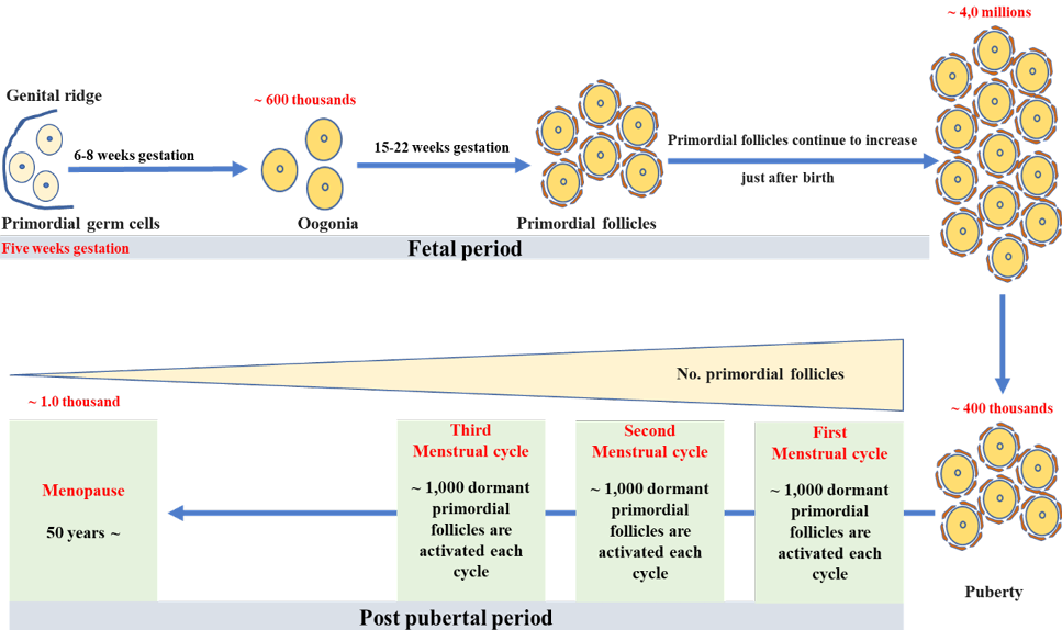
Development of primordial follicles during fetal and cycling periods in human.
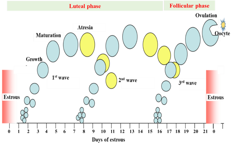
Ovarian cyclic waves during estrous cycle in cattle.
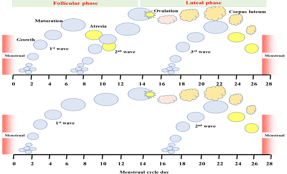
Ovarian cyclic waves during menstrual cycle in human.
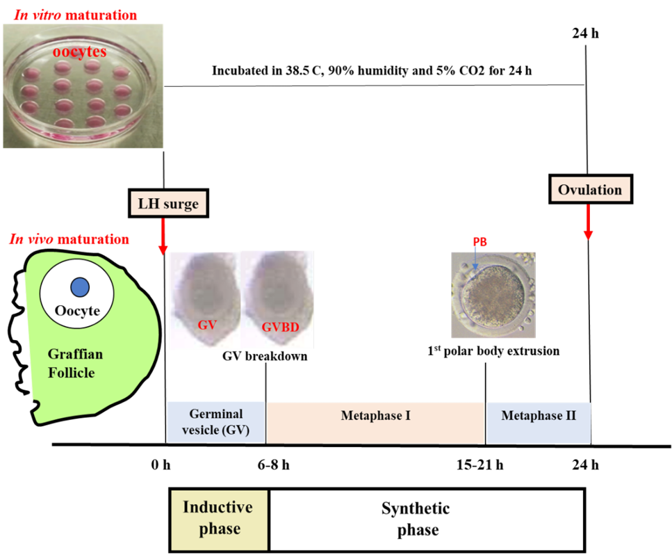
Chronology of events during the maturation of bovine oocytes.
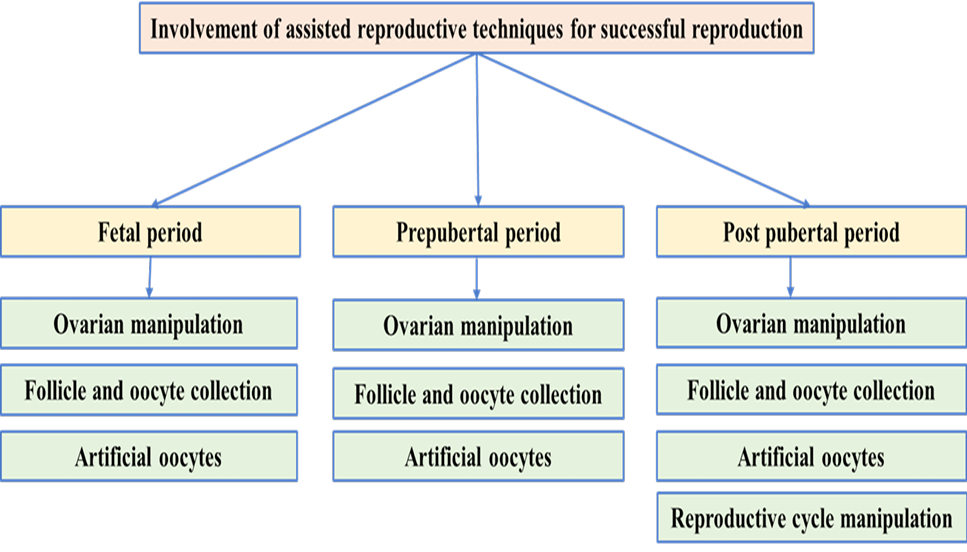
Application of assisted reproductive techniques during fetal, prepubertal and post pubertal periods.
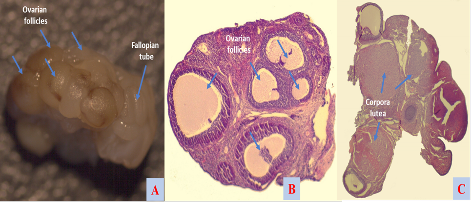
Ovarian morphology and histology upon ovarian transplantation in rats. A, morphology of active transplanted ovary; B, antral follicles of active transplanted ovary; C, corpora lutea of active transplanted ovary.
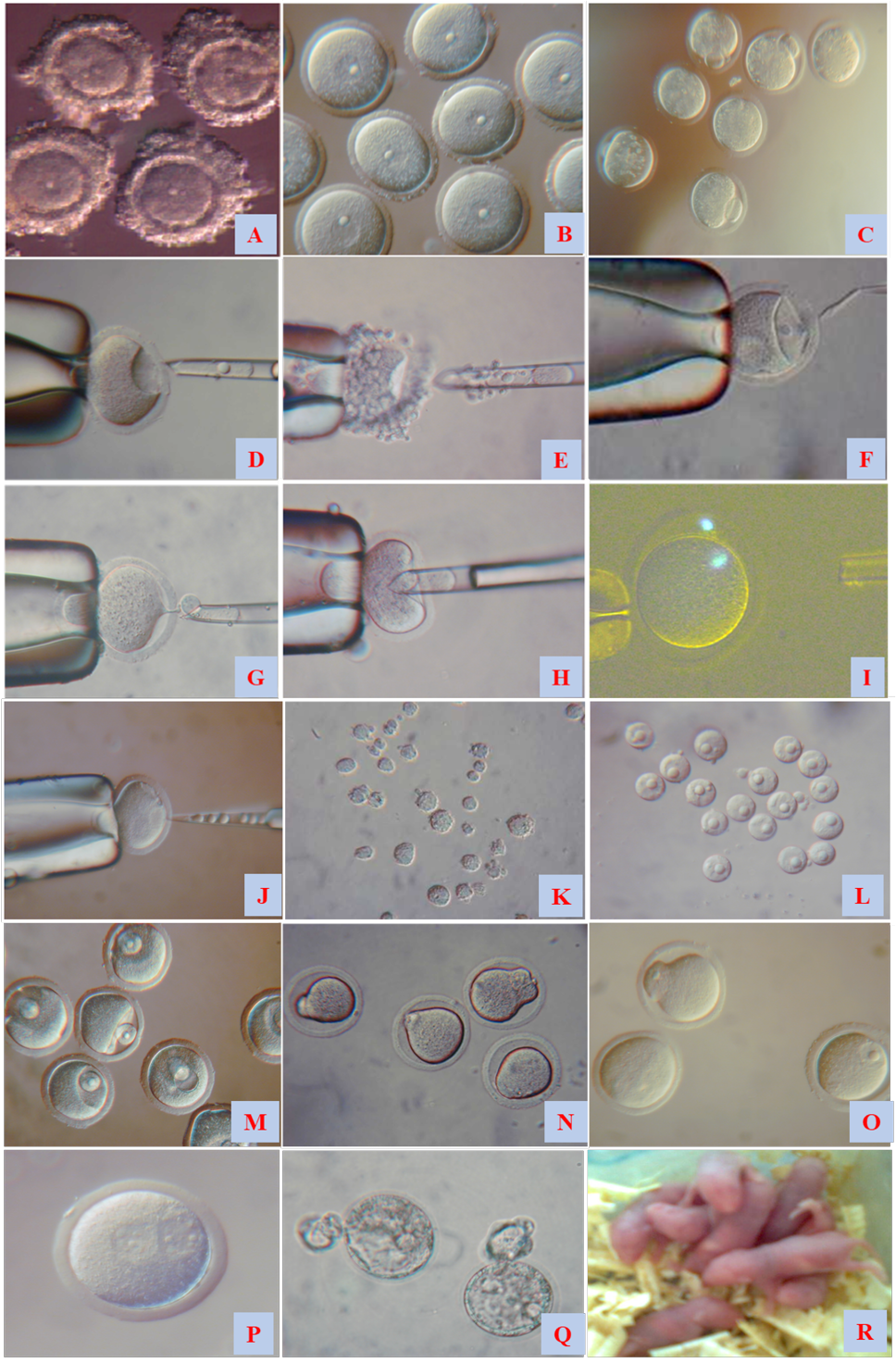
Maturation and manipulation of mouse oocytes and the resulting outcomes: cumulus-enclosed germinal vesicle (GV) oocytes (A), denuded GV oocytes (B), matured oocytes (C), complete enucleation of germinal vesicle oocyte (D), complete enucleation of cumulus-enclosed germinal vesicle oocyte (E), selective enucleation of g germinal vesicle oocyte (F), enucleation of pro-metaphase I oocyte (G), enucleation of metaphase I oocyte (H), enucleation of metaphase II oocyte (I) enucleolation of germinal vesicle oocyte (J), fetal fibroblast (K), germinal vesicle karyoplasts (L) GV karyoplasts placed under the zona pellucida of enucleated GV oocytes (M) fused with cytoplasts (N, O) developed zygote upon embryonic nuclear transfer (P), developed hatching blastocyst upon embryonic nuclear transfer (Q) offspring upon fallopian embryo transfer.
References
- Adhikari, D., and Liu, K., 2009. Molecular mechanisms underlying the activation of mammalian primordial follicles. Endocr. Rev., 30: 438–464. https://doi.org/10.1210/er.2008-0048[CrossRef]
- Albertini, D.A., 2015. The mammalian oocyte (Chapter 2). In: Knobil and Neill’s Physiology of reproduction, Fourth Edition. pp. 59-97. https://doi.org/10.1016/B978-0-12-397175-3.00002-8[CrossRef]
- Ali, M.A., Alshaheen, T., Senosy, W., Kassab, A., and Mohammed, A.A., 2021. Effects of feeding green microalgae and Nigella sativa on productive performance and metabolic profile of Boer goats during peripartum period in subtropics. Fresenius environ. Bull., 30: 8203-8212.
- Baby, T.E. and Bartlewski, P.M., 2011. Circulating concentrations of ovarian steroids and follicle-stimulating hormone (FSH) in ewes with 3 or 4 waves of antral follicle emergence per estrous cycle. Reprod. Biol., 11: 19-36. https://doi.org/10.1016/S1642-431X(12)60061-8[CrossRef]
- Baerwald, A., and Pierson, R., 2020. Ovarian follicular waves during the menstrual cycle: physiologic insights into novel approaches for ovarian stimulation. Fertil. Steril., 114: 443-457. https://doi.org/10.1016/j.fertnstert.2020.07.008[CrossRef]
- Baird, D., and Mitchell, A., 2013. Hormonal control of folliculogenesis: The key to successful reproduction. The future of the oocyte: Basic and clinical aspects. Ernst Schering Research Foundation Worshop. pp. 41.
- Bavister, B.D., 1995. Culture of preimplantation embryos: Facts and artifacts. Hum. Reprod. Update, 1: 91–148. https://doi.org/10.1093/humupd/1.2.91[CrossRef]
- Benc, M., Fulka J., Strejček F., Morovič M., Murín M., Martínková S., Jettmarová D., and Laurinčík J., 2019. Enucleolation and nucleolus transfer in mammalian oocytes and zygotes. Int. J. Dev. Biol., 63: 253-258. https://doi.org/10.1387/ijdb.190002mb[CrossRef]
- Bouniol-Baly, C., Hamraoui L., Guibert J., Beaujean N., Szollosi M.S. and Debey P., 1999. Differential transcriptional activity associated with chromatin configuration in fully grown mouse germinal vesicle oocytes. Biol. Reprod., 60: 580–587. https://doi.org/10.1095/biolreprod60.3.580[CrossRef]
- Campbell, B.K., Scaramuzzi R.J., and Webb, R., 1995. Control of antral follicle development and selection in sheep and cattle. J. Reprod. Fertil. Suppl., 49: 335-350.
- Campbell, B.K., Souza C., Gong J., Webb R., Kendall N., Marsters P., Robinson G., Mitchell A., Telfer E.E., and Baird D.T. 2003. Domestic ruminants as models for the elucidation of the mechanisms controlling ovarian follicle development in humans. Reprod. Suppl., 61: 429-443.
- Cavalieri, F.L.B., Morotti, F., Seneda, M.M., Colombo, A.H.B., Andreazzi, M.A., Emanuelli, I.P., and Rigolon, L.P., 2018. Improvement of bovine in vitro embryo production by ovarian follicular wave synchronization prior to ovum pick-up. Theriogenology, 117: 57-60. https://doi.org/10.1016/j.theriogenology.2017.11.026[CrossRef]
- Chang, H.C., Liu H., Zhang J., Grifo J. and Krey L.C., 2005. Developmental incompetency of denuded mouse oocytes undergoing maturation in vitro is ooplasmic in nature and is associated with aberrant Oct-4 expression. Hum. Reprod., 20: 1958-1968. https://doi.org/10.1093/humrep/dei003[CrossRef]
- Chelenga, M., Sakaguchi, K., Kawano, K., Furukawa, E., Yanagawa, Y., Katagiri, S., and Nagano, M., 2022. Low oxygen environment and astaxanthin supplementation promote the developmental competence of bovine oocytes derived from early antral follicles during 8 days of in vitro growth in a gas-permeable culture device. Theriogenology, 177: 116-126. https://doi.org/10.1016/j.theriogenology.2021.10.014[CrossRef]
- Chesnel, F. and Eppig, J.J., 1995. Synthesis and accumulation of p34cdc2 and cyclin B in mouse oocytes during acquisition of competence to resume meiosis. Mol. Reprod. Dev., 40: 503–508. https://doi.org/10.1002/mrd.1080400414[CrossRef]
- Chesnel, F., Wigglesworth, K. and Eppig, J.J., 1994. Acquisition of meiotic competence by denuded mouse oocytes: Participation of somatic-cell products and cAMP. Dev. Biol., 161: 285–295. https://doi.org/10.1006/dbio.1994.1028[CrossRef]
- Cho, E., Kim, Y.Y., Noh, K., Ku, S.Y., 2019. A new possibility in fertility preservation: The artificial ovary. J. Tissue Eng. Regen. Med., 13: 1294-1315. https://doi.org/10.1002/term.2870[CrossRef]
- Christianson, M.S., Lukish D.A., McCarterJr R., Pryor H., and Lukish J.R., 2021. Ovarian tissue cryopreservation in young females with cancer and its impact on ovarian follicle density. J. Pediatr. Surg., 56: 2354-2359. https://doi.org/10.1016/j.jpedsurg.2021.05.005[CrossRef]
- Condorelli, M., Bruzzone, M., Ceppi, M., Ferrari, A., Grinshpun, A., Hamy, A.S., de Azambuja, E, Carrasco, E., Peccatori, F.A., Di Meglio, A., Paluch-Shimon, S., Poorvu, P.D., Venturelli, M., Rousset-Jablonski, C., Senechal, C., Livraghi, L., Ponzone, R., De Marchis, L., Pogoda, K., Sonnenblick, A., Villarreal-Garza, C., Córdoba, O., Teixeira, L., Clatot, F., Punie, K., Graffeo, R., Dieci, M.V., Pérez-Fidalgo, J.A., Duhoux, F.P., Puglisi, F., Ferreira, A.R., Blondeaux, E., Peretz-Yablonski, T., Caron, O., Saule, C., Ameye, L., Balmaña, J., Partridge, AH, Azim, H.A., Demeestere, I. and Lambertini, M., 2021. Safety of assisted reproductive techniques in young women harboring germline pathogenic variants inBRCA1/2 with a pregnancy after prior history of breast cancer. Eur. Soc. Med. Oncol. Open, 6: 100300. https://doi.org/10.1016/j.esmoop.2021.100300[CrossRef]
- Cui, L.B., Huang X.Y. and Sun F.Z., 2005. Transfer of germinal vesicle to ooplasm of young mice could not rescue ageing-associated chromosome misalignment in meiosis of oocytes from aged mice. Hum. Reprod., 20: 1624-1631. https://doi.org/10.1093/humrep/deh826[CrossRef]
- Dath, C., Eyck, A.S.V., Dolmans, M.M., Romeu L., Vigne, L.D., Donnez, J. and Van Langendonckt, A., 2010. Xenotransplantation of human ovarian tissue to nude mice: comparison between four grafting sites. Hum. Reprod., 25: 1734–1743. https://doi.org/10.1093/humrep/deq131[CrossRef]
- de Vantery, C., Stutz, A., Vassalli, J.D. and Schorderet-Slatkine, S., 1997. Acquisition of meiotic competence in growing mouse oocytes is controlled at both translational and posttranslational levels. Dev. Biol., 187: 43–54. https://doi.org/10.1006/dbio.1997.8599[CrossRef]
- Dolmans, M., Wolff M., Poirot, C., Diaz-Garcia, C., Cacciottola, L., Boissel, N., Liebenthron, J., Pellicer, A., Donnez, J., and Andersen C.Y., 2021. Transplantation of cryopreserved ovarian tissue in a series of 285 women: A review of five leading European centers. Fertil. Steril., 115: 1102–1115. https://doi.org/10.1016/j.fertnstert.2021.03.008[CrossRef]
- Dawson, K.M., Collins J.L. and Baltz J.M., 1998. Osmolarity dependent glycine accumulation indicates a role for glycine as an organic osmolyte in early preimplantation mouse embryos. Biol. Reprod., 59: 225–232.
- Edwards, L.J., Williams, D.A., and Gardner, D.K., 1998. Intracellular pH of the mouse preimplantation embryo: Amino acids act as buffers of intracellular pH. Hum. Reprod., 13: 3441–3448. https://doi.org/10.1093/humrep/13.12.3441[CrossRef]
- Eppig, J.J., Chesnel, F., Hirao, Y., O’Brien, M.J., Pendola, F.L., Watanabe, S. and Wigglesworth, K., 1997. Oocyte control of granulosa cell development: How and why. Hum. Reprod. 12: 127–132.
- Eppig, J.J., Schultz, R.M., O’Brien, M. and Chesnel, F., 1994. Relationship between the developmental programs controlling nuclear and cytoplasmic maturation of mouse oocytes. Dev. Biol., 164: 1–9. https://doi.org/10.1006/dbio.1994.1175[CrossRef]
- Escrich, L., Grau, N., Meseguer, M., Pellicer, A., and Escribá M., 2010. Morphologic indicators predict the stage of chromatin condensation of human germinal vesicle oocytes recovered from stimulated cycles. Fertil. Steril., 93: 2557-2564. https://doi.org/10.1016/j.fertnstert.2009.05.077[CrossRef]
- Eyck, A.S.V., Bouzin, C., Feron, O., Romeu, L., Langendonckt, A.V., Donnez, J. and Dolmans, M.M., 2010. Both host and graft vessels contribute to revascularization of xenografted human ovarian tissue in a murine model. Fertil. Steril., 93: 1676–1685. https://doi.org/10.1016/j.fertnstert.2009.04.048[CrossRef]
- Ferré, L.B., Kjelland, M.E., Strøbech, L.B., Hyttel, P., Mermillod, P., and Ross, P.J., 2020. Review: Recent advances in bovine in vitro embryo production: reproductive biotechnology history and methods. Animal, 14: 991-1004. https://doi.org/10.1017/S1751731119002775[CrossRef]
- Findlay, J.K., Hutt, K.J., Hickey, M., and Anderson, R.A., 2015. How is the number of primordial follicles in the ovarian reserve established? Biol. Reprod., 93: 111. https://doi.org/10.1095/biolreprod.115.133652[CrossRef]
- Fulka, H., Mrazek, M., and Fulka, J. Jr., 2004. Nucleolar dysfunction may be associated with infertility in humans. Fertil. Steril., 82: 486-487. https://doi.org/10.1016/j.fertnstert.2003.12.042[CrossRef]
- Gordon, I., 2003. Establishing pregnancies with IVP Embryos. In: Laboratory production of cattle embryos (2nd). CABI Publishing UK. pp. 303–321. https://doi.org/10.1079/9780851996660.0303[CrossRef]
- Guérin, P., Ménézo Y., and Ménézo, Y., 2001. Oxidative stress and protection against reactive oxygen species in the pre-implantation embryo and its surroundings. Hum. Reprod. Update, 7: 175–189. https://doi.org/10.1093/humupd/7.2.175[CrossRef]
- Ikeda, S., and Yamada, M., 2014. Midkine and cytoplasmic maturation of mammalian oocytes in the context of ovarian follicle physiology. Br. J. Pharmacol., 171: 827–836. https://doi.org/10.1111/bph.12311[CrossRef]
- Kang, T., Zhao, S., Shi, L., and Li, J., 2021. Glucose metabolism is required for oocyte maturation of zebrafish. Biochem. biophys. Res. Commun., 559: 191-196. https://doi.org/10.1016/j.bbrc.2021.04.059[CrossRef]
- Lechowska, A., Bilinski, S., Choi, Y., Shin, Y., Kloc, M., and Rajkovic, A., 2011. Premature ovarian failure in nobox-deficient mice is caused by defects insomatic cell invasion and germ cell cyst breakdown. J. Assist. Reprod. Genet., 28: 583–589. https://doi.org/10.1007/s10815-011-9553-5[CrossRef]
- Lee, S.H., Oh, H.J., Kim, M.J., Kim, G.A., Choi, Y.B., Jo, Y.K., Setyawan, E.M.N., and Lee, B.C., 2018. Effect of co-culture canine cumulus and oviduct cells with porcine oocytes during maturation and subsequent embryo development of parthenotes in vitro. Theriogenology, 106: 108-116. https://doi.org/10.1016/j.theriogenology.2017.09.015[CrossRef]
- Liu, Z., and Foote, R.H., 1995. Effects of amino acids on the development of in-vitro matured/ in-vitro fertilization bovine embryos in a simple protein-free medium. Hum. Reprod., 10: 2985–2991. https://doi.org/10.1093/oxfordjournals.humrep.a135834[CrossRef]
- Mattson, B.A. and Albertini, D.F., 1990. Oogenesis, chromatin and microtubule dynamics during meiotic prophase. Mol. Reprod. Dev., 25: 374–383. https://doi.org/10.1002/mrd.1080250411[CrossRef]
- Mazoochi, T. and Ehteram, M., 2018. Apoptosis in the ovary and follicular atresia. Feyz, J. Kashan Univ. med. Sci., 22: 112-119.
- McGee, E.A., and Hsueh, A.J.W., 2000. Initial and cyclic recruitment of ovarian follicles. Endocr. Rev., 21: 200–214. https://doi.org/10.1210/edrv.21.2.0394[CrossRef]
- Meirow, D., Baum, M., Yaron, R., Levron, J., Hardan, I., Schiff, E., Nagler, A., Yehuda, D.B., Raanani, H., Hourvitz, A. and Dor, J., 2007. Ovarian tissue cryopreservation in hematologic malignancy, Ten years’ experience. Leuk. Lymphoma., 48: 1569–1576. https://doi.org/10.1080/10428190701471957[CrossRef]
- Mitra, J. and Schultz, R.M., 1996. Regulation of the acquisition of meiotic competence in the mouse, Changes in the subcellular localization of cdc2, cyclin b1, cdc25C and wee1, and in the concentration of these proteins and their transcripts. J. Cell Sci., 109: 2407–2415. https://doi.org/10.1242/jcs.109.9.2407[CrossRef]
- Mohammed, A.A. and Al-Hozab A., 2016. Preselection of offspring sex at the time of conception in mammals. Aust. J. Basic appl. Sci., 10: 17-23.
- Mohammed, A.A. and Al-Hozab, A.A., 2020. + (-) catechin raises body temperature, changes blood parameters, improves oocyte quality and reproductive performance of female mice. Indian J. Anim. Res., 54: 543-548. https://doi.org/10.18805/ijar.B-981[CrossRef]
- Mohammed, A.A. and Attaai, A.H., 2011. Effects of dietary urea on timing of embryo cleavages and blood components in mice. Vet. World, 4: 360-363. https://doi.org/10.5455/vetworld.2011.360-363[CrossRef]
- Mohammed, A.A. and Kassab, A.Y., 2014. Biochemical changes of blood and ovarian follicular fluid after time storage in goats. Egypt. J. Anim. Prod., 52: 47-54. https://doi.org/10.21608/ejap.2015.93638[CrossRef]
- Mohammed, A.A., 2006. Developmental competence of mouse oocytes reconstructed with G2/M somatic nuclei. Biotechnology, Scientific Redagogical Publishing, C. Budejovice, pp. 1117-1120.
- Mohammed, A.A., 2008. Contributions of cumulus cells on timing of oocytes maturation and developmental potential. Assiut. J. agric. Sci., 39: 43-50.
- Mohammed, A.A., 2014a. Is nucleo-cytoplasmic incompatibility the reason of acceleration polar body extrusion? Int. J. Curr. Eng. Technol., 4: 287-291.
- Mohammed, A.A., 2014b. Maturation and developmental competence of selectively enucleated germinal vesicle oocytes of mammals upon nuclear transfer. Int. J. Curr. Eng. Technol., 4: 292-299.
- Mohammed, A.A., 2018a. Ovarian tissue transplantation in mice and rats: Comparison of ovaries age. Pakistan J. Zool., 50: 481-486.
- Mohammed, A.A., 2018b. Development of oocytes and preimplantation embryos of mice fed diet supplemented with Dunaliella salina. Adv. Anim. Vet. Sci., 6: 33-39. https://doi.org/10.17582/journal.aavs/2018/6.1.33.39[Full text][CrossRef]
- Mohammed, A.A., 2019. Nigella sativa oil improves physiological parameters, oocyte quality after ovarian transplantation, and reproductive performance of female mice. Pakistan J. Zool., 51: 2225-2231. https://doi.org/10.17582/journal.pjz/2019.51.6.2225.2231[Full text][CrossRef]
- Mohammed, A.A., Abd El-Hafiz G.A., and Ziyadah H.M.S., 2012a. Effect of dietary urea on ovarian structures in Saidi ewes during follicular and luteal phases. Egypt. J. Anim. Prod., 49: 29-35. https://doi.org/10.21608/ejap.2012.94345[CrossRef]
- Mohammed, A.A., Abdelnabi, M.A. and Modlinski, J.A., 2012b. Evaluation of anesthesia and reproductive performance upon diazepam and xylazine injection in rats. Anim. Sci. Pap. Rep., 30: 285-292.
- Mohammed, A.A., Al-Hizab, F., Al-Suwaiegh, S., Alshaheen, T., Kassab, A., Hamdon, H., and Waleed, S.W., 2021. Effects of propylene glycol on ovarian restoration, reproductive performance, Metabolic status and milk production of Farafra ewes in subtropics. Fresen. Environ. Bull., 30: 8192-8202.
- Mohammed, A.A., Al-Suwaiegh, S., and Al-Shaheen, T., 2020. Changes of follicular fluid composition during estrous cycle, The effects on oocyte maturation and embryo development in vitro. Indian J. Anim. Res., 54: 797-804. https://doi.org/10.18805/ijar.B-1030[CrossRef]
- Mohammed, A.A., Al-Suwaiegh S., and Al-Shaheen, T., 2019b. Effects of follicular fluid components on oocyte maturation and embryo development in vivo and in vitro. Adv. Anim. Vet. Sci., 7: 346-355. https://doi.org/10.17582/journal.aavs/2019/7.5.346.355[Full text][CrossRef]
- Mohammed, A.A., Al-Suwaiegh, S., and Al-Shaheen, T., 2019a. Do the cytoplast and nuclear material of germinal vesicle oocyte support developmental competence upon reconstruction with embryonic/ somatic nucleus. Cell Reprogram, 21: 163-170. https://doi.org/10.1089/cell.2019.0032[CrossRef]
- Mohammed, A.A., and Farghaly, M.M., 2018. Effect of Nigella sativa seeds dietary supplementation on oocyte maturation and embryo development in mice. Egypt. J. Anim. Prod., 55: 195-201. https://doi.org/10.21608/ejap.2018.93241[CrossRef]
- Mohammed, A.A., Karasiewicz, J., and Modlinski, J.A., 2008. Developmental potential of selectively enucleated immature mouse oocytes upon nuclear transfer. Mol. Reprod. Dev., 75: 1269-1280. https://doi.org/10.1002/mrd.20870[CrossRef]
- Mohammed, A.A., Karasiewicz, J., Kubacka, J., Greda, P., and Modlinski, J.A., 2010. Enucleated GV oocytes as recipients of embryonic nuclei in the G1, S, or G2 stages of the cell cycle. Cell Reprogram, 12: 427-435. https://doi.org/10.1089/cell.2009.0107[CrossRef]
- Mohammed, A.A., Karasiewicz, J., Papis K., Modlinski, J.A., 2005. Oocyte maturation in the presence of randomly pooled follicular fluid increases bovine blastocyst yield in vitro. J. Anim. Feed Sci., 14: 501-512. https://doi.org/10.22358/jafs/67048/2005[CrossRef]
- Mohammed, A.A., Sayed M.A.M., Abdelnabi, M.A., 2011a. A new protocol of anesthesia using thiopental, diazepam and xylazine in white New Zealand rabbits. Aust. J. Basic Appl. Sci., 5: 1296-1300.
- Mohammed, A.A., Ziyad, H.M., and Abd El-Hafiz, G.A., 2011b. Changes of follicular fluid composition in relation to dietary urea level and follicle size during follicular and luteal phases in Saidi ewes. Theriogenol. Insight, 1: 31-42.
- Parfenov, V., Potchukalina, G., Dudina, L., Kostyuchek, D. and Gruzova, M., 1989. Human antral follicles, oocytes, nucleus and the karyosphere formation: electron microscopic and autoradiographic data. Gamete Res., 22: 219–231. https://doi.org/10.1002/mrd.1120220209[CrossRef]
- Pepling, M.E., and Spradling, A.C., 2001. Mouse ovarian germ cell cysts undergo programmed breakdown to form primordial follicles. Dev. Biol., 234: 339–351. https://doi.org/10.1006/dbio.2001.0269[CrossRef]
- Pinyopummintr, T., and Bavister, B.D., 1996. Effects of amino acids on development in vitro of cleavage-stage bovine embryos into blastocysts. Reprod. Fertil. Dev., 8: 835–841. https://doi.org/10.1071/RD9960835[CrossRef]
- Reader, K.L., Stanton, J.L. and Juengel, J.L., 2017. The role of oocyte organelles in determining developmental competence. Biology, 6: 35. https://doi.org/10.3390/biology6030035[CrossRef]
- Richards, J.S. and Pangas, S.A., 2010. The ovary, basic biology and clinical implications. J. clin. Invest., 120: 963-972. https://doi.org/10.1172/JCI41350[CrossRef]
- Rieger, D., 1992. Relationship between energy metabolism and development of early mammalian embryos. Theriogenology, 37: 75–93. https://doi.org/10.1016/0093-691X(92)90248-P[CrossRef]
- Senosy, W., Kassab, A.Y., and Mohammed, A.A., 2017. Effects of feeding green microalgae on ovarian activity, reproductive hormones and metabolic parameters of Boer goats in arid subtropics. Theriogenology, 96: 16-22. https://doi.org/10.1016/j.theriogenology.2017.03.019[CrossRef]
- Senosy, W., Kassab, A.Y., Hamdon, H.A., and Mohammed, A.A., 2018. Influence of organic phosphorus on reproductive performance and metabolic profiles of anoestrous Farafra ewes in subtropics at the end of breeding season. Reprod. Domest. Anim., 53: 904-913. https://doi.org/10.1111/rda.13183[CrossRef]
- Shores, E.M., and Hunter, M.G., 1999. Immunohistochemical localization of steroidogenic enzymes and comparison with hormone production during follicle development in the pig. Reprod. Fertil. Dev., 11: 337-344. https://doi.org/10.1071/RD99085[CrossRef]
- Sinclair, K.D., Lunn, L.A., Kwong, W.Y., Wonnacott, K., Linforth, R.S.T., and Craigon, J., 2008. Amino acid and fatty acid composition of follicular fluid as predictors of in-vitro embryo development. Reprod. BioMed. Online, 16: 859-868. https://doi.org/10.1016/S1472-6483(10)60153-8[CrossRef]
- Sorrenti, M., Klinger, F.G., Iona, S., Rossi, V., Marcozzi, S., and De-Felici, M., 2020. Expression and possible roles of extracellular signal-related kinases 1-2 (ERK1-2) in mouse primordial germ cell development. J. Reprod. Dev., 66: 399-409. https://doi.org/10.1262/jrd.2019-141[CrossRef]
- Tang, K., Yang W., Li, X., Wu, C., Sang, L., and Yang, L., 2012. GDF-9 and bFGF enhance the effect of FSH on the survival, activation, and growth of cattle primordial follicles. Anim. Reprod. Sci., 131: 129-134. https://doi.org/10.1016/j.anireprosci.2012.03.009[CrossRef]
- van den Hurk, R., and Santos, R., 2009. Development of fresh and cryopreserved early-stage ovarian follicles, with special attention to ruminants. Anim. Reprod., 6: 72-95.
- Webb, R., Nicholas, B., Gong, J.G., Campbell, B.K., Gutierrez, C.G., Garverick, H.A., and Armstrong, D.G., 2003. Mechanisms regulating follicular development and selection of the dominant follicle. Reprod. Suppl., 61: 71-90.
- Wickramasinghe, D., Ebert, K.M. and Albertini, D.F., 1991. Meiotic competence acquisition is associated with the appearance of M-phase characteristics in growing mouse oocytes. Dev. Biol., 143: 162–172. https://doi.org/10.1016/0012-1606(91)90063-9[CrossRef]
- Wu, Z., Liu, K., and Zhang, C., 2021. Clinical applications of in vitro primordial follicle activation. Curr. Opin. Endocr. Metab. Res., 18:194–200. https://doi.org/10.1016/j.coemr.2021.03.020[CrossRef]
- Xiang, D., Liu, Y., Zhou, E., Yanfeng, and Wang, Y., 2021. Advances in the applications of polymer biomaterials for in vitro follicle culture. Biomed. Pharmacother., 140: 111422. https://doi.org/10.1016/j.biopha.2021.111422[CrossRef]
- Yang, M.Y., and Fortune, J.E., 2008. The capacity of primordial follicles in fetal bovine ovaries to initiate growth in vitro develops during mid-gestation and is associated with meiotic arrest of oocytes. Biol. Reprod., 78: 1153–1161. https://doi.org/10.1095/biolreprod.107.066688[CrossRef]
- Youm, H.Y., Lee, J.R., Lee, J., Jee, B.C., Suh, C.S. and Kim, S.H., 2015. Transplantation of mouse ovarian tissue: Comparison of the transplantation sites. Theriogenology, 83: 854–861. https://doi.org/10.1016/j.theriogenology.2014.11.026[CrossRef]
- Yousefian, I., Zare-Shahneh, A., Goodarzi, A., Baghshahi, H., and Fouladi-Nashta, A.A., 2021. The effect of Tempo and MitoTEMPO on oocyte maturation and subsequent embryo development in bovine model. Theriogenology, 176: 128-136. https://doi.org/10.1016/j.theriogenology.2021.09.016[CrossRef]
- Zhang, H., Risal, S., Gorre, N., Busayavalasa, K., Li, X., Shen, Y., Bosbach, B., Brannstrom, M., and Liu, K., 2014. Somatic cells initiate primordial follicle activation and govern the development of dormant oocytes in mice. Curr. Biol., 24: 2501-2508. https://doi.org/10.1016/j.cub.2014.09.023[CrossRef]
- Zuccotti, M., Piccinelli, A., Giorgi, R.P., Garagna, S. and Redi, C.A., 1995. Chromatin organization during mouse oocyte growth. Mol. Reprod. Dev., 41: 479–485. https://doi.org/10.1002/mrd.1080410410[CrossRef]
Cited by 2 articles
In journals published by ResearchersLinks.
- Cytoplasmic and Nuclear Maturation of Intact and Reconstructed Oocytes Controlling the Developmental Competence of EmbryosPakistan Journal of Zoology · 2025
- Roles of Assisted Reproductive Techniques in Mammals: Developmental Competence of Oocytes and EmbryosAdvances in Animal and Veterinary Sciences · 2023
Services
Article usage statistics
Loading…