Changes in Nucleus and Cytoplast During Oocyte Maturation: Involvement in Embryo Production
· · · ·
Affiliations & Notes ⌄
1Department of Clinical Studies, College of Veterinary Medicine, King Faisal University, P.O. Box 400, Al-Hassa, Kingdom of Saudi Arabia
2Department of Public Health and Animal Care, College of Veterinary Medicine, King Faisal University, P.O. Box 400, Al-Hassa, Kingdom of Saudi Arabia
3Department of Animal and Fish Production, College of Agriculture and Food Sciences, King Faisal University, P.O. Box 400, Al-Hassa, Kingdom of Saudi Arabia
4Faculty of Human Medicine, Assiut University, Egypt, 71526. *Correspondence | Al-Hassan Mohammed, Faculty of Human Medicine, Assiut University, Egypt, 71526
The higher the oocyte maturation the higher embryo development and offspring obtained after transfer.
Therefore, the success in oocyte maturation would be helpful in assisted reproductive techniques.
Further studies are still required to make development of oocyte matured in vitro comparable to oocytes matured in vivo.
Selected from the article's own text.
Abstract
Embryo production steps either in vivo or in vitro include oocyte meiotic maturation, fertilization and culture processes. Oocyte maturation is the first step for in vivo or in vitro embryo production. Oocyte maturation appears to be a simple step, but it is a very complex process. Maturation of oocytes include several changes at the cytoplasmic and nuclear levels that makes the oocytes able to undergo fertilization, pre-implantation and post-implantation embryo development. Oocyte meiotic maturation in vivo starts after LH hormone surge whereas it starts in vitro upon release of oocyte from the ovarian follicle. Rodent oocyte maturation lasts 15-17hr. whereas that of ruminant oocyte lasts 24hr. Several factors affect on oocytes maturation either in vitro or in vivo and therefore affect development of the resulting embryos. Because of the significance of oocyte maturation stage on further embryonic development, this review aims to discuss the knowledge of oocyte maturation and its effect on further development of embryos in vivo and in vitro.
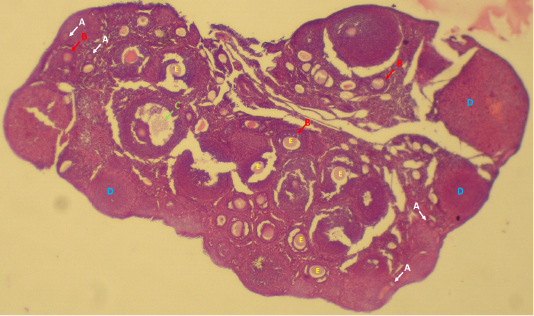
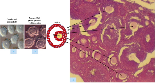
Ovarian primordial follicles’ growth to the preovulatory follicles is dependent on multiple intra-follicular and extra-follicular factors including stage-specific growth as growth and differentiation factor-9 (GDF-9) or basic fibroblast growth factor (bFGF) and hormonal factors ; . Ovarian follicle development occurs during prenatal and postnatal periods through initial and cyclic recruitments ; 2003). Initial recruitment characterizes dormant primordial follicles’ recruitment continuously to the growing follicles’ pool whereas cyclic recruitment characterizes antral follicles’ recruitment per estrous cycle (Figure 1; . At the onset of each estrous cycle, there is a group of early antral follicles developed to one or more ovulatory follicle(s) continuously through follicles’ selection and dominance. Recent interest has grown in the use of aspirated oocytes from antral ovarian follicles of live animals or slaughter-house ovaries for embryo production in vitro (Figure 2; ; ; ; ; ; ; , which could be used for different purposes.
Follicle stimulating hormone (FSH), equine chorionic gonadotropin (eCG), luteinizing hormone (LH) and human chronic gonadotropin (hCG) were used for regulating ovarian follicle growth and ovulation in vivo ;; ; ; . In addition, culture conditions including gases, culture media and additives to culture media were used in vitro for regulating ovarian follicles’ growth and development and oocytes’ maturation ; ; ; . Collectively, the embryos’ developmental competence resulting from oocytes in vivo matured is higher than those resulting from oocytes in vitro matured, reflecting the inefficiency of maturation culture conditions. Because of the significance of oocyte maturation stage on further embryonic development, this review aims to discuss the knowledge of oocyte maturation and its effect on further development of embryos in vivo and in vitro in addition to the meiotic maturation and development of reconstructed GV cytoplasts.
Growing and Fully-Grown Germinal Vesicle Oocytes
Nuclei of germinal vesicle oocytes are stopped in the diplotene of 1st meiotic prophase stages ; ; . Germinal vesicle oocytes are in a long G2 stage because the last DNA replication round occurred at the 12th to 13th days of fetal stage in mice . Germinal vesicle oocyte must grow and develop to be able to undergo meiotic maturation.
A complex bidirectional attachment between the enclosing cumulus cells and germ cell occurs during oocyte growth and differentiation ; ; . Oocyte within primordial follicle stage is stopped at prophase of 1st meiotic stages and enclosed by a single layer-squamous cells. Follicles’ growth and their containing oocytes are regulated through paracrine factors secreted from both the surrounding cells and germ cell of juvenelle ovary . Formation of antral follicles in mice occurs after birth approximately at day 14th. Only a small proportion of oocytes at day 14th of age is competent to resume meiosis. At day 22nd after birth, oocytes aspirated from the large antral follicles have acquired the ability of meiotic maturation and pre-implantation embryonic development .
During oocyte growth, meiotic competence is acquired progressively in mice ; ; and necessitates an accumulation of cell cycle regulatory molecules including p34cdc2 and cyclin B ; Viveiros and De La Fuente, 2019). In addition, meiotic resumption is also associated with further translational and post translational modification of mitotic kinases ; . The growing oocyte undergoes dynamic changes in chromatin and microtubule configurations . The nuclear morphology undergoes dynamic modification from a decondensed chromatin configuration (not surrounded nucleolus; NSN) to a condensed chromatin around the nucleolus (surrounded nucleolus; SN) . Both SN and NSN chromatin configurations are found in the fully grown germinal vesicle oocytes . Synthesis and storage of transcripts during oocytes’ growth are essential constructs in oocytes for further embryo development before transcriptional repression takes place at germinal vesicle stage.
Gonadotropins stimulation to mature females resulted in increase of oocytes with the SN configuration . Gonadotropin stimulation in vitro increased the proportion of SN oocytes that have compact enclosed-cumulus cells whereas those oocytes with loosely or denuded cumulus cells upon stimulation have a similar proportion of NSN and SN configurations (De La Fuente and Eppig, 2001).
Oocyte communication with surrounding cumulus cells is probably essential for both oocyte growth and acquisition of meiotic competency . However, both p34cdc2 and cyclin B components accumulate in cumulus-enclosed and denuded germinal vesicle oocytes . In addition, neither oocyte growth nor oocyte competence to undergo germinal vesicle breakdown (GVBD) occurs at the same rate in cumulus-enclosed and denuded germinal vesicle oocytes. This suggests that meiotic competence is regulated by an oocyte-intrinsic program and granulosa cells .
Meiotic Maturation of Germinal Vesicle Oocytes
Meiosis occurs in mammalian germ cell up to the diplotene stage during the fetal period. Germinal vesicle oocyte remains blocked at the diplotene stage of the 1st meiotic division in growing and dominant ovarian follicles. The germinal vesicle oocytes resume meiosis after removing from the antral follicles or LH surge ; . Meiosis resumption in vivo is initiated by LH surge and occurs only in fully-grown germinal vesicle oocyte of pre-ovulatory follicle. The oocyte is surrounded before LH surge by layers of compact cumulus-enclosed cells, numerous projections of cells penetrate the zona pellucida and end on the oocyte oolemma with gap junctions. Disruption of these junctions occurs shortly after the LH hormone surge. Between the period of LH surge and ovulation, the fully grown germinal vesicle oocyte of pre-ovulatory follicle undergoes several changes in its GV nucleus and cytoplast known as oocyte maturation. The first sign of resumption of meiotic maturation is GVBD, which occurred approximately 2h of starting maturation in mouse oocytes ; ; ; Mohammed et al., 2019; , and thereafter the chromosomes condense, microtubules pull the chromosomes to form the metaphase I (MI) plate. The 2nd meiotic division occurs immediately without chromosome replication and the oocyte reaches the MII stage. The oocytes remain arrested at the MII stage until fertilization occur and the oocyte complete the second meiotic division.
Cytoplasmic maturation involves transformations that prepare oocyte to support fertilization and development of resulting embryo. Both nuclear and cytoplasmic maturation are needed for subsequent embryo cleavages and development . Cytoplasmic and nuclear maturation are required after fertilization to block polyspermy, to decondense fertilized spermatozoa and to form male and female pronuclei. The cytoplasmic maturation changes include organelles redistribution and mitochondrial migration to a perinuclear position. The nuclear maturation includes the changes from GV to MII stage. Furthermore, ultrastructural changes occurred including changes in maturation-promoting factor (MPF), mitogen-activated protein kinase (MAP kinase) and cyclic adenosine monophosphate (cAMP) levels.
Maturation-Promoting Factor
Meiosis resumption in oocyte is regulated by MPF. MPF is composed of two subunits: p34cdc2 and cyclin B . p34cdc2 is the catalytic component and cyclin B is the regulatory component. Activity of MPF appears shortly before GVBD, maintains at high level during MI stage, decreases prior to the 1st polar body extrusion, and rises again throughout the metaphase II stage .
Maturation-promoting factor seems to be the universal regulator of meiotic cell cycles . MPF phosphorylates number of proteins. MPF is believed to be responsible for GVBD, chromatin condensation and microtubular relocation . The oocytes then acquire the ability to form a MI plate and a spindle. The next step is progression to the second metaphase plate and 1st polar body extrusion. The last meiotic event is arrest at the MII stage through cytostatic factor (CSF) . Inactivation of MPF by degradation of the cyclin component occur after sperm or parthenogenic activation to meiosis resume .
Mitogen-Activated Protein Kinases
Mitogen-activated protein kinases (MAPKs) are known to be involved in maturation processes of oocytes. Two isoforms of mitogen-activated protein kinases (MAPKs) in mammalian oocytes are presented including ERK1 and ERK2 . MAPKs appears activated after GVBD in mouse oocytes . MAPKs activity during oocyte maturation is associated with cytoplasmic events including regulation of microtubule dynamics, spindle assembly and chromosomal condensation .
Regulating growth and development of ovarian follicles in vivo and in vitro is considered the most important process for successful reproductive performance. The great importance extends to oocyte maturation where essential cytoplasmic and nuclear changes occurs for successful fertilization and further developmental competence of embryos. The potential regulation of ovarian follicles and oocytes’ maturation in vitro and in vivo occur through nutrition and feed additives, hormonal supplementation, conditions of in vitro culture system including gases, culture media, additive to culture media including fetal calf serum, cumulus cells and follicular fluid (FF), hormones and amino acids ; ; ; . The importance of oocyte maturation confirmed through gene expression, fertilization, timing of embryo cleavage, stage of embryo development and offspring obtained after transfer to the surrogate mothers. The higher the oocyte maturation the higher embryo development and offspring obtained after transfer. Therefore, the success in oocyte maturation would be helpful in assisted reproductive techniques. Further studies are still required to make development of oocyte matured in vitro comparable to oocytes matured in vivo.
Factors Affecting Oocyte Maturation
Oocyte maturation is the most important step for pre- and post-implantation development of embryos . The in vitro maturation of mouse oocyte needs approximately 15-17 hr. (Mohammed and Farghaly, 2018; ; ; . The cytoplasmic and nuclear changes that occur during maturation of oocyte are important for successful fertilization and embryo development ; ; ; ; . Changes of maturation promoting and cytostatic factors in addition to two asymmetrical meiotic divisions resulting in a single oocyte and polar bodies for oocyte maturation.
The oocyte resumes meiotic division to the metaphase II (MII) stage in vivo after the LH hormone surge and in vitro when it removes from antral ovarian follicle and cultured in favorable medium within 5.0% CO2 incubator ; . Development of in vivo matured oocytes to embryos is higher than in vitro matured ones ; . This might be attributed to insufficient nucleus and/or cytoplasmic maturity .
Follicle size ; , nutrition and feed additives ; ; ; ; ;; , the media for maturation and their enrichments (follicular fluid, bovine serum albumin, amino acids and hormones: ; ;, incubator conditions (humidity, CO2 and oxygen concentrations) were found to affect oocyte maturation ; .
Enrichment of maturation media with cumulus cells , growth factors and hormones ; ; ; , and other factors ; ; ; improves oocyte maturation and embryo development thereafter ; ;. In addition, super-stimulation of ovarian follicles via gonadotropin injections resulted in changes in small, medium, and large follicles according to the number and dose of gonadotropin injections and side of the ovary . FSH stimulates transcription and translation in ovarian granulosa cells, which are essential for female reproductive endocrine regulation . Collectively, it could be concluded that the in vivo and in vitro conditions or factors effect on oocytes’ maturation and their developmental competence to embryos and fetus.
Germinal Vesicle Cytoplast For Creation “Artificial Gamete”
Fully-grown germinal vesicle (GV) cytoplasts could be divided through two consecutive meiotic divisions; first and second meiotic divisions if cultured in vitro. The hypothesis that the GV cytoplasts are able to divide G0/G1 or G2/M nucleus through first and second meiotic divisions, respectively (Mohammed, 2006; ; ; Mohammed et al., 2010; ; ;. Therefore, GV nuclei, male germ cells, embryonic and somatic cells at G0/G1 or G2/M stage might divide correctly in the GV cytoplasts through 1st and 2nd meiotic divisions, respectively to solve the problem of infertility as ageing-associated chromosome misalignment in meiosis of oocytes from the aged mice or studying reprogramming of the introduced nuclei or nucleolar dysfunction ; ; . In case of male infertility due to complete absence of the germline, GV cytoplast could be reconstructed with germ cell of male in order to create “artificial oocyte” containing the male haploid genome of the fused germ cell after in vitro maturation.
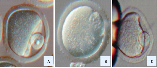
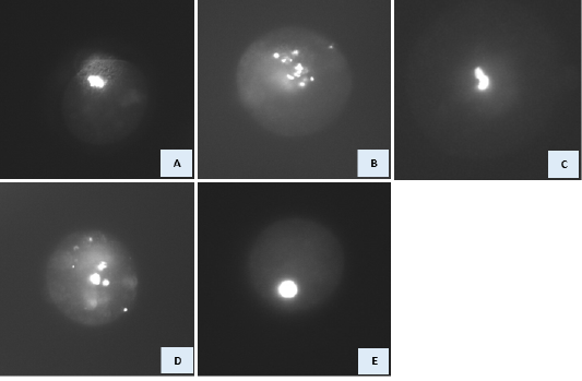
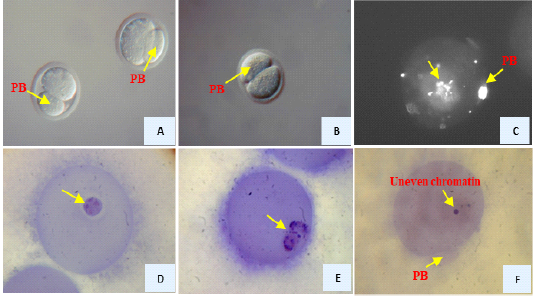
Unfortunately, the few trials so far concerning the GV cytoplast reconstructed with embryonic/somatic nuclei resulted in abnormalities in maturation compared to normal maturation if the GV cytoplast reconstructed with GV nucleus (Figures 3, 4 & 5; . One of the first studies in which GV oocytes were reconstructed with G0/G1 somatic nucleus performed by Kubelka and Moor et al. (1997) and Fulka et al. (2002). These studies failed to obtain 1st meiotic maturation of cell-oocyte complex. However, Polanski et al. (2005) reported meiotic maturation of G0/G1 cumulus cells after their transfer into GV cytoplasts. Reports thereafter described the meiotic mat
uration of the enucleated GV oocytes after nuclear transfer of G2/M stage embryonic or somatic ; ; ; ; ; ; cell nuclei. The previous results demonstrated that meiotic maturation of GV cytoplasts reconstructed with embryonic/somatic cells (G0/G1 or G2/M stage) were associated with abnormalities in earlier extrusion of 1st polar body, chromosomal alignment over spindle and condensation, and cytokinesis (Figures 4 and 5; . Our trials to overcome and to explore such abnormalities have been reported ; ; . Such previous trials improved the competence of enucleated GV cytoplast after embryonic/somatic nuclear transfer followed by maturation and activation/fertilization. The proper pronuclei of the introduced embryonic/somatic nucleus were formed in addition to the male pronucleus in case of fertilization. The zygotes proceed development to the blastocyst and hatching/hatched stage. This was occurred through a technique called “selective enucleation” of GV oocyte surrounding with cumulus cells. In this technique. the nucleolus and nuclear sap were left in the GV cytoplast in addition to cumulus attachment with zona pellucida. Such selective enucleation technique reached the reconstructed GV cytoplasts to the embryonic stages of blastocysts and hatched blastocysts compared to complete enucleation of denuded GV oocyte. The germinal vesicle cytoplasts obtained with technique called “complete enucleation” where the whole GV nucleus removed upon enucleation.
Over reconstruction with embryonic/somatic nuclei, the GV cytoplasts were blocked at one cell-stage embryos. Hence, further studies are still needed required for improvement embryos development, which obtained through GV cytoplast reconstructed with embryonic somatic nuclei in addition to obtaining offspring over embryo transfer to surrogate mothers.
CONCLUSION
Regulating ovarian follicles’ growth and development either in vitro or in vivo is considered necessitate process for successful reproductive performance. The great importance extends to oocyte maturation where essential cytoplasmic and nuclear changes occurs for subsequent fertilization and development of the resulting embryos. The potential regulation of maturation of ovarian follicles and oocytes either in vitro or in vivo occurs through nutrition and feed additives, hormonal supplementation, conditions of in vitro culture system including gases, culture media, additive to culture media including fetal calf serum, cumulus cells and follicular fluid (FF), hormones and amino acids. The importance of oocyte maturation confirmed through gene expression, fertilization, timing of embryo cleavage, stage of embryo development and offspring obtained after transfer to the surrogate mothers. The higher the oocyte maturation the higher embryo development and offspring obtained after transfer. Therefore, the success in oocyte maturation would be helpful in assisted reproductive techniques. Further studies are still required to make development of oocyte matured in vitro comparable to oocytes matured in vivo.
ACKNOWLEDGEMENTS
This work was supported through the Annual Funding track by the Deanship of Scientific Research, Vice Presidency for Graduate Studies and Scientific Research, King Faisal University, Saudi Arabia [GRANT 1015].
CONFLICT OF INTEREST
There is no conflict of interest for authors to declare.
Authors contribution
Mohammed wrote and submit manuscript. Al Mufarji, Haitham Al Masruri, Rashid Al Zeidi, and Al-Hassan Mohammed collected references and prepared figures.
Figures
Figure 1. Figure 1.
Ovarian follicle structures in mice; pre-antral follicles (A), antral follicles (B), preovulatory follicles (C), corpora lutea (D) and oocytes inside the follicles (E).
Figure 2.
Aspiration of oocytes from follicles (A), antral follicle (B), the collected cumulus-enclosed germinal vesicle oocytes (C) stripped-off cumulus cells from the cumulus-enclosed germinal vesicle oocytes (D) in mice.
Figure 3.
Germinal vesicle cytoplast used for germinal vesicle (A) embryonic (B) and somatic (C) nuclear transfer
Figure 4.
Nuclear morphology at the expected MI stage of GV cytoplasts reconstructed with GV, embryonic and somatic nuclei. DNA was stained with Hoechst 33342. A) GV cytoplast reconstructed with GV nucleus. Various abnormal nuclear morphology was observed in manipulated GV cytoplast reconstructed with embryonic/somatic at the expected MI stage. These include: condensed, scattered chromosomes (B), partially condensed chromosomes (C), formation of micronuclei and «pycnotic» nuclei (D), and interphase-like nuclei (E).
Figure 5.
Reconstructed GV oocytes with somatic cells after 17 hr. of maturation. Extruded large-sized PBs (A, B), DNA was stained with Hoechst 33342 where the oocyte had scattered chromosomes (C), DNA was stained haematoxyline where the oocyte had interphase-like nucleus (D), the oocyte had partially condensed chromosomes (E) the oocyte had uneven distribution of chromatin (micronuclei)
References
Abdelnaby EA, El-Maaty AMA, El-Badry DA (2021). Evaluation of ovarian hemodynamics by color and spectral Doppler in cows stimulated with three sources of follicle-stimulating hormone. Reprod. Biol. 21(1): 100478. https://doi.org/10.1016/j.repbio.2020.100478[CrossRef]
Al Zeidi R., Al Masruri H., Al Mufarji A., Al-Hassan Mohammed A.A. (2022). Role of cumulus cells and follicular fluid on oocyte maturation and developmental competence of embryos: intact and reconstructed oocytes. Adv. Anim. Vet. Sci. 10(6): 1219-1226. https://doi.org/10.17582/journal.aavs/2022/10.6.1219.1226[Full text][CrossRef]
Baerwald A., Pierson R. (2020). Ovarian follicular waves during the menstrual cycle: physiologic insights into novel approaches for ovarian stimulation. Fertil. Steril. 114 (3): 443-457. https://doi.org/10.1016/j.fertnstert.2020.07.008[CrossRef]
Bahrami M., Cottee P.A. (2022). Culture conditions for in vitro maturation of oocytes – A review, Reproduction and Breeding, Volume 2, Issue 2, 2022, Pages 31-36. https://doi.org/10.1016/j.repbre.2022.04.001[CrossRef]
Baruffi R.L., Avelino K.B., Petersen C.G., Mauri A.L., Garcia J.M., Franco J.G. (2004). Nuclear and cytoplasmic maturation of human oocytes cultured in vitro, Fertil. Steril. 82: S265-S266.
Blondin P., Coenen K., Guilbault L. A., Sirard M.-A. (1997). In vitro production of bovine embryos: developmental competence is acquired before maturation. Theriogenol. 47(5): 1061–1075. https://doi.org/10.1016/S0093-691X(97)00063-0[CrossRef]
Bouniol-Baly C., Hamraoui L., Guibert J., Beaujean N., Szollosi M.S. Debey P. (1999). Differential transcriptional activity associated with chromatin configuration in fully grown mouse germinal vesicle oocytes. Biol. Reprod. 60: 580–587. https://doi.org/10.1095/biolreprod60.3.580[CrossRef]
Bunel A., Nivet A.L., Blondin P., Vigneault C., Richard F.J., Sirard M.A. (2020). The effects of LH inhibition with cetrorelix on cumulus cell gene expression during the luteal phase under ovarian coasting stimulation in cattle. Domest. Anim. Endocrinol. 72: 106429. https://doi.org/10.1016/j.domaniend.2019.106429[CrossRef]
Caballero J., Blondin P., Vigneault C., Sirard M-A., Richard F.I. (2020). The use of adenosine to inhibit oocyte meiotic resumption in Bos taurus during pre-IVM and its potential to improve oocyte competence, Theriogenol.142: 207-215. https://doi.org/10.1016/j.theriogenology.2019.10.001[CrossRef]
Campbell K.H.S., Loi P., Otaegui P.J., Wilmut I. (1996). Cell cycle co-ordination in embryo cloning by nuclear transfer. Rev. Reprod. 1: 40–46. https://doi.org/10.1530/ror.0.0010040[CrossRef]
Campbell B.K., Scaramuzzi R.J., Webb R. (1995). Control of antral follicle development and selection in sheep and cattle. J. Reprod. Fertil. Suppl. 49: 335-50.
Campbell B.K., Souza C., Gong J., Webb R., Kendall N., Marsters P., Robinson G., Mitchell A., Telfer E.E., Baird D.T. (2003). Domestic ruminants as models for the elucidation of the mechanisms controlling ovarian follicle development in humans. Reprod. Suppl. 61: 429-43.
Cavalieri F.L.B., Morotti F., Seneda M.M., Colombo A.H.B., Andreazzi M.A., Emanuelli I.P., Rigolon L.P. (2018). Improvemen+t of bovine in vitro embryo production by ovarian follicular wave synchronization prior to ovum pick-up. Theriogenol. 117: 57-60. https://doi.org/10.1016/j.theriogenology.2017.11.026[CrossRef]
Chang C.C., Nagy Z.P., Abdelmassih R., Yang X, Tian X.C. (2004). Nuclear and microtubule dynamics of G2/M somatic nuclei during haploidization in germinal vesicle-stage mouse oocytes. Biol. Reprod. 70: 752-758. https://doi.org/10.1095/biolreprod.103.024497[CrossRef]
Chang H.C., Liu H., Zhang J., Grifo J., Krey L.C. (2005). Developmental incompetency of denuded mouse oocytes undergoing maturation in vitro is ooplasmic in nature and is associated with aberrant Oct-4 expression. Hum. Reprod. 20: 1958-1968. https://doi.org/10.1093/humrep/dei003[CrossRef]
Chelenga M, Sakaguchi K, Kawano K., Furukawa E., Yanagawa Y., Katagiri S., Nagano M. (2022). Low oxygen environment and astaxanthin supplementation promote the developmental competence of bovine oocytes derived from early antral follicles during 8 days of in vitro growth in a gas-permeable culture device. Theriogenol. 177: 116-126. https://doi.org/10.1016/j.theriogenology.2021.10.014[CrossRef]
Chesnel F., Eppig J.J. (1995). Synthesis and accumulation of p34cdc2 and cyclin B in mouse oocytes during acquisition of competence to resume meiosis. Mol. Reprod. Dev. 40: 503–508. https://doi.org/10.1002/mrd.1080400414[CrossRef]
Dai X.-X., Jiang Z.-Y., Wu Y.-W., Sha Q.-Q., Liu Y., Ding J.-Y., Xi W.-D., Li J., Fan H.-Y. (2021). CNOT6/6L-mediated mRNA degradation in ovarian granulosa cells is a key mechanism of gonadotropin-triggered follicle development. Cell Rep. 37(7): 110007. https://doi.org/10.1016/j.celrep.2021.110007[CrossRef]
De La Fuente R, Eppig J.J. (2001). Transcriptional Activity of the Mouse Oocyte Genome: Companion Granulosa Cells Modulate Transcription and Chromatin Remodeling. Dev. Biol. 229: 224–236. https://doi.org/10.1006/dbio.2000.9947[CrossRef]
de Senna Costa J.A., Cezar G.A., Monteiro P.L.J., Silva D.M.F., Silva R.A.J.A., Bartolomeu C.C., Filho A.S.S., Wischral A., Batista A.M. (2022). Leptin improves in-vitro maturation of goat oocytes through MAPK and JAK2/STAT3 pathways and affects gene expression of cumulus cells. Reprod. Biol. 22(1): 100609. https://doi.org/10.1016/j.repbio.2022.100609[CrossRef]
de Vantery C., Stutz A., Vassalli J.D., Schorderet-Slatkine S. (1997). Acquisition of meiotic competence in growing mouse oocytes is controlled at both translational and posttranslational levels. Dev. Biol. 187: 43–54. https://doi.org/10.1006/dbio.1997.8599[CrossRef]
Dedieu T., Gall L., Crozet N., Sevellec C., Ruffini S. (1996). Mitogen-activated protein kinase during goat oocyte maturation and the acquisition of meiotic competence. Mol. Reprod. Dev. 45 351-358. https://doi.org/10.1002/(SICI)1098-2795(199611)45:3%3C351::AID-MRD12%3E3.0.CO;2-1[CrossRef]
Dieleman S.J., Kruip T.A.M., Fontijne P., de Jong W.H.R., van der Weyden G.C. (1983). Changes in oestradiol, progesterone and testosterone concentration in follicular fluid and in micromorphology of preovulatory bovine follicles relative to the peak of luteinizing hormone. J. Endocrinol. 97: 31-42. https://doi.org/10.1677/joe.0.0970031[CrossRef]
Doherty C.A., Amargant F., Shvartsman S.Y., Duncan F.E., Gavis E.R. (2022). Bidirectional communication in oogenesis: a dynamic conversation in mice and Drosophila. Trends Cell Biol., 32(4): 311-323. https://doi.org/10.1016/j.tcb.2021.11.005[CrossRef]
Eppig J.J. (1991). Intercommunication between mammalian oocytes and companion somatic cells. BioEssays. 13: 569-574. https://doi.org/10.1002/bies.950131105[CrossRef]
Eppig J.J. (2001). Oocyte control of ovarian follicular development and function in mammals. Reprod. 122: 829-838. https://doi.org/10.1530/rep.0.1220829[CrossRef]
Eppig J.J., Schroeder A.C. (1989). Capacity of mouse oocytes from preantral follicles to undergo embryogenesis and development to live young after growth, maturation and fertilization in vitro. Biol. Reprod. 41: 268–276. https://doi.org/10.1095/biolreprod41.2.268[CrossRef]
Eppig J.J., Schultz R.M., O’Brien M., Chesnel F. (1994). Relationship between the developmental programs controlling nuclear and cytoplasmic maturation of mouse oocytes. Dev. Biol. 164: 1–9. https://doi.org/10.1006/dbio.1994.1175[CrossRef]
Eppig J.J., Chesnel F., Hirao Y., O’Brien M.J., Pendola F.L., Watanabe S., Wigglesworth K. (1997). Oocyte control of granulosa cell development: How and why. Hum. Reprod. 12 (11 Supplement): 127–132.
Ferré L.B., Kjelland M.E., Strøbech L.B., Hyttel P., Mermillod P., Ross P.J. (2020). Review: Recent advances in bovine in vitro embryo production: reproductive biotechnology history and methods. Anim. 14(5): 991-1004. https://doi.org/10.1017/S1751731119002775[CrossRef]
Fulka J., Martinez F., Tepla O., Mrazek M., Tesarik J. (2002). Somatic and embryonic cell nucleus transfer into intact and enucleated immature mouse oocytes. Hum. Reprod. 17: 2160-2164. https://doi.org/10.1093/humrep/17.8.2160[CrossRef]
Fulka H., Mrazek M., Fulka J. Jr. (2004). Nucleolar dysfunction may be associated with infertility in humans. Fertil. Steril. 82: 486-487. https://doi.org/10.1016/j.fertnstert.2003.12.042[CrossRef]
Gao S., Gasparrini B., McGarry M., Ferrier T., Fletcher J., Harkness L., De Sousa P., Wilmut I. (2002). Germinal vesicle material is essential for nucleus remodeling after nuclear transfer. Biol. Reprod. 67: 928-934. https://doi.org/10.1095/biolreprod.102.004606[CrossRef]
Gautier J., Norbury C., Lohka M., Nurse P., Maller J. (1988). Purified maturation promoting factor contains the product of a Xenopus homolog of the fission yeast cell cycle control gene cdc2. Cell. 54: 433-439. https://doi.org/10.1016/0092-8674(88)90206-1[CrossRef]
Gavin A.C., Cavadore J.C., Schorderet-Slatkine S. (1994). Histone H1 kinase activity, germinal vesicle breakdown and M phase in mouse oocytes. J. Cell Sci. 107: 275-283. https://doi.org/10.1242/jcs.107.1.275[CrossRef]
Ge L., Sui H., Lan G., Liu N., Wang J., Tan J. (2008). Coculture with cumulus cells improves maturation of mouse oocytes denuded of the cumulus oophorus: observations of nuclear and cytoplasmic events, Fertil. Steril. 90(6): 2376-2388. https://doi.org/10.1016/j.fertnstert.2007.10.054[CrossRef]
Gordon I. (2003). Establishing Pregnancies with IVP Embryos, In, Laboratory Production of Cattle Embryos (2nd) page 303 – 321. CABI Publishing UK. https://doi.org/10.1079/9780851996660.0303[CrossRef]
Grabarek J.B., Plusa B., Modlinski J.A., Karasiewicz J. (2004). Reconstruction of enucleated mouse germinal vesicle oocytes with blastomere nuclei. Zygote. 12: 163-172. https://doi.org/10.1017/S0967199404002746[CrossRef]
Gutiérrez-Añez J.C., Lucas-Hahn A., Hadeler K., Aldag P., Niemann H. (2021). Melatonin enhances in vitro developmental competence of cumulus-oocyte complexes collected by ovum pick-up in prepubertal and adult dairy cattle. Theriogenol. 161: 285-293. https://doi.org/10.1016/j.theriogenology.2020.12.011[CrossRef]
Kang T., Zhao S., Shi L., Li J. (2021). Glucose metabolism is required for oocyte maturation of zebrafish. Biochem. Biophys. Res. Commun. 559: 191-196. https://doi.org/10.1016/j.bbrc.2021.04.059[CrossRef]
Ko Y., Kim J.H., Lee S.R., Kim S.H., Chae H.D. (2021). Influence of pretreatment of insulin on the phosphorylation of extracellular receptor kinase by gonadotropin-releasing hormone and gonadotropins in cultured human granulosa cells. Eur. J. Obstet. Gynecol. Reprod. Biol. 262: 113-117. https://doi.org/10.1016/j.ejogrb.2021.05.016[CrossRef]
Koloda Y., Korsak V., Rozenson O., Anshina M., Sagamonova K., Baranov I., Yakovenko S., D’Hooghe T., Ershova A., Lispi M. (2022). Use of a recombinant human follicle-stimulating hormone: recombinant human luteinizing hormone (r-hFSH:r-hLH) 2:1 combination for controlled ovarian stimulation during assisted reproductive technology treatment: A real-world study of routine practice in the Russian Federation. Best. Pract. Res. Clin. Obstet. Gynaecol, 2022: 1521-6934. https://doi.org/10.1016/j.bpobgyn.2022.01.009[CrossRef]
Kubelka M., Moor R.M. (1997). The behaviour of mitotic nuclei after transplantation to early meiotic ooplasts or mitotic cytoplasts. Zygote. 5: 219-227. https://doi.org/10.1017/S0967199400003658[CrossRef]
Kumar S., Singla S.K., Manik R., Palta P., Chauhan M.S. (2020). Effect of basic fibroblast growth factor (FGF2) on cumulus cell expansion, in vitro embryo production and gene expression in buffalo (Bubalus bubalis). Reprod. Biol. 20(4): 501-511. https://doi.org/10.1016/j.repbio.2020.08.003[CrossRef]
Lee S.H., Oh H.J., Kim M.J., Kim G.A., Choi Y.B., Jo Y.K., Setyawan E.M.N., Lee B.C. (2018). Effect of co-culture canine cumulus and oviduct cells with porcine oocytes during maturation and subsequent embryo development of parthenotes in vitro. Theriogenol. 106: 108-116. https://doi.org/10.1016/j.theriogenology.2017.09.015[CrossRef]
Lima-de-Faria A., Borum K. (1962). The period of DNA synthesis prior to meiosis in the mouse. J. Cell Biol. 14: 381–388. https://doi.org/10.1083/jcb.14.3.381[CrossRef]
Liu Z., Foote R.H. (1995). Effects of amino acids on the development of in-vitro matured/in-vitro fertilization bovine embryos in a simple protein-free medium. Hum. Reprod. 10: 2985–2991. https://doi.org/10.1093/oxfordjournals.humrep.a135834[CrossRef]
Llano E., Iyyappan R., Aleshkina D., Masek T., Dvoran M., Jiang Z., Pospisek M., Kubelka M., Susor A. (2022). SGK1 is essential for meiotic resumption in mammalian oocytes. European J. Cell Biol. 101(2): 151210. https://doi.org/10.1016/j.ejcb.2022.151210[CrossRef]
Margalit T., Ben-Haroush A., Garor R., Kotler N., Shefer D., Krasilnikov N., Tzabari M., Oron G., Shufaro Y., Sapir O. (2019). Morphokinetic characteristics of embryos derived from in-vitro-matured oocytes and their in-vivo-matured siblings after ovarian stimulation. Reprod. Biomed. Online. 38(1): 7–11.
Martínez-Quezada R., González-Castañeda G., Bahena I., Domínguez A., Domínguez-López P., Casas E., Betancourt M., Casillas F., Rodríguez J.J., Álvarez L., Mateos R.A., Altamirano M.A., Bonilla E. (2021). Effect of perfluorohexane sulfonate on pig oocyte maturation, gap-junctional intercellular communication, mitochondrial membrane potential and DNA damage in cumulus cells in vitro. Toxicol. In vitro. 70: 105011. https://doi.org/10.1016/j.tiv.2020.105011[CrossRef]
Masui Y., Markert C.L. (1971). Cytoplasmic control of nuclear behavior during maturation of frog oocytes. J. Exp. Zool. 177: 129-146. https://doi.org/10.1002/jez.1401770202[CrossRef]
Mattson B.A., Albertini D.F. (1990). Oogenesis: Chromatin and microtubule dynamics during meiotic prophase Mol. Reprod. Dev.. 25: 374–383. https://doi.org/10.1002/mrd.1080250411[CrossRef]
Mitra J., Schultz R.M. (1996). Regulation of the acquisition of meiotic competence in the mouse: Changes in the subcellular localization of cdc2, cyclin b1, cdc25C and wee1, and in the concentration of these proteins and their transcripts. J. Cell Sci. 109: 2407–2415. https://doi.org/10.1242/jcs.109.9.2407[CrossRef]
Mohammed A.A. (2006). Developmental competence of mouse oocytes reconstructed with G2/M somatic nuclei. Ph.D. Institute of Genetics and Animal Breeding, Polish Academy of Sciences, Wolka Kosowska, Poland.
Mohammed A.A. (2008). Contributions of cumulus cells on timing of oocytes maturation and developmental potential. Assiut J. Agric. Sci. 39: 43-50.
Mohammed A.A. (2014a). Is nucleo-cytoplasmic incompatibility the reason of acceleration polar body extrusion? Int. J. Curr. Eng. Technol. 4 (1): 287-291.
Mohammed A.A. (2014b). Maturation and Developmental Competence of Selectively Enucleated Germinal Vesicle Oocytes of Mammals upon nuclear transfer. Int. J. Curr. Eng. Technol 4 (1): 292-299.
Mohammed A.A. (2018). Development of oocytes and preimplantation embryos of mice fed diet supplemented with dunaliella salina. Adv. Anim. Vet. Sci. 6: 33-39. https://doi.org/10.17582/journal.aavs/2018/6.1.33.39[CrossRef]
Mohammed A.A. (2019). Nigella Sativa oil improves physiological parameters, oocyte quality after ovarian transplantation, and reproductive performance of female mice. Pak. J. Zool. 51 (6): 2225-2231. https://doi.org/10.17582/journal.pjz/2019.51.6.2225.2231[CrossRef]
Mohammed A.A., Karasiewicz J., Papis K., Modlinski J.A. (2005). Oocyte maturation in the presence of randomly pooled follicular fluid increases bovine blastocyst yield in vitro. J. Anim. Feed. Sci. 14: 501-512. https://doi.org/10.22358/jafs/67048/2005[CrossRef]
Mohammed A.A., Abd El-Hafiz G.A., Ziyadah H.M.S. (2012a). Effect of dietary urea on ovarian structures in Saidi ewes during follicular and luteal phases. Egypt. J. Anim. Prod. 49 (1): 29-35. https://doi.org/10.21608/ejap.2012.94345[CrossRef]
Mohammed A.A., Abdelnabi M.A., Modlinski J.A. (2012b). Evaluation of anesthesia and reproductive performance upon diazepam and xylazine injection in rats. Anim. Sci. Pap. Rep., 30: 285-292.
Mohammed A.A., Al-Hozab A. (2020). +(-)catechin raises body temperature, changes blood parameters, improves oocyte quality and reproductive performance of female mice. Indian J. Anim. Res. 54(5): 543-548. https://doi.org/10.18805/ijar.B-981[CrossRef]
Mohammed A.A., Al-Suwaiegh S., Al-Shaheen T. (2019a). Do the Cytoplast and Nuclear Material of Germinal Vesicle Oocyte Support Developmental Competence Upon Reconstruction with Embryonic/Somatic Nucleus. Cell. Reprogram. 21(4):163-170. https://doi.org/10.1089/cell.2019.0032[CrossRef]
Mohammed A.A., Al-Suwaiegh S., Al-Shaheen T. (2019b). Effects of follicular fluid components on oocyte maturation and embryo development in vivo and in vitro. Adv. Anim. Vet. Sci. 7 (5): 346-355. https://doi.org/10.17582/journal.aavs/2019/7.5.346.355[CrossRef]
Mohammed A.A., Al-Suwaiegh S., Al-Shaheen T. (2020). Changes of follicular fluid composition during estrous cycle, The effects on oocyte maturation and embryo development in vitro. Indian J. Anim. Res. 54(7): 797-804. https://doi.org/10.17582/journal.aavs/2019/7.5.346.355[CrossRef]
Mohammed A.A., Farghaly M.M. (2018). Effect of Nigella sativa seeds dietary supplementation on oocyte maturation and embryo development in mice. Egypt. J. Anim. Prod. 55 (3): 195-201. https://doi.org/10.21608/ejap.2018.93241[CrossRef]
Mohammed A.A., Karasiewicz J., Kubacka J., Greda P., Modlinski J.A. (2010). Enucleated GV oocytes as recipients of embryonic nuclei in the G1, S, or G2 stages of the cell cycle. Cell. Reprogram. 12(4): 427-435. https://doi.org/10.1089/cell.2009.0107[CrossRef]
Mohammed A.A., Karasiewicz J., Modlinski J.A. (2008). Developmental potential of selectively enucleated immature mouse oocytes upon nuclear transfer. Mol. Reprod. Dev. 75(8): 1269-1280. https://doi.org/10.1002/mrd.20870[CrossRef]
Mohammed A.A., Al Mufarji A., Alawaid S. (2022). Developmental potential of ovarian follicles in mammals: involvement in assisted reproductive techniques. Pak. J. Zool. 1-11. https://doi.org/10.17582/journal.pjz/20220128140127[CrossRef]
Němcová L., Hulínská P., Ješeta M., Kempisty B., Kaňka J., Machatková M. (2019). Expression of selected mitochondrial genes during in vitro maturation of bovine oocytes related to their meiotic competence. Theriogenol. 133: 104-112. https://doi.org/10.1016/j.theriogenology.2019.05.001[CrossRef]
Patton B. K., Madadi S., Pangas S. A. (2021). Control of ovarian follicle development by TGF-β family signaling. Curr. Opin. Endocr. Metab. Res.18: 102–110. https://doi.org/10.1016/j.coemr.2021.03.001[CrossRef]
Pincus G., Enzmann E.V. (1935). The comparative behavior of mammalian eggs in vitro and in vivo. J. Exp.Med. 62: 665-675. https://doi.org/10.1084/jem.62.5.665[CrossRef]
Polanski Z., Hoffmann S., Tsurumi C. (2005). Oocyte nucleus controls progression through meiotic maturation. Dev. Biol. 281: 184-195. https://doi.org/10.1016/j.ydbio.2005.02.024[CrossRef]
Polanski Z., Ledan E., Brunet S., Louvet S., Verlhac M.H., Kubiak J.Z., Maro B. (1998). Cyclin synthesis controls the progression of meiotic maturation in mouse oocytes. Development. 125: 4989– 4997. https://doi.org/10.1242/dev.125.24.4989[CrossRef]
Pournaghi M., Khodavirdilou R., Saadatlou M.A.E., Nasimi F.S., Yousefi S., Mobarak H., Darabi M., Shahnazi V., Rahbarghazi R, Mahdipour M. (2021). Effect of melatonin on exosomal dynamics in bovine cumulus cells. Process Biochem. 106: 78-87. https://doi.org/10.1016/j.procbio.2021.03.008[CrossRef]
Saini S., SharmaV., Ansari S., Kumar A., Thakur A., Malik H., Kumar S., Malakar D. (2022). Folate supplementation during oocyte maturation positively impacts the folate-methionine metabolism in pre-implantation embryos. Theriogenol. 182: 63-70. https://doi.org/10.1016/j.theriogenology.2022.01.024[CrossRef]
Sakaguchi K., Nagano M. (2020). Follicle priming by FSH and pre-maturation culture to improve oocyte quality in vivo and in vitro. Theriogenol. 150: 122–129.
Salhab M., Tosca L., Cabau C., Papillier P., Perreau C., Dupont J., Mermillod P., Uzbekova S. (2011). Kinetics of gene expression and signaling in bovine cumulus cells throughout IVM in different mediums in relation to oocyte developmental competence, cumulus apoptosis and progesterone secretion. Theriogenol. 75(1): 90-104. https://doi.org/10.1016/j.theriogenology.2010.07.014[CrossRef]
Somfai T., Inaba Y., Watanabe S., Geshi M., Nagai T. (2012). Follicular fluid supplementation during in vitro maturation promotes sperm penetration in bovine oocytes by enhancing cumulus expansion and increasing mitochondrial activity in oocytes. Reprod. Fertil. Dev. 24(5), 743–752. https://doi.org/10.1071/RD11251[CrossRef]
Sun Q.Y., Blumenfeld Z., Rubinstein S., Goldman S., Gonen Y., Breitbart H. (1999). Mitogen-activated protein kinase in human eggs. Zygote. 7: 181–185. https://doi.org/10.1017/S0967199499000556[CrossRef]
Tang K., Yang W., Li X., Wu C., Sang L., Yang L. (2012). GDF-9 and bFGF enhance the effect of FSH on the survival, activation, and growth of cattle primordial follicles. Anim. Reprod. Sci. 131(3-4): 129-34. https://doi.org/10.1016/j.anireprosci.2012.03.009[CrossRef]
Tian H., Qi Q., Yan F., Wang C., Hou F., Ren W., Zhang L., Hou J. (2021). Enhancing the developmental competence of prepubertal lamb oocytes by supplementing the in vitro maturation medium with sericin and the fibroblast growth factor 2 - leukemia inhibitory factor - Insulin-like growth factor 1 combination. Theriogenol. 159: 13-19. https://doi.org/10.1016/j.theriogenology.2020.10.019[CrossRef]
Verde F., Dogterom M., Stelzer E., Karsenti E., Leibler S. (1992). Control of microtubule dynamics and length by cyclin A- and cyclin B-dependent kinases in Xenopus egg extracts. J. Cell Biol. 118: 1097-1108. https://doi.org/10.1083/jcb.118.5.1097[CrossRef]
Viveiros M.M., De La Fuente R. (2019). Chapter 11 - Regulation of Mammalian Oocyte Maturation, Editor(s): Peter C.K. Leung, Eli Y. Adashi, The Ovary (Third Edition), Academic Press, 2019, Pages 165-180. https://doi.org/10.1016/B978-0-12-813209-8.00011-X[CrossRef]
Wickramasinghe D., Ebert K.M., Albertini D.F (1991). Meiotic competence acquisition is associated with the appearance of M-phase characteristics in growing mouse oocytes. Dev. Biol. 143: 162–172. https://doi.org/10.1016/0012-1606(91)90063-9[CrossRef]
Wolff M., Eisenhut M., Stute P., Bersinger N.A. (2022). Gonadotrophin stimulation reduces follicular fluid hormone concentrations and disrupts their quantitative association with cumulus cell mRNA. Reprod. BioMed. Online. 44 (1): 193-199. https://doi.org/10.1016/j.rbmo.2021.08.018[CrossRef]
Wu B., Ignotz G., Currie W.B., Yang X. (1997). Dynamics of maturation-promoting factor and its constituent proteins during in vitro maturation of bovine oocytes. Biol. Reprod. 56: 253-259. https://doi.org/10.1095/biolreprod56.1.253[CrossRef]
Yousefian I., Zare-Shahneh A., Goodarzi A., Baghshahi H., Fouladi-Nashta A.A. (2021). The effect of Tempo and MitoTEMPO on oocyte maturation and subsequent embryo development in bovine model. Theriogenol. 176: 128-136. https://doi.org/10.1016/j.theriogenology.2021.09.016[CrossRef]
Zabihi A., Shabankareh H.K., Hajarian H., Foroutanifar S. (2021). In vitro maturation medium supplementation with resveratrol improves cumulus cell expansion and developmental competence of Sanjabi sheep oocytes. Livest. Sci. 243: 104378. https://doi.org/10.1016/j.livsci.2020.104378[CrossRef]
Zhang M. (2018). Oocyte Meiotic Arrest. Editor(s): Michael K. Skinner, Encyclopedia of Reproduction (Second Edition), Academic Press, 2018, Pages 153-158. https://doi.org/10.1016/B978-0-12-801238-3.64443-4[CrossRef]
Zuccotti M, Piccinelli A, Giorgi Rossi P, Garagna S and Redi CA (1995) Chromatin organization during mouse oocyte growth. Mol. Reprod. Dev. 41: 479–485.