
Topographical Anatomical Studies of the Internal Organs of the Blue Swimmer Crab (Portunus pelagicus) in Conjunction with its External Features


Topographical Anatomical Studies of the Internal Organs of the Blue Swimmer Crab (Portunus pelagicus) in Conjunction with its External Features
Reem R. Tahon, Nora A. Shaker*


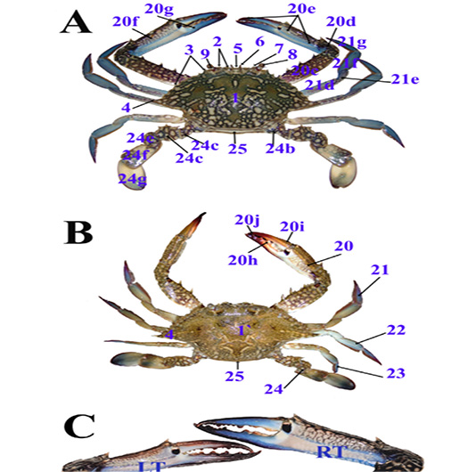
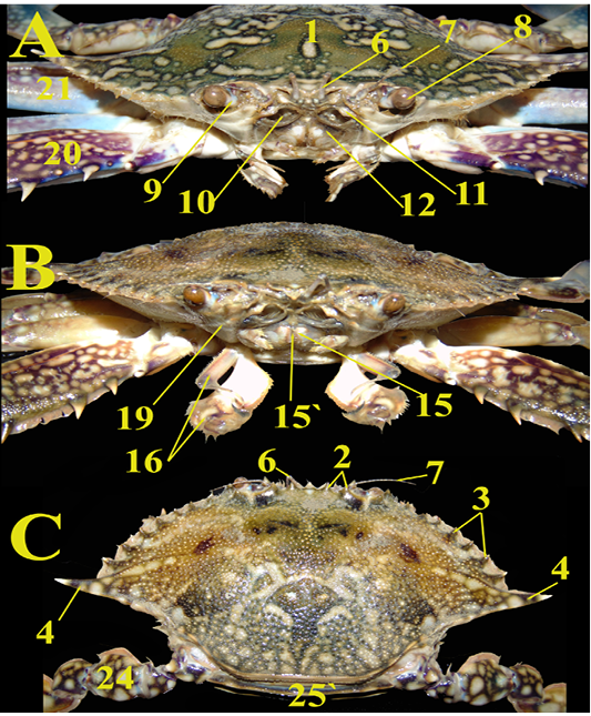
A & B; Photographs showing the dorsal aspect of the male and female blue swimmer crab (P. pelagicus), respectively, C; Magnified view of the right (RT) and left (LT) male chelipeds.

A & B; Photographs showing the frontal view and mouth parts of the male and female blue swimmer crab (P. pelagicus), respectively, C; Photograph showing the female crab spines on the carapace after removal of the first four pereiopods.
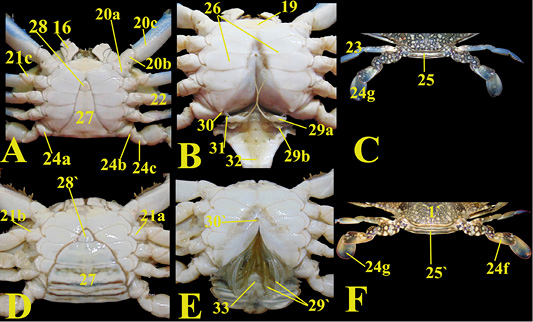
A & D; The ventral view of the male and female blue swimmer crab (P. pelagicus) respectively. B & E; The reflection of the abdominal segments in both male and female crabs, respectively. C & F; The caudodorsal view of the male and female crab, respectively.

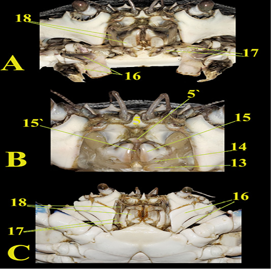
A; Fontal view of the male crab showing the three maxillipeds. B; Reflected maxillipeds showing the second and first maxillae and mandible underneath. C; Magnified ventral view of the maxillipeds of the male crab.

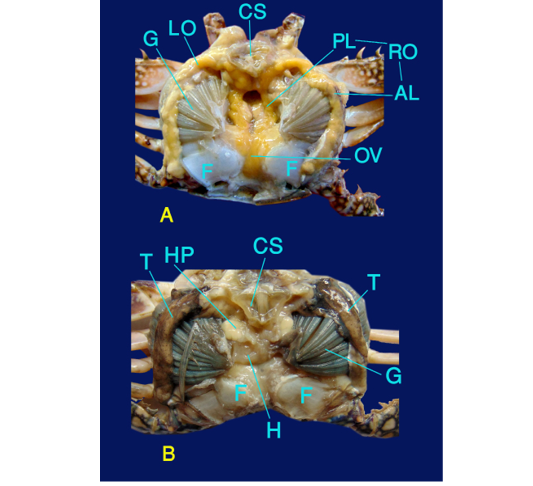
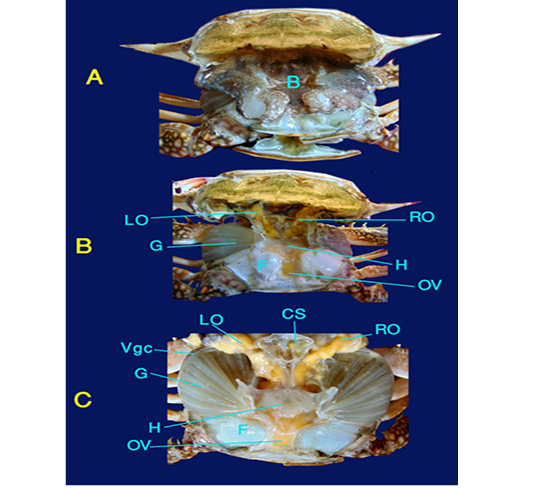
Photographs showing the internal organs of a female blue swimmer crab (P. pelagicus), A; The carapace reflected, B; Following removal of branchiostegite, C; Reflected carapace with view of internal organs.
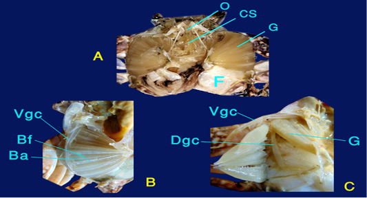
A, B & C; Photographs showing the gills of the crab
A & B; Photographs showing the internal organs of a female and male blue swimmer crab (P. pelagicus), respectively

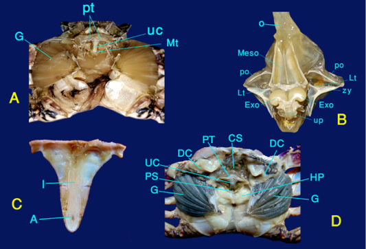
Photographs showing the digestive system of the crab, A, D; Dorsal view of the internal organs after carapace removal, B; Magnified view of the stomach, C. Inverted male abdominal segments


