
Serological, Molecular and Histopathological Study of Brucella melitensis Infection in Ewes


Serological, Molecular and Histopathological Study of Brucella melitensis Infection in Ewes
Ihab G. AL-Shemmari1*, Ali Hussein Fadhil1, Mohammed Assad S. Alkabi1, Eman Jawad Jabber2


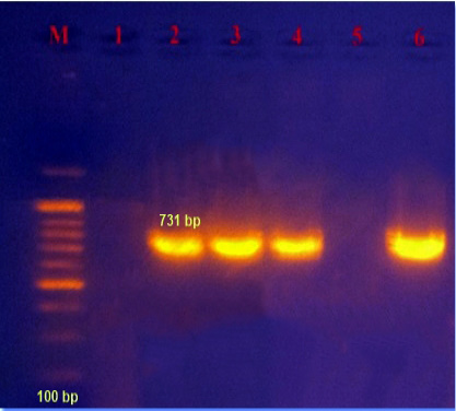
Figure 1
Agarose gel analysis of PCR products, the lane (M) is the DNA ladder; lane 1 is the control negative; lanes numbers 2,3,4 and 6 represent 731 bp for Brucella melitensis.

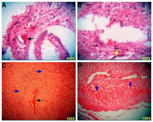
Figure 2
Section in the uterus post infection shows (A) severe congested blood vessels (black arrow) in the subserosal area. (B) Congested blood vessels with few mononuclear cells (yellow arrow) infiltration in the endometrium. (C and D) Congested of blood vessel (black arrow) and vacuolation (blue arrows) of muscle cells. H & E stain, 100X and 400X.


October 2023
Vol. 11, Iss. 10, pp. 1597-1756
