

Recombinant Slit2 Requires Heparin Sulphate to Inhibit TGF- β Induced Tumor Proliferation in Lung Cancer and Glioblastoma


Recombinant Slit2 Requires Heparin Sulphate to Inhibit TGF- β Induced Tumor Proliferation in Lung Cancer and Glioblastoma
Quratulain Amjad1,2 and Abdul Rauf Shakoori1,2*

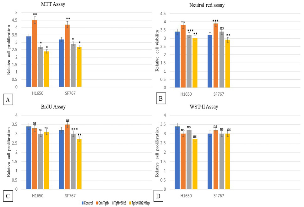
Cell functional assays in H1650 and Sf767 cells. MTT assay (A) showed decreased metabolic activity in presence of Slit2 and heparin. Neutral red assay (B) showed decreased cell viability with Slit2 and heparin treatment. BrdU assay (C) and WST-II assay (D) showed slight down-regulation in cell proliferation with Slit2 and heparin.
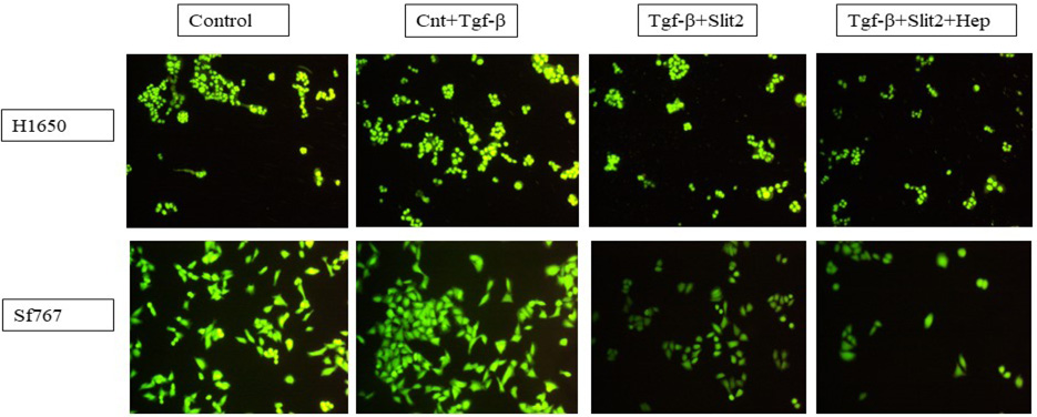
FDA stained viable/live H1650 cells and SF767 cells. The number of viable cells decreased with Slit2 addition and with heparin even in presence of TGF-β.
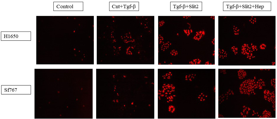
PI stained dead/non-viable H1650 and SF767 cells. The number of non-viable PI-stained cells increased after addition of Slit2 along with heparin.
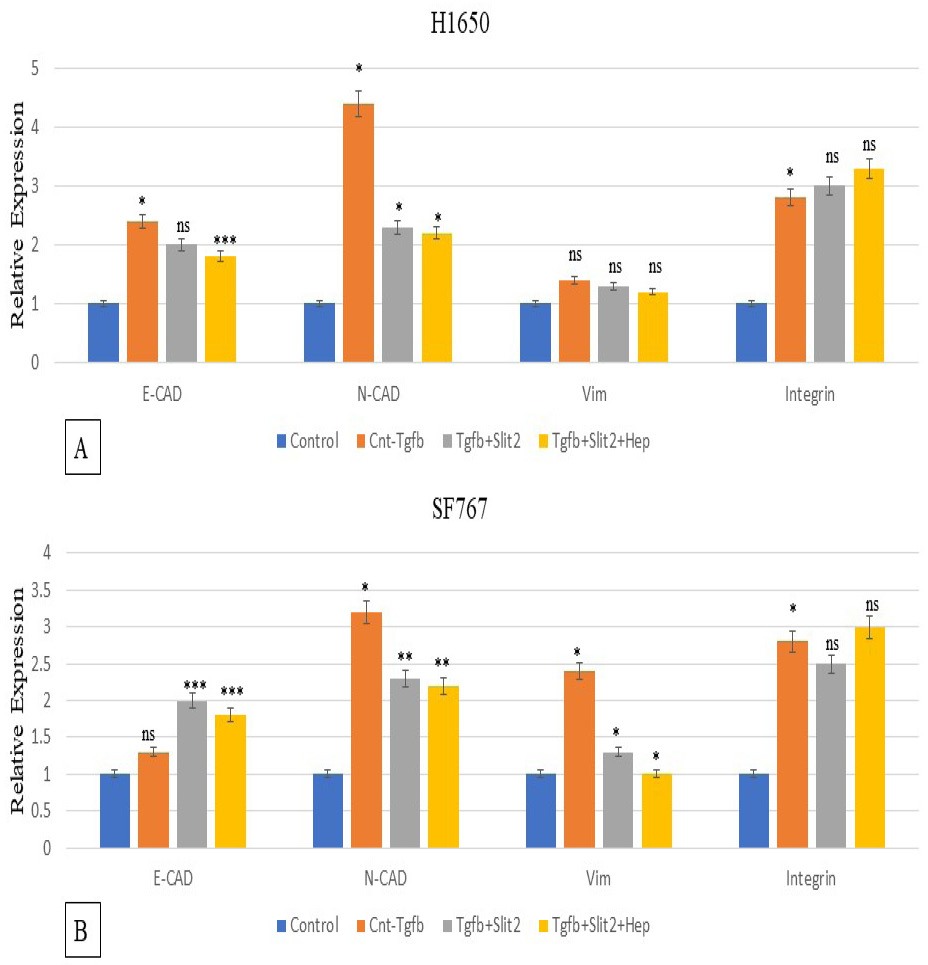
Expression analysis of cell adhesion genes in H1650 (A) and SF767 cells (B). There was significant down-regulation in expression of vimentin and N-Cad in presence of Slit2 that further amplifies in presence of heparin along with Slit2. Integrin beta 1 expression remained unchanged in H1650 and Sf767 cells.
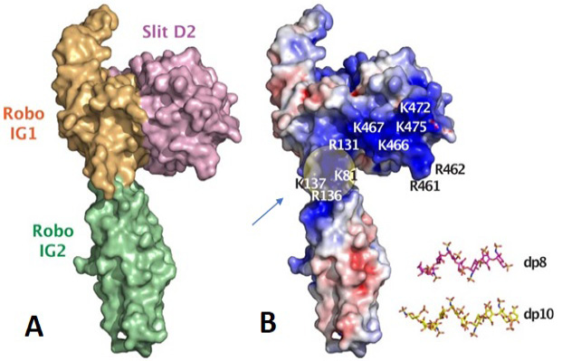
A model of human Slit2 (Domain 2) and Robo1 (IG1) complex. (A) shows the surface representation. (B) shows the electrostatic representation. The position of heparin binding to Slit2 and Robo is shown. The location of heparin oligosacharides in Robo1 is indicated by arrow. Figure taken from Fakuhara et al. (2008).


