
Preventive Role of Propolis and L-Arginine ® Supplement on Dna Damage for Sperm and Male Reproductive Hormones Values in Selenium Nanoparticles-Remedied Rats


Preventive Role of Propolis and L-Arginine ® Supplement on Dna Damage for Sperm and Male Reproductive Hormones Values in Selenium Nanoparticles-Remedied Rats
Zainab Sattar Ali* , Safa Azhar Razzaq , Layla Hammody Hashem


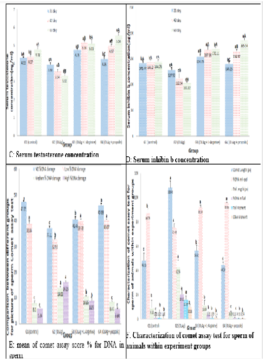
Impact of Black Tea selenium nanoparticles (BTSe), L-Arginine and propolis were taken it orally on serum FSH, LH, testosterone and inhibin B for triple times (after 3, 6 and 9 week) to each experimental group and mean score of DNA damage and the value of comet assay criteria % to sperm for male rats.
Numbers represent mean ± standard error.
Different capital leters signify to significant difference (P<0.05) between periods in same group.
Different small leters signify significant difference (P<0.05) between periods all groups.
Control (G1) group: rats of this group were received distilled water. Black Tea selenium nanoparticles (BTSe) (G2) group: animals in this group were intubated 200mg/kg body weight of BTSe.
Black Tea selenium nanoparticles (BTSe) + L-Arginine (G3) group: animals in this group were intubated 200 mg/kg body weight of (BTSe) + 200 mg/kg body weight of propolis.
Black Tea selenium nanoparticles (BTSe) and propolis (G4) group: animals in this group were intubated 200 mg/kg body weight of BTSe + 200 mg/kg body weight of L-Arginine.
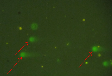
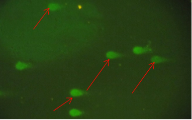
Comet assay for sperms of rats in G1
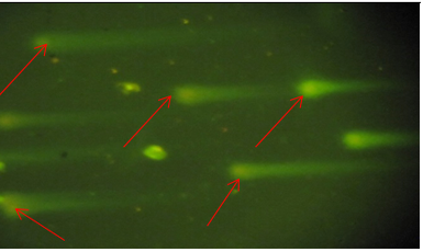
Comet assay for sperms of rats in G2
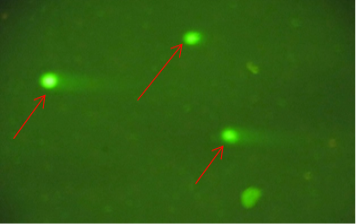
Comet assay for sperms of rats in G3

Comet assay for sperms of rats in G4
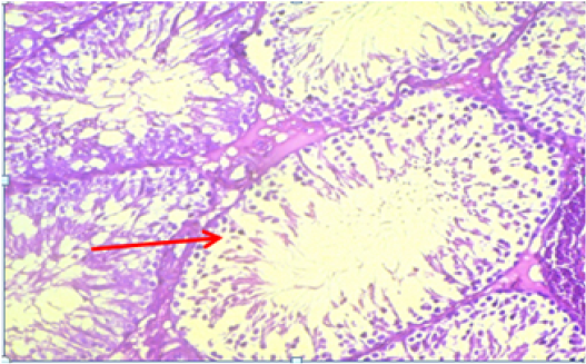
Histopathogical section for rat testes of control group, no clear lesions was observed.

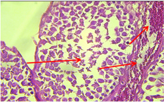
Histopathogical section for rats testes of group 2, was observed congestion and inflammatory cell and showed germ cells degeneration and Leydig cells, and absence of spermatogenesis.
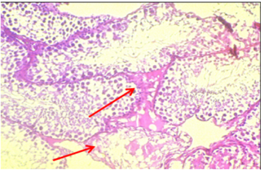
Histopathogical section for rat testes of group 3, was observed extension of interstitial space, cellular debris and derangement of spermatogonia anywhere in seminiferous tubule lumen.
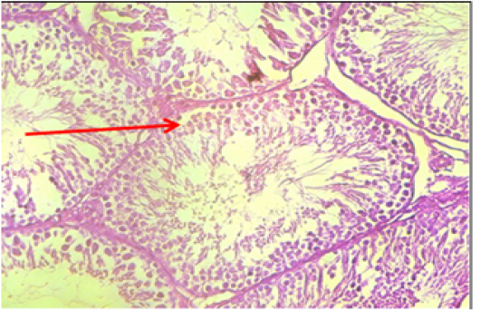
Histopathogical section for rat testes of group 4, no clear lesions was observed.


