
Prevalence of Nematode Parasites in Different Birds with Histopathological Changes in the Intestinal Tissue of Common Quail (Coturnix coturnix L.) with Special Reference to Heterakis gallinarum Schrank, 1788
Prevalence of Nematode Parasites in Different Birds with Histopathological Changes in the Intestinal Tissue of Common Quail (Coturnix coturnix L.) with Special Reference to Heterakis gallinarum Schrank, 1788
Rubab Malik, Nasira Khatoon* and Samina Waheed
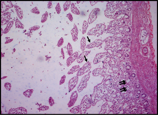
Figure 1:
Photomicrograph of intestine showing congestion and fibroid formation of muscularis mucosa, extreme distortion of villi (arrow), crypt glands can be seen emptied (double arrow).
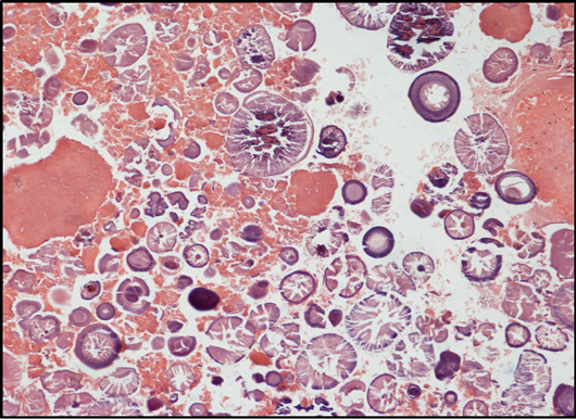
Figure 2:
Photomicrograph showing a section of intestine with heavy infiltration of mononuclear inflammatory cells.

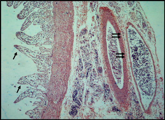
Figure 3:
Photomicrograph of intestine showing an increase in apparent size of villi with blunt and pointed ends (arrow), shrinkage of serosa and muscularis mucosa along with necrotic patches can be seen visible and nerve plexus (double arrow).

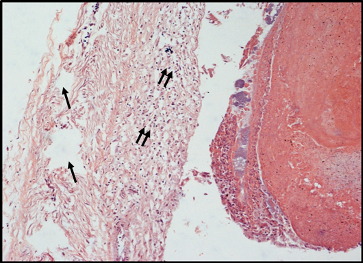
Figure 4:
Photomicrograph showing the vacuoles (arrow), large number of inflammatory cells (double arrow) and complete destruction of villi.
December 2022
Pakistan Journal of Nematology, Vol. 40, Iss. 2, Pages 86-173

