
Pathological Status of Fascioliasis of Bovine Liver Collected from Slaughter Houses in Dinajpur City of Bangladesh


Pathological Status of Fascioliasis of Bovine Liver Collected from Slaughter Houses in Dinajpur City of Bangladesh
Yonis Abukar Mohamed1, Md. Nazrul Islam1, Md. Haydar Ali1, Mahfuza Akther1 and Abdiaziz Idiris Mohamud2*

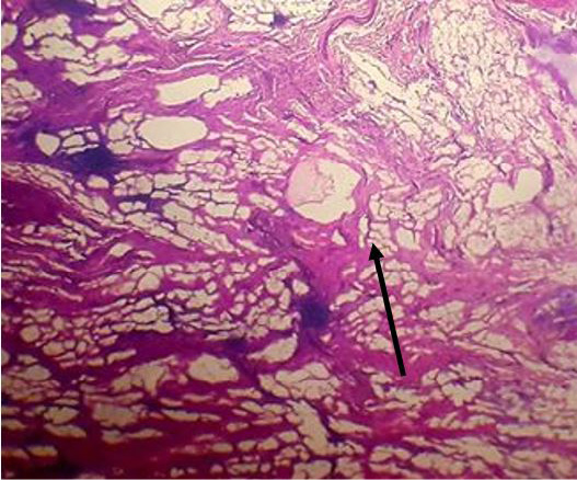
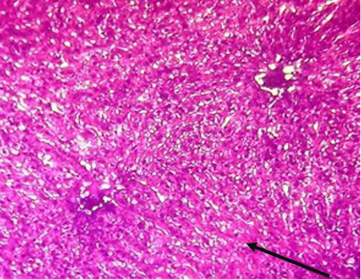
Swelling of individual hepatocytes by increase in size and characterized by opaque cytoplasm (Black arrow).
Abscesses including necrotic debris surrounded by a large numbers of inflammatory cells (Black arrow).

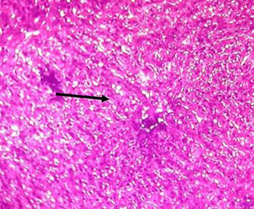
Congestion due to dilation of central vein and sinusoids and engorged with a large number of RBCs (Black arrow).
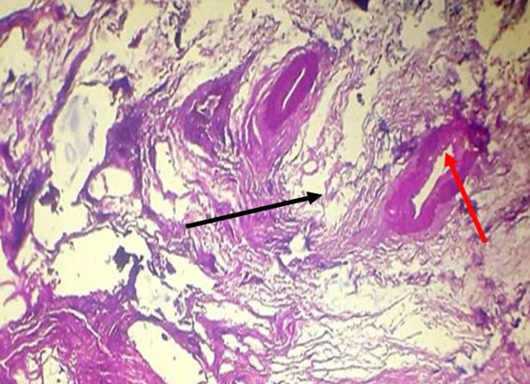
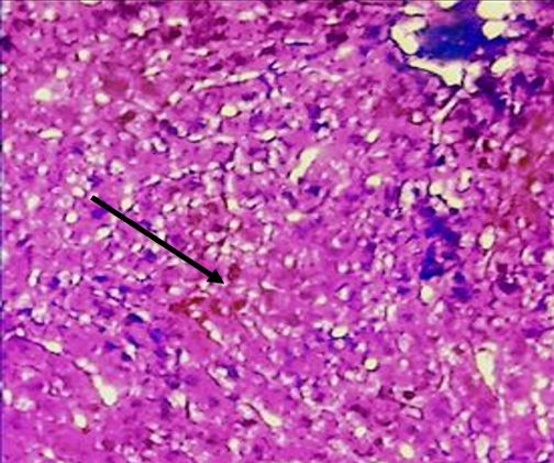
Migratory tract with lymphocytic infiltration (Black arrow) and cross section of mature liver fluke with heavy infiltration of inflammatory cells (Red arrow).
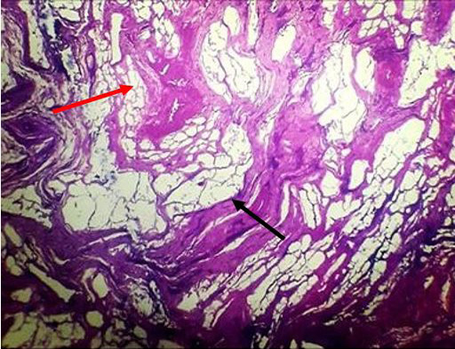
Fibroblastic proliferation in the previous migratory tract (Black arrow) and biliary cirrhosis with extensive proliferation of fibrous connective tissue around the intra-hepatic bile ductules (Red arrow)
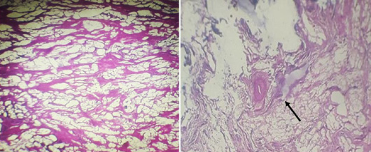
Liver cirrhosis, indicated by different dark and light spots and proliferated fibrous connective tissue around the regenerative hepatic lobules (Black arrow) was infiltrated with inflammatory cells.

Fatty change with clear vacuoles appeared in the cytoplasm with peripherally located nuclei (Black arrow).


