Pathological Effects of Candida Auris Yeast on Liver of White Male Mice Pre-Treated with Alcoholic Extract of Syzygium Aromaticum
Pathological Effects of Candida Auris Yeast on Liver of White Male Mice Pre-Treated with Alcoholic Extract of Syzygium Aromaticum
Anas A. Humadi1*, Samer I. Sabeeh2, Ahmed Talib Yassen Aldossary3, Bushra I. Al-Kaisei2
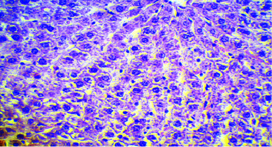
Photomicrograph of liver in control group depicting normal liver tissue (H and E stain; 20x).
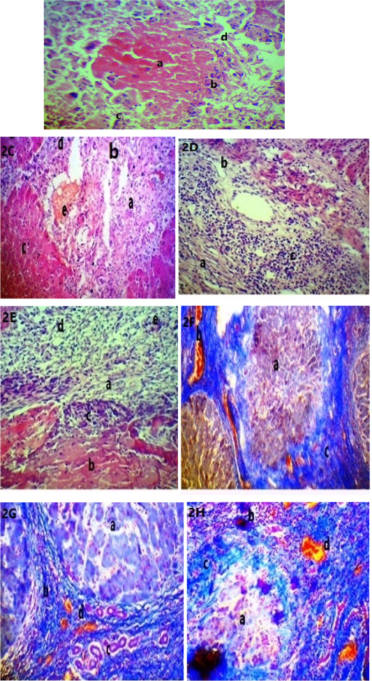
A: Photomicrograph of the liver in 2nd group showed: (a) center of necrosis, (b) mononuclear cells, (c) foreign body giant cell, (d) fibrosis (H and E stain; X40). B: (a) fibrosis, (b) newly central vein, (c) necrotic hepatocytes (H and E stain; X20). C: (a) fibrosis, (b) newly central vein, (c) necrotic hepatocytes, (d) mononuclear cells, (e) hemorrhage (H and E stain; X20). D: (a) fibrosis, (b) newly blood vessels, (c) severe infiltration of inflammatory cells (H and E stain; X20). E: (a) liver fibrosis, (b) necrosis, (c) mononuclear cells infiltration, (d) newly blood vessels, (e) newly bile duct (H and E stain; X20). F: (a) cirrhotic liver, (b) newly blood vessels, (c) fibrosis (Masson trichrome stain; X20). G: (a) pseudolobulated liver, (b) fibrosis, (c) newly bile duct, (d) congested blood vessels (Masson trichrome stain; X40). H: (a) pseudonecrotic lobules, (b) clotted blood vessels, (c) mononuclear cells infiltration, (d) newly bile duct (Masson trichrome stain; X20).
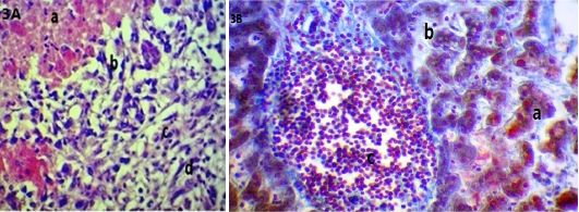
A: Photomicrograph of the liver in 3rd group showed: (a) acute cellular swelling, (b) increase width of sinusoid, (c) infiltration of mononuclear cells, (d) newly bile duct (H and E stain; X40). B: Photomicrograph of the liver in 3rd group showed: (a) acute cellular swelling, (b) width sinusoid, (c) mononuclear cells infiltration and eosinophil (Masson trichrome stain; X40).


