
Immune-Pathological Studies on the Distribution and Localization of Bovine Viral Diarrhea Virus


Immune-Pathological Studies on the Distribution and Localization of Bovine Viral Diarrhea Virus
Mahmoud S. Sirag1*, Effat L. El Sayed2, Mahmoud M. Hussein3, Khalid A. El-Nesr1, Mahmoud B. El-Begawey1

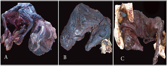
Mummified fetus showing. A) Four legs arched back (sclerosis of vertebrae); complete resorbed eyes; remnant of umbilical cord; collapsed skull as a result of resorption of the brain tissues. B) Hard on texture; It has neither legs nor eyes; The skull was very hard and had the pyriform shape; This fetus belong to the current or acute infected group. C) Hard on texture, absence of one fore legs and presence of two hind legs. The skull was pyriform in shape and has only eye fissures without eye balls, this fetus also belongs to the current infected group.
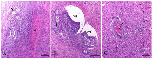
Photomicrograph from ovary of PI cow showing. A) Disrupted primordial follicles (arrow) and obliterative atretic follicle. (AF). H&E. Bar.200µm. B) disrupted secondary and tertiary follicles (2ndf, 3rf). Minute focal granulosa cell tumor (arrow) could be observed. H&E. Bar.200 µm. C) Active corpus luteum with large (L), small(S) regressing cells (Rc). The blood vessel showing congestion and degenerated wall (BVs) (arrow).H&E. Bar.200 µm.
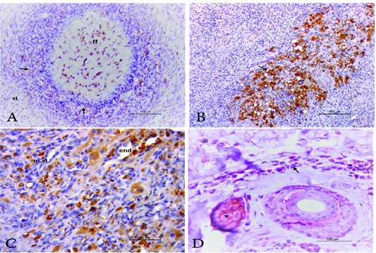
Photomicrograph of ovary and ear notch from PI animal stained by IHC showing, A) growing follicles where the positive cells appear at follicular fluid (ff); granulosa cells (arrow) and at the stromal tissue (st). Stained by IHC. Bar.100µm. B) BVDV antigen positive cells (macrophages) at the atretic follicles (arrow) in the Bar.100µm. C) BVDV antigen positive cells (macrophage) in the ovarian medullar stroma (m.st) as well as endothelium of the blood vessels (end). Bar.50µm. D) ear notch revealed positivity against BVDV antigen in dermal cells (arrow) and sebaceous gland (arrow head); IHC. Bar.100µm.

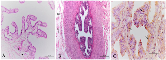
Photomicrograph of uterine tube of PI animal showing. A) Infundibulum with moderate folding of mucosa –submucosa layer and congestion (arrow) together with edema and thrombosis of the vasculature (arrow head) at the tunica serosa and musculosa. H&E. Bar.200µm. B) Isthmus suffer from edema (ed) and congestion (arrow) in stratum vascular. H&E. Bar.200µm. C) positive reaction as brown color granules in the cytoplasm of epithelium (arrow) IHC labeling. Bar.50µm.

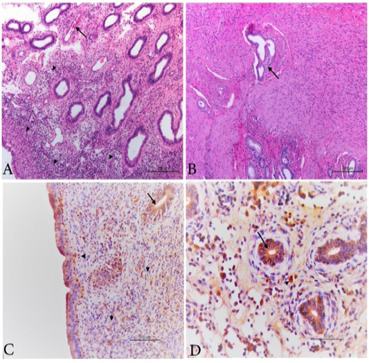
Photomicrograph from uterus of PI animal showing, A) endometritis with submucosal and interglandular infiltration with inflammatory cells (arrow head) as well as congestion of the vasculature (arrow). H&E Bar.200µm. B) Islets from endometrial glands between bundles of uterine muscles (endometriosis) (Arrow). H&E Bar.200µm. C) positive BVDV antigen in the cytoplasm of the glandular epithelium (arrow) and in macrophages at sub epithelial, inter -glandular tissue and in the endometrial glands (arrow head). IHC labelled. Bar.100µm. D) positive brown granules in the cytoplasm of the glandular epithelium (arrow) and in macrophages at the inter -glandular tissue (arrow head). IHC labelled. Bar.50µm.

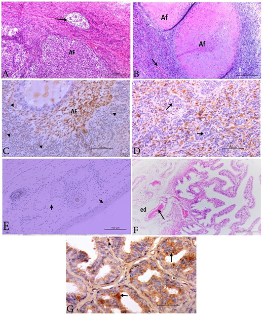
Photomicrograph from ovary, ear notch and uterine tube of acute infected animals. A) Ovary with collapsed atretic follicle (AF) and focal granulosa cell tumor (arrow) .H&E. Bar. 200µm. B) Ovary with collapsed atretic follicles (AF) and congested stromal blood vessels (arrow).H&E. Bar. 200µm. C) Ovary revealed positive BVDV antigen in the collapsed atretic follicle (AF) and in macrophages at the ovarian stroma. IHC. Bar.100µm. D) Ovary with positive macrophages at the ovarian stroma while the blood vessels appear clear negative (arrow). H&E. Bar.50 µm. E) Ear notch showing negative reaction for BVDV antigen (arrow). IHC. Bar.100 µm. F) Infundibulum showing marked folding of mucosa –submucosa layer and severe congestion of the vasculature (arrow). also edema (ed) at the tunica serosa and musculosa. H&E. Bar.200µm. G) Uterine tube showing positive BVDV antigen in the epithelium in focal manner (arrow) and in macrophages at the sub mucosa (arrow head). IHC. Bar.50µm.
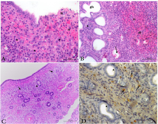
Photomicrograph from uterus of acute infected animals showing, A) Necrosis (N); sloughing of mucosa; diffuse infiltration of mononuclear cells (arrow head); congestion (arrow) and hemorrhages (H). H&E. Bar.50µm. B) Necrosis; hyperplasia of the endometrial gland (arrow head) associated with Cystically dilated glands (gls) beside congestion (arrow) and hemorrhages (H). H&E. Bar.100µm. C) Chronic endometritis presented by granulation tissue (arrow) and periglandular fibrosis at the endometrial stroma (arrow head).H&E. Bar.200µm. D) brown granules represent the positive BVDV antigen (arrow). All uterine glands are free from BVDV antigen (arrow head).H&E. Bar.50µm.

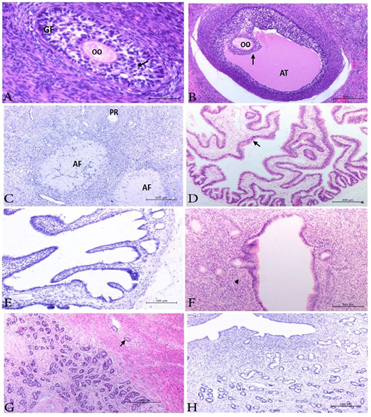
Photomicrograph from ovary, uterine tube and uterus of negative cows showing, A) ovary with growing follicles (GF) had intact oocyte(OO) and corona radiata (arrow). H&E. Bar.50µm. B) ovary with mature follicles (MF) had intact oocyte (OO) and corona radiata (arrow) as well as antrum (AT). H&E. Bar.200µm. C) Ovary with primordial (PR) and collapsed atretic follicles (AF). No positive reaction could be detected. IHC. Bar.200µm. D) uterine tubes showing normal mucosa and sub mucosa. H&E. Bar.200µm. E) uterine tubes appear free from BVDV antigen. labelled by IHC Bar.100 µm. F) Uterus showing mild chronic endometritis (arrow head). H&E. Bar .200 µm. G) Uterus with endometrial gland hyperplasia associated with endometriosis (arrow). H&E. Bar.200µm. H) Uterus showing negative BVDV; antigen. IHC. Bar.200 µm.

