
Histopathological Modifications in Musa cavendishii Roots Induced by Radopholus similis and Meloidogyne incognita

Histopathological Modifications in Musa cavendishii Roots Induced by Radopholus similis and Meloidogyne incognita
Atef M. El-Sagheer1*, Aline F. Barros2, El-Sayed M. Abd El-Aal3, Mohamed M. Gad4, Doaa S. Mahmoud4 and Amr M. El-Marzoky3
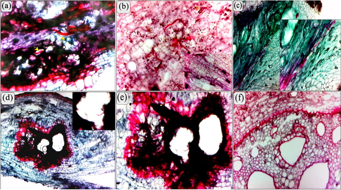
(a, b, and c) Longitudinal section of a banana root showing pre-adult stages and adult stage of Radopholus similis (arrow) coiled in medial parenchyma. (b) High power represents the lignification in sub-epidermal cortical cells. (c) High power showing the pathway of pre-adult stages of R. similis in the cortical parenchyma. (d and e) showing the overlap infection between pre-adult stages of Radopholus similis through the gall formed by M. incognita. (f) Transverse section of a banana root showing the modified xylem, phloem vessels and parenchyma caused by Radopholus similis. Note a few of phenolic and lignified cells in vascular cylinder (b). (Scale bars: a = 70 μm; b-f = 200 μm).
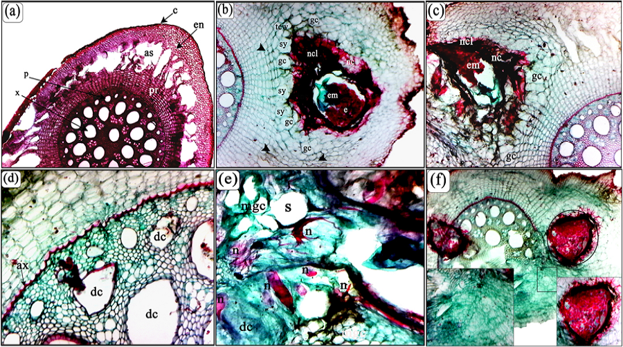
(a). Transverse section of healthy banana cv. Hindi roots; Central cylindrical or stele [Phloem (p), xylem (x)], Prosenchyma (pr), Endodersmis (en), Mesodermis (parenchyma) (m), Cortex (Epidermis and exodermis) (c), Air space (as). (b) Transverse section through the gall and cortex cells of the infested banana root with Meloidogyne incognita showed the feeding site of adult female with egg masses (em), note the multinucleate cells, giant cells and lateral expansion in necrotic cell (nc) within layers (lnc) in the inner cortical tissue is beginning to spread to the stele with compressed of cells size in various layers of the of the root (head arrow) with thickness in cell walls (tcw). (c) showing lateral expansion of giant cells (gc) necrotic cell (nc) within layers (lnc) in the inner cortical tissue is beginning to spread to the stele through a passage cell in the endodermis (e). (d) The deformed cells in the parenchyma and abnormal xylem (ax). (e) Fourth stage of M. incognita (n) near of the cambium (ca). It notes a large laceration in neighboring cells of feeding site; multinucleate giant cells (mgc), note dark tissue elements and thickness of the cell wall in neighbor cells of multinucleate cells and dense cytoplasm. (f) Adult female and eggs of M. incognita near of the vascular cylinder, notes a large laceration in neighboring cells of feeding site. (Scale bars: a-f = 200 μm).
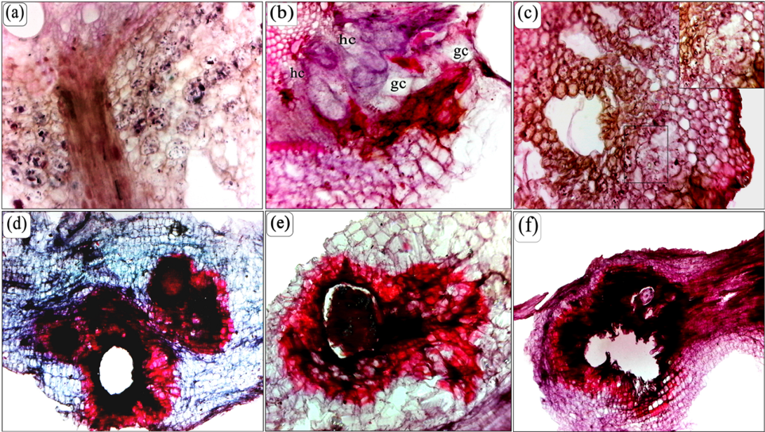
(a) Transverse section of a banana root showing the granular structures in the cells of cortex layer near the feeding site of M. incognita. (b) Note the nuclei in the vicinity of nematodes either enlarged or disintegrating and either cytoplasm contracting from cell walls still intact or contracted and cell walls beginning to rupture. In addition, giant cell transforming from undifferentiated hypertrophied cells. (c) Transverse section through the cortex noted the extensive cavity in the cortex, the nematodes at the periphery of the lesion and some cell fragments towards the center necrotic and lignified cells. (d, e and f) Longitudinal section of a banana root through the gall showing the laceration and deformation area and phenolic and lignified cells of damaged cortex extended towards the pericycle on the old root and the root hair. (Scale bars: a-f = 200 μm).
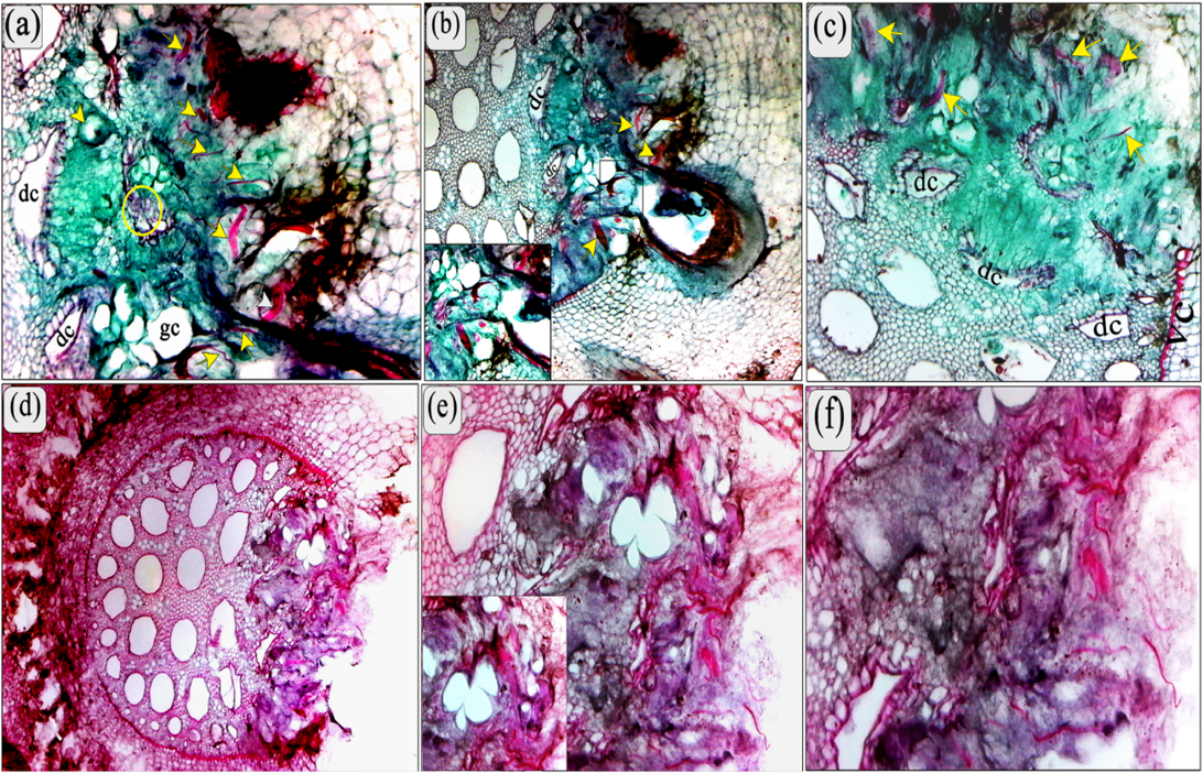
(a) Transverse section of banana roots showing the adult female of M. incognita (arrows) inducing the clear multinucleate cells (cycle) and with fused syncytial cells presenting dense cytoplasm and hypertrophic nuclei, and shows some of adult and larval stages of R. similis (arrows). (b) Showing deformed cells (dc) and noted that the extension of the feeding site to the vascular cylinder. High power showing the most damage in root cells as results of a combination of both nematodes. (c and d) root infested with pre-adult stages of both M. incognita and R. similis showing the abnormal xylem, clear dissolution of endodermis and central cylinder (arrows) and note the thickness of endodermis layer compared to the normal size in the control. Also, note all the modification symptoms associated with the feeding process; multinucleate cells, giant cells, and modified cells. (e, f and d) The laceration and dissipation of the central cylinder (xylem and phloem vessel) as a result of the feeding of both species. Note the extent of the feeding sites inside the vascular cylinder. (Scale bars: a-f = 200 μm).
