Experimental Evaluation for Graviola Extracts and Eucalyptus Extract’s Capacity to Induce Healing of Hepatocytes in Rabbits Model
Experimental Evaluation for Graviola Extracts and Eucalyptus Extract’s Capacity to Induce Healing of Hepatocytes in Rabbits Model
Heba Saleh Shaheed1, Zainab J. Malik2*, Ghusoon A.A. Alneamah1, Salih T.H.2
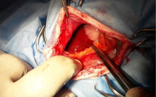
Gross section showed a midline laparotomy. In the upper abdomen, exposing the fourth lobe of the liver, a surgical incision was made to perform a partial liver resection in rabbits.
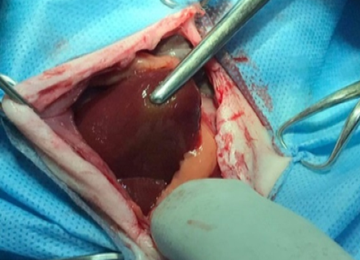
Gross section showed handling by use thump forceps and revelation of the third lobe of liver a for doing partial hepatoctomy in rabbit.
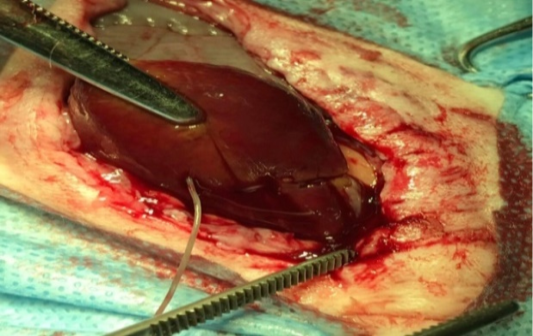
Gross section showed the base of the liver and near the inferior vena cava, sutures were tied over the liver lobe and surgical removed done for doing partial hepatoctomy in rabbit.
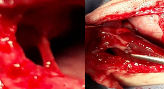
Gross section showed grade II adhesion (MSAS) between the liver, omentum, and abdominal wall in the Graviola group (A) and Eucalyptus camaldulensis group (B).
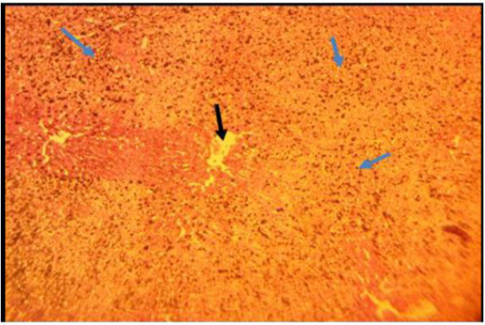
Histopathological section of liver of rabbit of Graviola group showed heavy inflammatory reaction infiltration in entire hepatic parenchyma (blue arrows) with dilation of central veins (black arrow) (staining with H and E stain 10X).
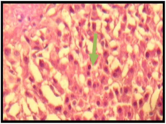
Histopathological section of a rabbit liver from Eucalyptus camaldolensis leaf group extract showed mild regeneration of hepatocyte of liver (arrow) (staining with H and E stain 10X).
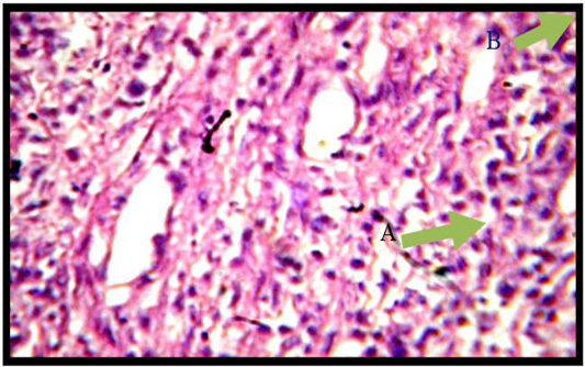
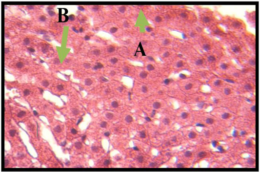
Histopathological section of liver related to treated group to extract of eucalyptus camaldulensis leaves, shows infiltrated mononuclear cells (A) and appear of myofibroblast (B), with granulation tissue formation (staining with H and E stain 10X.).
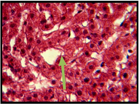
Histopathological section The liver after returning to its normal structure, which is represented by hepatocytes cord around the central vein (arrow) (staining with H and E stain 100X).
Histopathological section of liver related to Graviola treated group, shows normal hepatic architecture which represented by normal lobules, CV (staining with H and E stain 10X).
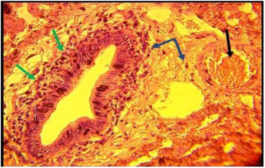
Histopathological section of liver of rabbit of eucalyptus camaldulensis extract of leaves group showed infiltration of inflammatory cells (blue arrows) and the bile duct is dilated and surrounded by inflammatory cells (green arrows) in addition to the dilation there is congested of hepatic artery (black arrow), (staining with H and E stain 40X.)


