
Chronic Submandibular Lymphadenopathy due to Cryptococcosis in Cat


Chronic Submandibular Lymphadenopathy due to Cryptococcosis in Cat
Soedarmanto Indarjulianto1*, Alfarisa Nururrozi1, Yanuartono1, Sitarina Widyarini2, Dhasia Ramandani3


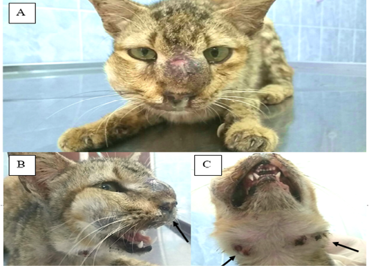
Figure 1
Nasal deformity, swollen nasal bridge and maxillary sinus (A). Mucopurulent discharge were observed from both nostrils (B). Bilateral enlargement of submandibular lymph nodes (arrow) (C).

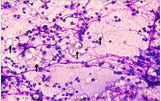
Figure 2
The nasal swab stained with Giemsa showed Cryptococcus spp. intracellular in the nasal epithelium. Fungi were rounded with a variably thick non-staining capsule and pale on center (arrow) (Giemsa stain 400 X).

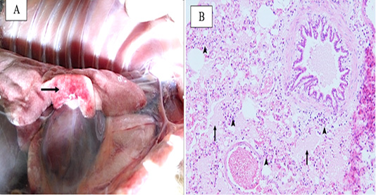
Figure 3
Postmortem findings on lung show focal consolidation (A). Histopathological examination showed neutrophils infiltration in broncheolus and interstitial area of the lungs (arrow). Also, mild edema as homogen eosinophilic mass in alveoli (arrow) (B).

March 2023
Vol. 11, Iss. 1, Pages 1-98
