
Bacteriological Identification and Molecular Detection of Klebsiella pneumoniae from Pneumonic Camels in Al-Muthanna Province


Bacteriological Identification and Molecular Detection of Klebsiella pneumoniae from Pneumonic Camels in Al-Muthanna Province
Hayder M. Watban1,2, Nabeel M.H. Al-Maaly2*

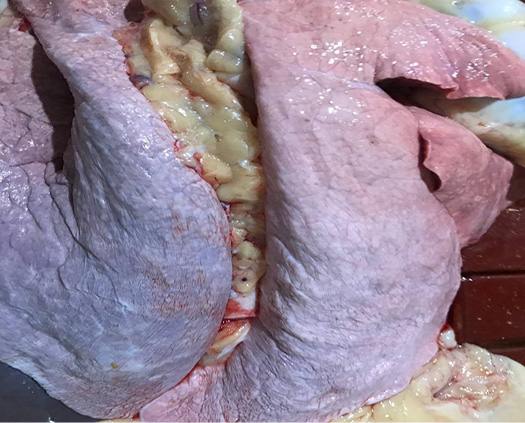
Figure 1:
A post-mortem examination of the left lung in the camels unveiled a pneumonic lesion characterized by a firm, blunt end, along with petechial hemorrhaging and red consolidation.
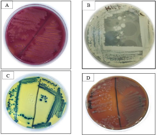
Figure 2:
K. pneumoniae on MacConkey, nutrient, blood and CHROMagar orientation agars. (A) K. pneumoniae on MacConkey agar. (B) K. pneumoniae on nutrient agar. (C) K. pneumoniae on CHROMagar orientation. (D) K. pneumoniae is a non-hemolytic on blood agar.
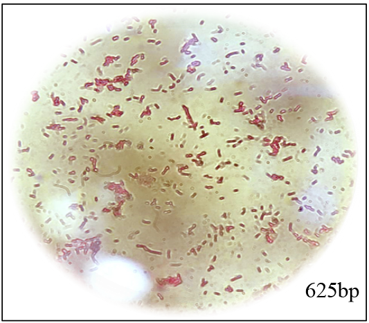
Figure 3:
Gram stain of K. pneumoniae.

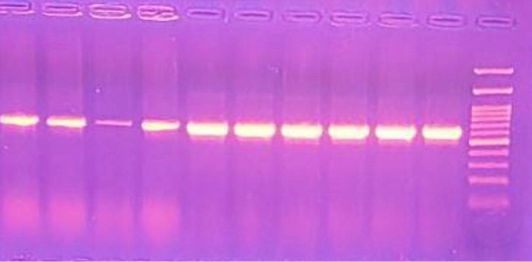
Figure 4:
PCR Gel electrophoresis positive results of K. pneumoniae with red stained for amplification size 625bp of 16SrRNA K. pneumoniae.

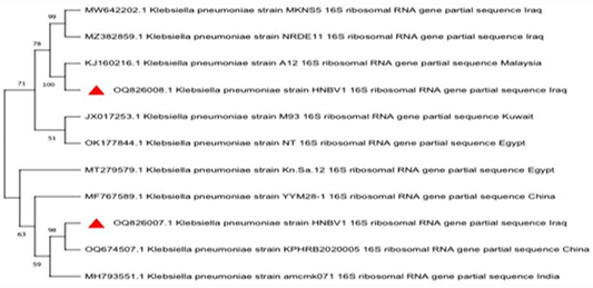
Figure 5:
Phylogenetic tree of K. pneumonia in comparison with local and international isolates.

November 2023
Vol. 11, Iss. 11, pp. 1757-1910
