
Antidiarrheal Effects of Prosopis farcta L. Fruit Extract: An Enteropooling and Histopathological Study in Rats


Antidiarrheal Effects of Prosopis farcta L. Fruit Extract: An Enteropooling and Histopathological Study in Rats
Sara H. Zughayyar*, Amer H. Gyad

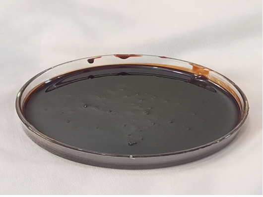
Fruits of Prosopis farcta L. appearance in ethanolic extract.
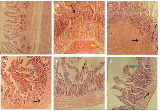
Histopathological comparison of the duodenal layers in male rats from different treatment groups (H&E stain 20×). A) Histopathlogical section of the duodenum for one rat in the first group (negative control) showing no abnormal lesion that each layers of mucosa, submucosa, muscularis and serosa give normal appearance. B) Histopathological section of the duodenum for one rat in the second group (positive control) showing thickening in each layers of duodenum and infiltration of infilammatory cells (Mononuclear cells) (Blue arrow) and hyper plastic proliferation of goblet cells (Black arrow) and glandular of sub mucosal (Green arrow) and thickening of muscular layer (Yellow arrow). C) Histopathological section of the duodenum for one rat in the third group (treated by Prosopis farcta L. fruits extract 300mg /kg.BW) Sub mucosa also showing infiltration of inflammatory cells (Blue arrow) with edema (Black arrow) and destruction of glands, muscularis showing thicking with slight infiltration of inflammatory cells and thickening of all layers. D) Histopathological section of the duodenum for one rat in the fifth group (treated by Prosopis farcta L. fruits extract 400mg /kg.BW) showing sloughing and infiltration of inflammatory cells of mucosa (Blue arrow) and sub mucosa slight edema in mucosa (Black arrow). E) Histopathological section of the duodenum for one rat in the fourth group (treated by Prosopis farcta L. fruits extract 500mg /kg.BW) All layers of this group showing slight lesion which involve infiltration of mono nuclear cells especially in mucosal layer (Blue arrow) with very little sloughing amount of epithelia lining mucosa (Black arrow) also slight edema and inflammatory cells in sub mucosa (Green arrow) with normal appearance of muscular layer. F) Histological section of the duodenum for one rat in the sixth group (treated by loperamide 2.5mg /kg.BW) showing sloughing and infiltration of inflammatory cells of mucosa (Blue arrow) and sub mucosa slight edema in mucosa (Black arrow).


