An In Vitro Assessment of the Cytotoxic and Apoptotic Potency of Silymarin and Silymarin Loaded Solid Lipid Nanoparticles on Lung and Breast Cancer Cells
An In Vitro Assessment of the Cytotoxic and Apoptotic Potency of Silymarin and Silymarin Loaded Solid Lipid Nanoparticles on Lung and Breast Cancer Cells
Canan Vejselova Sezer*
The shape and morphology of silymarin (a, arrows-silymarin particles) and silymarin loaded SLN (b, arrows-silymarin-loaded SLN particles).
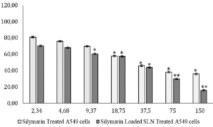
Viability percentages of silymarin and silymarin-loaded SLN treated A549 cells for 24 h. IC50 value was detected to be 33 µM (for silymarin) and 25 µM (for silymarin-loaded SLN). *p<0.05, **p<0.01
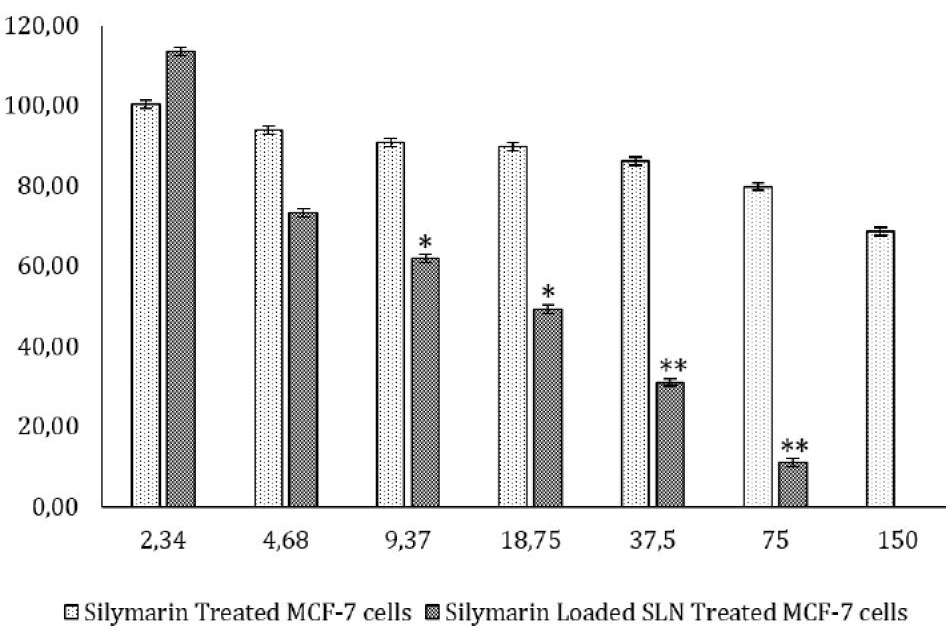
Viability percentages of silymarin and silymarin-loaded SLN treated MCF-7 cells for 24 h. IC50 value for silymarin treated MCF-7 cells was not detected in this concentration range of the agent for 24 h. IC50 value for silymarin-loaded SLN treated MCF-7 cells was detected to be 18 µM for 24 h. *p<0.05, **p<0.01
Untreated A549 cells (a), astersk-nucleus, arrow-cytoskeleton, rectangle-fusiform cell shape. A549 cells treated with IC50 value of silymarin (b and c), arrow-shrunken (circular shaped) cells, arrow head-holes on cytoskeleton, circle-nuclei with condensed chromatin and A549 cells treated with IC50 value of silymarin-loaded SLN (d, e and f) for 24 h; circle-cells with fragmented cytoskeleton, arrow-pyknotic nucleus, asterisk-chromatin condensation, arrow head-holes on cytoskeleton.
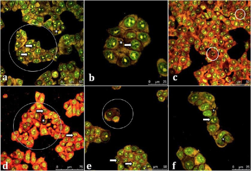
Untreated MCF-7 cells (a and b); asterisk-cytoskeleton, arrow-nuclei, circle-typical cell cluster. MCF-7 cells treated with IC50 value of silymarin (c and d); arrow-condensed chromatin, asterisk-condensed cell, circle-lacerated cell cluster, small circle-fragmented nuclei and MCF-7 cells treated with IC50 value of silymarin-loaded SLN (e and f) for 24 h; circle-shrunken cell cluster with decreased number of cells, arrow- excessive chromatin condensation, asterisk-highly perforated cytoskeleton.
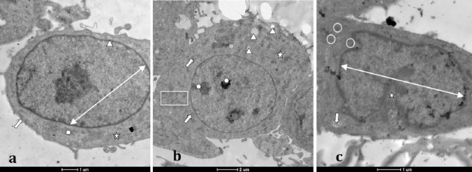
Untreated A549 cell (a), arrow-cell membrane, two-headed arrow-nucleus, circle-mitochondrion with compact cristae, asterisk-cytoskeleton. A549 cell treated with IC50 value of silymarin (b), Arrow-swollen mitochondria, rectangle-lacerated cisterna of endoplasmic reticulum, arrow head- holes on cytoskeletal disintegration, asterisk-granular cytoplasm, circle-condensed chromatin. A549 cell exposed to IC50 value of silymarin-loaded SLN for 24 h (c), two-headed arrow-pyknotic nucleus with extremely condensed chromatin, arrow-disintegrated mitochondrion with excessive loss of cristae, circle-highly swollen cisterna of endoplasmic reticulum and asterisk-fragmentation of nucleus.
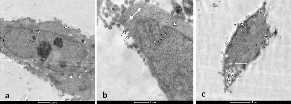
Untreated MCF-7 cell (a), arrow-nuclear membrane, circle-cell membrane, arrow head-compact mitochondrion. MCF-7 cell treated with IC50 value of silymarin (b), Arrow-secondary lysosomes, Arrow head-loss of cristae, two headed arrow-granulated cytoplasm, asterisk-cleavage of nuclear membrane (formation of pyknotic nucleus). Silymarin-loaded SLN treated MCF-7 cell (c), arrow-extreme chromatin condensation, rectangle-disintegrated cell membrane and asterisk-fragmentation of the cytoskeleton (huge holes on cytoskeleton).
Apoptosis profiles of untreated A549 cells (a) and A549 cells treated with IC50 values of silymarin (b) and silymarin-loaded SLN for 24 h (c). Apoptosis profiles of A549 control cells (a) showed 90.51% live, 2.16% early and 2.78% late apoptotic cells. In this group of cells percentage of dead cells was detected to be 4.55%. A549 cells treated with IC50 concentration of silymarin (b) percentage of live cells was detected 73.50. Early apoptotic and late apoptotic cells of this group were 6.21% and 19.61%, respectively. A negligible percentage (0.69%) of the same group of A549 cells were dead. Total percentage of cells that underwent apoptosis was 36.75 of that 32.19% were in late apoptosis in silymarin-loaded SLN treated A549 cells (c). The percentage of live cells in the same group was detected to 62.27 for 24 h.
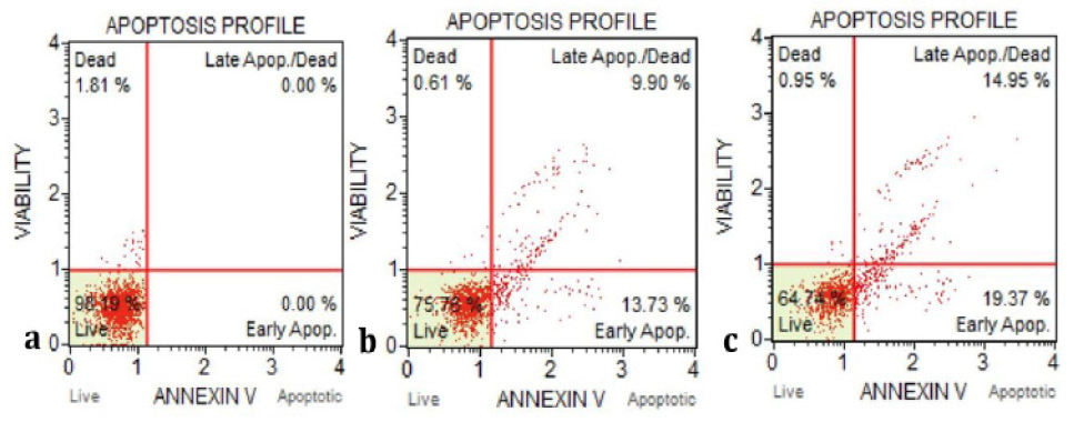
Apoptosis profiles of untreated MCF-7 cells (a) and MCF-7 cells treated with IC50 values of silymarin (b) and silymarin-loaded SLN (c) for 24 h. On the apoptosis profiles of untreated MCF-7 cells (a) were detected 98.19% live and 1.81% dead cells. In silymarin treated MCF-7 cells (b) the percentage of live cells was 75.76 and 13.73% and 9.90% of these cells were in early and late apoptotic stage, respectively. 0.61% of dead cells were detected in this group of cells. Early apoptotic and late apoptotic cells of this group were 6.21% and 19.61%, respectively. Live cells percentage in silymarin-loaded SLN treated MCF-7 cells (c) was detected as 64.74%. Early apoptotic and late apoptotic cell percentages were 19.37 and 14.95 for this cell group.


