
Gelatinase is a protease with potential applications in the food industry, particularly in the production of gelatin hydrolysates. Gelatinase-producing bacteria is the main source for industrial exploration. This study aimed to isolate gelatinase-producing bacteria from coastal sediments of Tegalpongo, Pasuruan, and identify them using molecular analyses. The research method was descriptive and exploratory, consisting of two steps. Bacterial isolation from coastal sediments was followed by identification using the morphology and molecular, 16S rRNA method, BLAST, and phylogenetic tree analysis. The results showed that all five isolates produced gelatinase. The highest gelatinase activity was observed in isolate A2.SD. The results of BLAST showed a query cover of 98% and an identity of 99%. Phylogenetic tree analysis demonstrated that isolate A2.SD had the closest relationship with Proteus penneri, with an E-value of 0.98%. Therefore, isolate A2.SD was designated as Proteus penneri, a Gram-negative rod-shaped bacterium. Hence, we named isolate A2.SD was to Proteus penneri UB-L024.
Special Issue: Innovations in Sustainable Fisheries, Marine Resources, and Food Systems for Food Security and Human Well-being · Volume 42, Issue 2, 22 October 2026 · Open Access
Molecular Identification of Gelatinase-Producing Bacteria from Tegalpongo Coastal Sediments
· · ·
Affiliations & Notes ⌄
- 1Department of Fisheries Product Technology, Faculty of Fisheries and Marine Sciences, Universitas Brawijaya, Jl. Veteran, Malang 65145, East Java, Indonesia
- 2Faculty of Food Science and Nutrition, Universiti Malaysia Sabah, Kinabalu 88400, Malaysia. *Correspondence | Asep Awaludin Prihanto, Department of Fisheries Product Technology, Faculty of Fisheries and Marine Sciences, Universitas Brawijaya. Jl. Veteran, Malang 65145, East Java, Indonesia
- 3Email: asep_awa@ub.ac.id
Article Info ⌄
- Journal:
- Sarhad Journal of Agriculture
- Published:
- 22 October 2026

From the article
- The species was identified by molecular identification of the 16S rRNA gene of isolate A2.SD indicated that the isolate had similarity or belonged to the same species as P. penneri with a different strain, with 99% similarity, and was designated as P. penneri UB JL024.
- Based on the results of this study, further research is needed for phenotypic identification using Microbact tests to strengthen the molecular identification of sediment bacteria capable of producing gelatinase enzymes.
- In addition, further research should be conducted on enzyme activity assays with the optimization of gelatinase enzyme production from P. penneri UB JL024 isolated from coastal sediments.
Selected from the article's own text.
Abstract
Introduction
Enzymes are biocatalysts in biochemical reactions, and each enzyme has specific capabilities to transform specific molecules. Enzymes are colloidal solutions or organic catalysts produced by microorganisms (Braun et al., 2025). They catalyze reactions without producing byproducts and are environmentally friendly, making them useful for targeted reactions or desired products. The use of enzymes has increased annually, with a growth rate of 10–15% per year . Enzymes are widely used in the food industry to improve texture, flavor, nutritional value, and efficiency . One such application is the use of gelatinase.
Gelatin is a linear polymer composed of repeating units of glycine–proline–proline and glycine–proline–hydroxyproline, forming polypeptide chains. Gelatinase acts as a compound that hydrolyzes gelatin into amino acids, glycine, and proline. In food industry applications, gelatin can be broken down into gelatin hydrolysates with the aid of gelatinases . Gelatin hydrolysates are a type of non-gelling gelatin produced by breaking protein chains from collagen raw materials through chemical, thermal, or biochemical processes ; . However, the availability of gelatin in Indonesia remains limited, leading to a dependence on imports. Therefore, exploration is needed to discover new sources of raw materials for the gelatin industry to meet the national demand.
Gelatinase is an extracellular protease, commonly classified as a metalloproteinase, that catalyzes the hydrolysis of gelatin and structurally related proteins, such as collagen, casein, and fibrinogen, resulting in the formation of oligopeptides and free amino acids. . Gelatinase enzymes are produced by gelatinolytic bacteria that utilize gelatin as a nutrient source. Several bacterial genera are known to produce gelatinases, including Micrococcus, Pseudomonas aeruginosa, Serratia marcescens, Bacillus subtilis, Corynebacterium, Salmonella, Vibrio comma, Chromobacterium violaceum, Staphylococcus aureus, and Proteus. . According to Song et al. (2023), bacteria are considered better sources of gelatinase because bacterial cultures are easier to extract and purify, production facilities are already well established, and bacterial growth is relatively fast.
Marine biological environments are unique sources of enzymes that are not found in terrestrial environments, offering novel properties for research ; . One such source is the bacteria present in coastal sediments. Coastal sediments contain high levels of nutrients, making them highly supportive of microbial growth . The bacterial population density in coastal sediments ranges from 101 to 10⁸ colonies per gram of sediment . The potential of gelatinase enzymes in food industry applications and the high demand for gelatin highlight the need to discover new bacterial strains capable of producing enzymes with novel properties. Therefore, this study aimed to meet the demand for gelatinase enzymes and explore new sources of gelatinase-producing bacteria from coastal sediments. Furthermore, this study was conducted to isolate and identify bacteria from the coastal sediments of Tegalpongo, Ngemplakrejo, and Pasuruan as producers of gelatinase enzymes. The main objective was to isolate bacteria capable of producing gelatinase and determine their identity based on 16S rRNA gene analysis.
Materials and Methods
Sample collection
Sediment samples were collected from the coastal area of Tegalpongo, Ngemplakrejo, Pasuruan (7°37’42.5”S, 112°54’35.5”E) during the rainy season in November 2024. The samples were collected at a depth of approximately 10 cm, placed in sterile polyethylene bags, and immediately transported in a cool box with ice (± 4°C) to preserve sample integrity. Upon arrival at the laboratory, all samples were maintained at 4°C until further processing.
Bacterial isolation and gelatinase screening
Isolation of bacteria from coastal sediments was carried out by drying the sediment samples on sterile tissue for 10 min (not completely dry), and then transferring them into test tubes for serial dilution . From each dilution, 200 µL was pipetted onto sterile Luria-Bertani (LB) (Sigma co.) agar plates and spread evenly using a sterile triangle spreader. The LB agar medium composition was as follows: yeast extract 0.5%, bacteriological peptone 1%, sodium chloride 1%, and bacteriological agar No.1 at 1.5%. Plates were incubated at 37 °C for 24 hours .
Gelatinase screening was performed according to Dela Cruz and Torres (2016) to determine whether the isolates produced gelatinase. The medium used was nutrient gelatin with the following composition: peptone, 5 g/L; beef extract, 3 g/L; and gelatin, 120 g/L, dissolved in 1 L of distilled water. Ten milliliters of nutrient gelatin medium was dispensed into each test tube. Pure bacterial isolates were inoculated into the medium using sterile toothpicks and inserted into the center of the medium. The tubes were incubated at room temperature (25°C) for at least 3 days and then stored in a refrigerator. A positive result for gelatinase production was indicated when the inoculated nutrient gelatin medium liquefied at room temperature and did not solidify again after refrigeration. Conversely, if the medium remained solid at room temperature and after refrigeration, the culture was considered negative for gelatinase production.
DNA isolation and purity measurement
DNA isolation was performed according to Fatchiyah et al. (2011) using the Wizard® Genomic DNA Purification Kit (Promega Corp, USA). Briefly, 0.5 g of the sample was placed in a 2 mL tube containing 1.5 mL of cell lysis solution buffer and homogenized. The samples were incubated at room temperature for 10 min and centrifuged at 2,000 rpm for 10 min. The supernatant was discarded, and the pellet was resuspended in 1 mL of nuclei lysis buffer and vortexed. Then, 0.3 mL of protein precipitation solution was added, vortexed for 20 s, and centrifuged at 2,000 rpm for 10 min. The supernatant was transferred into a new 1.5 mL tube containing 1 mL isopropanol, vortexed, and centrifuged at 2,000 rpm for 1 min. The supernatant was discarded, and the pellet was added to 1 mL of 70% ethanol and centrifuged at 2,000 rpm for 1 min. The supernatant was discarded, and the pellet was dried under vacuum to ensure that the ethanol was completely removed. The pellet was dissolved by adding 75 µL DNA rehydration solution, incubated at 65 °C for 1 hour, then stored at –20 °C until use.
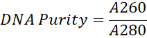
DNA purity was measured according to Fatchiyah et al. (2011) using a UV spectrophotometer (BioRad Laboratories Inc., USA). Briefly, 5 µL of DNA sample was added to 995 µL Tris-EDTA (TE) buffer and vortexed until the solution was homogeneous. The samples were then placed in a cuvette, and the absorbance was measured at 260 and 280 nm. The DNA purity level was determined using the following formula:
PCR amplification
Primers were designed using Oligo Analyzer version 1.0.2, Oligo Explore 1.1.0, and the Basic Local Alignment Search Tool (BLAST) accessible online at NCBI. The primers used were 27F and 1492R . Amplification according to Fatchiyah et al. (2011) was performed using the Polymerase Chain Reaction (PCR) technique. Genotyping amplification was performed using PCR with a thermal cycler. The PCR composition with a total volume of 20 µL/tube consisted of 6 µL ddH₂O, 10 µL PCR kit GoTaq® Green Master Mix (10× buffer, Taq polymerase, dNTP, MgCl₂, primer, Taq DNA polymerase, ddH₂O), 1 µL forward primer, 1 µL reverse primer, and 2 µL DNA sample from the isolation.
The amplification temperatures were as follow, pre-PCR at 95 °C for 5 minutes, denaturation at 94 °C for 1 minute, annealing at 50 °C for 1 minute, and extension at 72 °C for 2 minutes. PCR running was performed for 30 cycles and post-PCR at 72 °C for 10 minutes. The temperature was then lowered to 4 °C for 5 minutes.
Gene amplification results were confirmed using horizontal agarose gel electrophoresis at 1.5%. Agarose gel (1.5 %) was prepared by weighing 0.6 g of agarose powder, dissolving it in 40 mL TBE buffer, and heating until a transparent solution was formed. The agarose solution was allowed to cool until warm and then poured into an electrophoresis mold with 0.5 mL of ethidium bromide added. A comb was placed at the end of the electrophoresis tray and left until the agarose solution solidified (i.e., gel). The comb was removed from the agarose gel and transferred to an electrophoresis tray filled with TBE buffer until the gel surface was completely covered.
A total of 2 µL of DNA sample was mixed with 1 µL of loading dye and carefully loaded into the wells. Electrophoresis was performed at 65 volts for 1.5 h. The gel was visualized using a UV transilluminator and documented with a polaroid camera.
Purification of PCR amplicons
PCR amplicon samples, partially used for electrophoresis confirmation, were placed in tubes with a maximum volume of 1.5 mL. Sodium acetate (Merck) was added to the samples at 0.1% of the total volume of the amplicon solution in the tube to be purified. Next, cold absolute ethanol was added to twice the volume of the sodium acetate and amplicon solution. Spin down was performed using a centrifuge and stored at –20 °C for 60 minutes.
Subsequently, centrifugation was carried out at 13,000 rpm at 4 °C for 15 minutes. The supernatant was discarded, and 200 µL cold 70% ethanol was added, followed by centrifugation again at 13,000 rpm at 4 °C for 15 minutes. The resulting pellet was dried using an incubator at 37 °C. The dried pellet was then resuspended in 15 µL of ddH₂O.
DNA sequencing and phylogenetic analysis
Sequencing was performed using DNA samples from purified PCR products. In the confirmation of DNA amplification with agarose gel electrophoresis, the thickest band was selected as the sample for sequencing. The thickest band was chosen with the assumption that the DNA was recognized by specific primers and amplified with the appropriate sequence length. The sequencing composition consisted of 2 µL purified gene amplicon sample, 10 µL ddH₂O, 8 µL Big Dye® Terminator (using ABI PRISM® BigDye® Terminator v3.1 Cycle Sequencing Kit), 5 µL buffer solution with EDTA (Applied Biosystems), and 2 µL primer with a concentration of 20 pmol, resulting in a total volume of 25 µL.
PCR sequencing was performed at optimized temperatures according to the sequencing reagent kit instructions. The PCR sequencing products were purified again before being processed in the sequencing machine. The purified amplicons were mixed with a special buffer solution, Hi-Di™ Formamide (Genetic Analysis Grade, Applied Biosystems, USA), and sequenced using an ABI PRISM® 310 Genetic Analyzer (Applied Biosystems). Subsequently, bacterial isolates were confirmed to be gram-positive or gram-negative based on their morphology using Gram staining.
Forward and reverse sequences were assembled using the BioEdit program. Species identification was performed by comparing the tested sequences with species in GenBank using BLAST (Basic Local Alignment Search Tool) at http://www.ncbi.nlm.nih.gov. Phylogenetic analysis was conducted according to Dereeper et al. (2010) using the phylogeny.fr program.
Results and Discussion
Isolation of the candidate of gelatinase-producing bacteria
Based on the results of bacterial isolation, five bacterial isolates were obtained that grew as candidates for gelatinase-producing bacteria. Subsequently, streaking was performed in three quadrants to obtain pure cultures, and bacterial rejuvenation was performed periodically on slant agar. Bacterial rejuvenation aims to activate bacterial isolates and optimize their growth . The bacterial colonies had smooth surface morphology and appeared as milky-white spots. Five isolates were obtained with the code A1.SD, A2.SD, A3.SD, A4.SD, and A5.SD.
Screening of gelatinase-producing bacteria
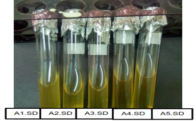
Based on the screening results, five bacterial isolates were found to be positive for gelatinase production, with varying levels of activity. As shown in Figure 1, the isolate had the largest liquefaction zone was A2.SD exhibited the highest gelatinase activity and showed good growth on nutrient gelatin medium. Gelatinase-producing bacteria are capable of hydrolyzing gelatin into simpler compounds such as amino acids .
Table 1: Screening results of gelatinase-producing bacteria isolates
|
No |
Bacterial isolate code |
The height of the medium when tilted |
Results of gelatinase production test |
|
1 |
A1.SD |
0-5.5 cm |
++ |
|
2 |
A2.SD |
0-4.4 cm |
+++ |
|
3 |
A3.SD |
0-5 cm |
++ |
|
4 |
A4.SD |
0-6.5 cm |
+ |
|
5 |
A5.SD |
0-7 cm |
+ |
Note: +: Low hydrolysis activity, ++: Moderate hydrolysis activity, +++: High hydrolysis activity
As shown in Table 1, the highest hydrolysis activity of the gelatinase enzyme was found in isolate A2.SD. Gelatinase enzyme activity was indicated by the change in gelatin consistency from solid to liquid after storage in the refrigerator. The more liquefied the gelatin medium, the higher was the gelatinase activity. Gelatinase enzyme activity can be used to illustrate the ability of bacterial isolates to hydrolyze gelatin. Gelatin testing is carried out by observing the changes that occur. If gelatin is hydrolyzed, the gelatin medium remains liquefied after cooling. The indication of gelatinase-producing bacteria growth is that the gelatin medium remains liquefied after storage in the refrigerator . The liquefaction of the gelatin medium was caused by the ability of bacteria to hydrolyze gelatin.
According to Mikhailov (2023), gelatine functions as a solidifying agent and substrate for gelatinase activity. When the gelatin medium in the tube is inoculated with gelatinase-positive bacteria, the secreted gelatinase hydrolyzes gelatin, resulting in medium liquefaction. Because gelatin is digested and is no longer able to gel, the medium will remain liquid when placed in the refrigerator or on ice. In contrast, gelatin medium inoculated with gelatinase-negative bacteria remains solid after cooling. Isolate A2.SD was subsequently identified using molecular identification of 16S rRNA to determine the bacterial species name and confirmed its morphology using Gram staining.
Isolation and purification of DNA samples
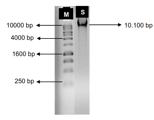
Based on Figure 2, the DNA band of bacterial isolate A2 is explained.SD was separated above the 10,000 bp DNA marker region, namely at 10,100 bp. The amplicon produced a clear and thick band, proving that the concentration of reagents during the process was optimal. This is consistent with the statement of Jaudou et al. (2022), that the electrophoresis pattern of bacterial chromosomal DNA using DNA markers of 250 – 10,000 bp will be separated in the region above 10,000 bp. Thus, bacterial chromosomal DNA has a low migration rate and is retained above the 10,000 bp region. The retention of molecular migration in the 10,000 bp region indicates that the molecule is bacterial chromosomal DNA. According to Demerdash (2012), bacterial chromosomal DNA molecules have a size >10,000 bp, namely 21,000–23,000 bp.
Based on the measurement of wavelength using NanoDrop, the 260/280 ratio was 0.66 (Table 2), indicating the purity of isolate A2.SD was below 1.8. The purity of the DNA ranged between 1.8 and 2.0. DNA with a purity value >2 at the 260/280 ratio indicates contamination by RNA, while DNA with a purity value <1.8 indicates contamination by protein. Based on the table above, the measurement of purity showed the presence of protein contamination; therefore, proteinase K was added to remove protein contamination. Purification of DNA from protein contamination can be carried out by adding proteinase K, which functions to degrade protein so that protein is released from DNA, and the sample can proceed to the PCR process.
Table 2: Result of DNA purification and concentration
|
Sample |
Abs 260 |
Abs 280 |
260/280 |
|
A2.SD |
32.00 nm |
48.16 nm |
0.66 |
PCR amplification
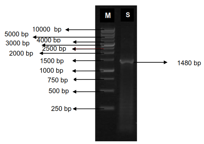
Gen amplification can be seen in Figure 3. The DNA band of the bacterial isolate A2.SD that underwent the PCR process was successfully amplified, producing a band in electrophoresis separated at 1480 bp. This indicated that the amplified gene fragment was approximately 1500 bp in size. The electrophoresis results are consistent with the theory of Jaudou et al. (2012), who stated that bacterial 16S rRNA gene fragments have a size of 1500 bp. Thus, it can be concluded that the amplification of the bacterial 16S rRNA gene fragment was successful.
The successful amplification of the 16S rRNA gene was due to PCR conditions that were able to amplify the amplicon properly according to the 16S rRNA target gene, resulting in a single band on the gel. The annealing temperature under these conditions
was highly influential. An inaccurate annealing temperature reduces primer sensitivity and can cause errors in primer attachment to the DNA template. Consequently, DNA amplification was not accurate, and a single band was not obtained.
BLAST analysis
Analysis of the 16S rRNA sequence using the BLAST program showed that isolate A2.SD had a query cover value of 100% and an E-value of 0.0, indicating that the level of homology between the sequences was identical. According to Lu et al. (2024), the E-value is an expected value that provides a statistically significant measure of similarity between two sequences. If the E-value is larger (greater than zero), it indicates low homology between the two sequences, whereas if the E-value is low or zero (0), it indicates high homology between the sequences. An E-value of 0 (zero) shows that the two sequences are identical.
The similarity or identity value of isolate A2.SD was 99% with Proteus penneri strain wf-1, with a maximum score of 2590. This explains that bacterial isolate A2.SD belongs to the same species as Proteus penneri strain wf-1. The higher the identity value, the greater the similarity to the reference sequence in GenBank. According to Wu et al. (2022), differences in DNA sequences within a species range from 0.2% to 1%. According to Yu and Wang (2024), a DNA sequence similarity value of 100% indicates that the tested isolate is the same strain, and a value of 99% indicates that it is the same species but a different strain. If the similarity value is between 89–98%, it indicates that the isolate belongs to the same genus.
Phylogenetic analysis
Based on Figure 4, the phylogenetic tree shows that P. penneri is located on the same branch as Proteus myxofaciens NCIMB 13273 and Proteus vulgaris 66P3. The bootstrap value on the first branch was 0.98%, indicating that P. penneri UB JL024 has a high level of relatedness to other species within the same genus. The bootstrap value for the next clade was 0.83%, indicating that P. penneri UB JL024 has a high level of relatedness within the same genus but a distant relationship with Streptococcus sp.
According to Nei (1987), bootstrap values ranging from 0.1–0.4 are low, 0.5–0.7 are moderate, and 0.8–1.0 are high. According to Chang et al. (2021), a bootstrap value of 95% or higher indicates that the branching topology is very accurate, consistent, and does not change even if the phylogenetic tree is reconstructed using other methods.
Gram staining and morphological characteristics
Based on Figure 5, the Gram staining results observed under the microscope at 1000× magnification indicated that the bacteria were Gram-negative because they appeared red, and their morphology was bacillus-shaped. The Gram staining results of P. penneri UB JL024 were consistent with the findings of Muhsal et al. (2025), who stated that P. penneri, belonging to the family Enterobacteriaceae, is Gram-negative, pink in color, and rod-shaped. Furthermore, their study explained that the isolate Proteus penneri UB JL024 is a gelatinase-producing bacterium because it tested positive for gelatin hydrolysis, indicating that the bacterium possesses extracellular gelatinase enzymes.
The consistency of these results confirms that P. penneri UB JL024 is Gram-negative and rod-shaped. P. penneri belongs to the family Enterobacteriaceae, Gram-negative bacilli, facultative anaerobes, occurring in groups or singly, with a size of 1–3 µm × 0.4–0.6 µm. This species is commonly found in the human digestive tract as part of the normal intestinal flora of humans. P. penneri can also be found in contaminated water, soil, and manure . P. penneri is a gelatinolytic bacterium that utilizes gelatin as a nutrient source and produces gelatinase enzymes that degrade gelatin .
According to Muhsal et al. (2025), Proteus species were positive for gelatin hydrolysis. Proteus species can produce hydrogen sulfide and lipase, hydrolyze gelatin, and show limited acid production from mannose. The optimal growth condition of this species is at 37 °C.
Therefore, among the five isolates obtained, A2.SD exhibited the strongest gelatinase activity, clearly surpassing that of the other strains. The identification of this isolate as P. penneri UB JL024 highlights two key novelties: its ecological origin from coastal sediments, which is rarely reported for this genus, and its functional capacity as a potent gelatinase producer. Together, these findings expand the known diversity of Proteus species and highlight their potential as new enzymatic resources for biotechnological applications.
Conclusions and Recommendations
The screening results of bacteria showed that five bacterial isolates with the potential to produce gelatinase enzymes were obtained from the coastal sediment of Tegalpongo Beach, Pasuruan, East Java. The species was identified by molecular identification of the 16S rRNA gene of isolate A2.SD indicated that the isolate had similarity or belonged to the same species as P. penneri with a different strain, with 99% similarity, and was designated as P. penneri UB JL024. Based on the results of this study, further research is needed for phenotypic identification using Microbact tests to strengthen the molecular identification of sediment bacteria capable of producing gelatinase enzymes. In addition, further research should be conducted on enzyme activity assays with the optimization of gelatinase enzyme production from P. penneri UB JL024 isolated from coastal sediments.
Acknowledgements
The authors would like to express their sincere gratitude to their colleagues at the Department of Fisheries Product Technology, Faculty of Fisheries and Marine Sciences, for their valuable support and collaboration throughout this research.
Novelty Statement
This study reports the first isolation of Proteus penneri UB-L024 from coastal sediments of Tegalpongo in Pasuruan, Indonesia. This strain exhibited clear gelatinase activity, further expanding the documented diversity of gelatinolytic bacteria in marine environments. Unlike previous reports that primarily focused on clinical or terrestrial sources, these findings highlight coastal sediments as untapped reservoirs of protease-producing microorganisms. The combination of molecular identification (16S rRNA, BLAST, and phylogenetic analysis) and gram staining provides robust evidence of its taxonomic placement, while its enzymatic potential underscores new opportunities for food biotechnology and environmental applications.
Authors’ Contribution
Indaka Juliana Siddiq: Was responsible for drafting the manuscript and processing the research data.
Asep Awaludin Prihanto: Developed the manuscript framework, contributed to data processing, and finalized the manuscript.
Happy Nursyam: Conceived the research concept and provided deeper analytical insights.
Sylvester Mantihal: Provided support in data interpretation and emphasized the novelty of the research.
All authors have read and approved the final version of the manuscript for publication.
Generative AI and AI-assisted technology statement
The authors declare that no generative AI and AI assisted technology was used in the creation of this manuscript.
Conflict of interest
The authors declare that they have no financial or personal relationships that could inappropriately influence the work reported in this paper.
Figures
Screening results of gelatinase-producing bacteria. A: A1.SD, B: A2.SD, C: A3.SD, D: A4.SD, E: A5.SD
Isolation result of A2.SD DNA isolate (M: DNA marker; S: DNA sample)
Agarose gel electrophoresis results. (M) DNA ladder; (S) bacterial DNA sample.
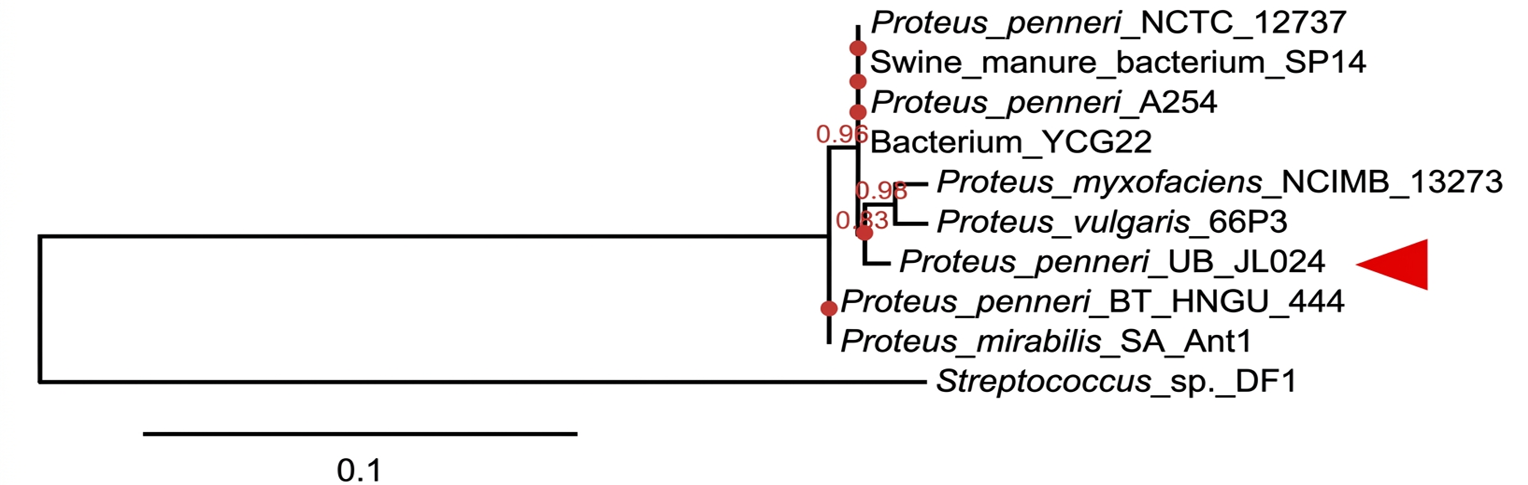
The phylogenetic tree of Proteus penneri UB JL024
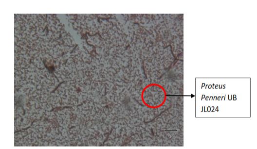
Gram staining result of Proteus penneri UB JL024
References
- Ahmed, D.A. 2015. Prevalence of Proteus spp. in Dome Hospital in Bagdad City. Iraqi J. Sci., 56: 665-672.
- Bellemare, A., T. John and S. Marqueteau. 2018. Fungal genomic DNA extraction methods for rapid genotyping and genome sequencing. Methods Mol. Biol., 1775: 11-20. https://doi.org/10.1007/978-1-4939-7804-5_2[CrossRef]
- Braun, M., A. Tripp, M. Chakatok, S. Kaltenbrunner, C. Fischer, D. Stoll, A. Bijelic, W. Elaily, M.G. Totaro, M. Moser, S.Y. Hoch, H. Lechner, F. Rossi, M. Aleotti, M. Hall and G. Oberdorfer. 2025. Computational enzyme design by catalytic motif scaffolding. Nature, 649(8095): 237-245. https://doi.org/10.1038/s41586-025-09747-9[CrossRef]
- Chang, J.M., E.W. Floden, J. Herrero, O. Gascuel, P. Di Tommaso and C. Notredame. 2021. Incorporating alignment uncertainty into Felsenstein’s phylogenetic bootstrap to improve its reliability. Bioinformat., 37(11): 1506-1514. https://doi.org/10.1093/bioinformatics/btz082[CrossRef]
- Debashish, G., S. Malay, S. Barindra and N. Joydeep. 2005. Marine enzymes. Adv. Biochem. Eng. Biotechnol., 96: 189-218.
- Dela Cruz, T.E.B. and J.M.O. Torres. 2016. Gelatin hydrolysis test protocol. Americ. Soc. Microbiol., 1-10.
- Demerdash, H.A.M. 2012. A simple and inexpensive procedure for chromosomal DNA extraction from Staphylococcus thermophilus strains. Middle-East J. Sci. Res., 11: 13-18.
- Demko, A.M., N.V. Patin and P.R. Jensen. 2021. Microbial diversity in tropical marine sediments assessed using culture-dependent and culture-independent techniques. Environ. Microbiol., 23(11): 6859-6875. https://doi.org/10.1111/1462-2920.15798[CrossRef]
- Dereeper, A., S. Audic, J.M. Claverie and G. Blanc. 2010. BLAST-EXPLORER helps you build datasets for phylogenetic analysis. BMC Evol. Biol., 10: 1-8.
- Fatchiyah, A.E.S., I.A. Estiri, W. Sri and S. Rahayu. 2011. Biologi molekular: Prinsip dasar analisis. Erlangga, Jakarta.
- Fredrikson, N.J., M. Hermansson and B.M. Ilén. 2013. The choice of PCR primers has great impact on assessments of bacterial community diversity and dynamics in a wastewater treatment plant. PLoS ONE., 8: 1-20.
- Ghattavi, S. and A. Homaei. 2023. Marine enzymes: Classification and application in various industries. Int. J. Biol. Macromol., 230: 123136. https://doi.org/10.1016/j.ijbiomac.2023.123136[CrossRef]
- Hicks, N., X. Liu, R. Gregory, J. Kenny, A. Lucaci, L. Lenzi, D.M. Paterson and K.R. Duncan. 2018. Temperature driven changes in benthic bacterial diversity influences biogeochemical cycling in coastal sediments. Front. Microbiol., 9: 1730. https://doi.org/10.3389/fmicb.2018.01730[CrossRef]
- Jaudou, S., M.L. Tran, F. Vorimore, P. Fach and S. Delannoy. 2022. Evaluation of high molecular weight DNA extraction methods for long-read sequencing of Shiga toxin-producing Escherichia coli. PLoS One., 17(7): e0270751. https://doi.org/10.1371/journal.pone.0270751[CrossRef]
- Kumar, A., S. Dhiman, B. Krishan, M. Samtiya, A. Kumari, N. Pathak, A. Kumari, R.E. Aluko and T. Dhewa. 2024. Microbial enzymes and major applications in the food industry: A concise review. Food Prod. Process. Nutr., 6(1). https://doi.org/10.1186/s43014-024-00261-5[CrossRef]
- Liu, D., M. Nikoo, G. Boran, P. Zhou and J.M. Regenstein. 2015. Collagen and gelatin. Annu. Rev. Food Sci. Technol., 6(1): 527-557. https://doi.org/10.1146/annurev-food-031414-111800[CrossRef]
- Lu, Y.Y., W.S. Noble and U. Keich. 2024. A BLAST from the past: Revisiting blastp’s E-value. Bioinformatics, 40(12): btae729. https://doi.org/10.1093/bioinformatics/btae729[CrossRef]
- Masurkar, S.A., G.R. Pathade and S.B. Gujar. 2025. Isolation, production and optimization of gelatinase from organic waste. J. Pharm. Bioallied Sci., 17(Suppl 3): S2280-S2282. https://doi.org/10.4103/jpbs.jpbs_1081_25[CrossRef]
- Mekonnen, M., S. Girma and A. Atnafu. 2022. Hydrolysis of gelatin from animal hoof using bacterial gelatinase. Int. J. Microbiol. Biotechnol., 7(3): 135-142. https://doi.org/10.11648/j.ijmb.20220703.14[CrossRef]
- Mikhailov, O.V. 2023. Gelatin as it is: History and modernity. Int. J. Mol. Sci., 24(4): 3583. https://doi.org/10.3390/ijms24043583[CrossRef]
- Muhsal, L.K., L. Theis, J. Overmann and A. Hamprecht. 2025. Proteus spp.-taxonomy, clinical significance, and antibiotic resistance of a highly versatile pathogen. Clin. Microbiol. Rev., 38(4): e0018124. https://doi.org/10.1128/cmr.00181-24[CrossRef]
- Nei, M. 1987. Molecular evolutionary genetics. Columbia Univ. Press, New York.
- O’Hara, C.M., F.W. Brenner and J.M. Miller. 2000. Classification, identification, and clinical significance of Proteus, Providencia, and Morganella. Clin. Microbiol. Rev., 13: 534-546.
- Okoth, B.A., H.M. Makonde, C.M. Bosire and C.M. Kibiti. 2025. Characterization and enzymatic potential of bacteria and fungi from Mwakirunge dumpsite, Kenya. Int. J. Microbiol., 2025(1): 7818433. https://doi.org/10.1155/ijm/7818433[CrossRef]
- Pelczar, M.J. and E.C.S. Chan. 2010. Dasar-dasar mikrobiologi. Vol. 2. UI Press, Jakarta.
- Perumal, L., S. Rengarajan and S. Natarajan. 2018. Isolation, identification and characterization of marine bacteria from the deep sea sediment of Bay of Bengal, India, and their antimicrobial and cytotoxicity potential. J. Microbiol. Biotechnol., https://doi.org/10.4014/jmb.1811.11010[CrossRef]
- Prihanto, A.A. and H. Nursyam. 2018. Screening and molecular identification of gelatinase-producing bacteria isolated from Indonesian mangrove ecosystem. Asian J. Agric. Biol., 6: 316-320.
- Prihanto, A.A., I. Yanti, M.A. Murtazam and Y.D. Jatmiko. 2020. Optimization of glutaminase-free L-asparaginase production using mangrove endophytic Lysinibacillus fusiformis B27. F1000 Res., 8: 1938.
- Razzaghi, M., A. Homaei, F. Vianello, T. Azad, T. Sharma, A.K. Nadda, R. Stevanato, M. Bilal and H.M.N. Iqbal. 2021. Industrial applications of immobilized nano-biocatalysts. Bioprocess Biosyst. Eng., 45(2): 237-256. https://doi.org/10.1007/s00449-021-02647-y[CrossRef]
- Schrieber, R. and H. Gareis. 2007. Gelatine handbook: Theory and industrial practice. Gelita AG, Germany.
- Shaik, S.P. and P. Thomas. 2019. In vitro activation of seed-transmitted cultivation-recalcitrant endophytic bacteria in tomato and host-endophyte mutualism. Microorgan., 7(5): 132. https://doi.org/10.3390/microorganisms7050132[CrossRef]
- Song, P., X. Zhang, S. Wang, W. Xu, F. Wang, R. Fu and F. Wei. 2023. Microbial proteases and their applications. Front. Microbiol., 14: 1236368. https://doi.org/10.3389/fmicb.2023.1236368[CrossRef]
- Tamura, K., J. Dudjey, M. Nei and S. Kumar. 2007. MEGA4: Molecular evolutionary genetics analysis software version 4.0. Mol. Biol. Evol., 24: 1596-1599.
- Veluchamy, C., N. Nallakaruppan, K. Nachiappan, M. Sekaran, R.B. Vartak, R.B. Zambare, S. Panneerselvam, R. Chandrasekaran, A. Sharma and K. Thiagarajan. 2025. Marine microbial bioprospecting and bioactive bioproducts: Seven decades of discovery and contemporary perspectives. Front. Mar. Sci., 12: 1698327. https://doi.org/10.3389/fmars.2025.1698327[CrossRef]
- Wang, H., Z. Sun, Y. Qi, Y. Hu, Z. Ni and C. Li. 2024. Update application of enzyme in food processing, preservation, and detection. Food Bioeng., 3(3): 380-394. https://doi.org/10.1002/fbe2.12105[CrossRef]
- Wu, J., T. Yonezawa and H. Kishino. 2022. Molecular evolutionary rate predicts intraspecific genetic polymorphism and species-specific selection. Genes., 13(4): 708. https://doi.org/10.3390/genes13040708[CrossRef]
- Yu, Z.L. and R.B. Wang. 2024. Revised taxonomic classification of the Stenotrophomonas genomes, providing new insights into the genus Stenotrophomonas. Front. Microbiol., 15: 1488674. https://doi.org/10.3389/fmicb.2024.1488674[CrossRef]
- Yulianti, N.M.A., A.A.G. Indraningrat and N.W. Widhidewi. 2024. Antibacterial activity screening of Bacillus sp. PCAR1 from seaweed Eucheuma spinosum against Staphylococcus aureus bacteria. 4(2): 165-171.
Services
Article usage statistics
Loading…