
The main aim of the current study was to investigate the Chinese patient having Primary ciliary dyskinesia (PCD). In the current genetic study, a Chinese patient was enrolled who was clinically diagnosed with PCD, and his family members were inquired for pedigree analysis. Different clinical and radiological tests were also performed. While genetic analysis, including Whole Exome (WE) and Sanger Sequencing for variant identification, was done. In silico functional analysis and Transmission electron microscopy (TEM) analysis were also performed. A 39 year old patient exhibited scoliosis and infertility. Genetic analysis of the patient identified a novel homozygous missense variant, NM_018139.3, c.899T>G, p.(Leu300Arg) in the DNAAF2 gene. Later, TEM analysis found that semen flagella showed defects in the outer dynamic arm (ODAs) and inner dynamic arms (IDAs). Sperm morphological analysis also showed abnormal, bent, coiled, and short-size flagella. His sperm motility was zero (0%). However, his hormonal profile was in reference range. Moreover, in silico analysis also confirmed the pathogenicity of the variant.The similarity index of superimpose 3D structure of wild-type and mutant DNAAF2 protein was just 14.81%. A current genetic study identified a homozygous variant of DNAAF2, which results in infertility in a Chinese male patient. This study will also assist in genetic counseling of Chinese families at risk of PCD.
Research Article · Volume 56, 8 March 2025 · Open Access
Identification of Novel Homozygous Varaint c.899T>G, p.(Leu300Arg) in DNAAF2 Gene Causing Infertility in Chinese Patient
· · ·
Affiliations & Notes ⌄
- 1Reproductive Hospital Affiliated to Shan Dong University, Jinan, 250000, China. * Corresponding author: lsming1110@163.com
Article Info ⌄
- Journal:
- Pakistan Journal of Zoology
- Published:
- 8 March 2025

From the article
- A current genetic study identified a homozygous variant NM_018139.3, c.899T>G, p.(Leu300Arg) in the DNAAF2 gene, which results in infertility in a single Chinese male patient.
- TEM analysis found that semen flagella showed defects in the outer dynamic arm (ODAs) and inner dynamic arms (IDAs).
- This study will also assist in genetic counselling of Chinese families which are at risk of PCD.
Selected from the article's own text.
Abstract
Introduction
Primary ciliary dyskinesia (PCD, MIM: 244400) is known as an orphan disease, which is a hereditary disease having major phenotypes of permanent lung damage, resulting in respiratory failure, congenital heart abnormalities, unusually positioned inner organs along with infertility ; . PCD is often inherited in an autosomal recessive form, however X-linked genes, such as RPGR and PIH1D3, have also been related to the disease. PCD is also a group of medically as well as inherently diverse disorders of cilia motility. The expected incidence of PCD is 1:5,000 to 1:20,000 around the world, but its exact prevalence is still unknown, which may be even higher .
Alterations in motile cilia result in PCD. Previous research has indicated that abnormality in ependymal motile cilia of zebrafish, may affect the movement of cerebrospinal fluid and may result in spinal dysplasia, ultimately leading to scoliosis . Scoliosis is a deformity of the spinal cord in the skeletally mature individual, having a Cobb angle of more than 12 degrees in the coronal plane . DNAAF2 protein is vital in the preassembly of early dynein in the cytoplasm , which is essential for the normal functioning of the motile cilia and results in adult scoliosis ; ; .
In the current study, WES and clinical data of a male Chinese patient diagnosed with PCD was analyzed, and a pathogenic variant was identified in the DNAAF2 gene. Moreover, the effect of the identified variant was investigated through in silico functional analysis, transmission electron microscopy (TEM) analysis, and linked pathogenic variant of the DNAAF2 gene to male infertility and likely scoliosis.
Materials and Methods
Family recruitment and clinical information
Non-consanguineous Chinese family, comprising a single male patient that was recruited to analyze the genetic cause of male sterility. A pedigree was drawn corresponding to the given data by the patient’s parents. A primary physical, as well as andrological investigation showed that patient has standard body mass index (BMI) and PCD symptoms. Different clinical and radiological tests including semen analysis, hormonal tests, karyotype analysis, Y chromosome microdeletion, and computed tomography scan (CT scan) of the chest and TEM, were performed and all parameters were recorded.
The patient’s fertile father was also enrolled to provide a positive control for semen analysis, while the parent’s DNA was examined for genetic analysis to check variant segregation in the current study.
DNA extraction and whole exome sequencing
Hepranized blood samples were used to obtain DNA by utilizing the DNeasy Blood Kit (Qiagen). Targeted panel sequencing of samples was prepared using IDT xGen Exome Research Panel V1.0. A number of sequencing libraries were evaluated by Qubit 2.0 fluorometer (Thermo Fisher Scientific). Condition, as well as the size of the libraries, were calculated by 2100 Bioanalyzer High Sensitivity DNA Assay (Agilent Technologies).
Next-generation sequencing was done using known libraries and was exposed to 2 × 150-bp paired-end sequencing on the Illumina NovaSeq platform (Illumina, San Diego, USA). FASTQ data were aligned with the human reference genome (hg19/ GRCh37) by using BWA v0.7.13 . Variant (missense, indels, and splice site) were genotyped by using recalibrated BAM files from GATK 4.0 and then annotated by using ANNOVAR (Acessed on 1st Jan 2023) alongside numerous databases, containing populace frequency, HGVS variant report, phenotype as well as variant functional prediction. The categorization of variants was done as disease-causing, likely to cause disease, variant having an unknown significance (VUS), probably benign, or benign after guidelines of the American College of Medical Genetics (ACMG). Copy number variants were called by DNAcopy R package (Acessed on 1st Jan 2023) , filtered and categorized according to ACMG guidelines , and then checked manually using the Integrative Genomics Viewer . PCD-reported genes acquired from the literature were utilized to classify pathogenic variants.
Sanger sequencing
Segregation of disease variants was checked through Sanger sequencing. To design primers, an online tool was used Primer3, Available at https://primer3.ut.ee (Acessed on 4th Jan 2023). The sequences of primers that were used for sequencing of the identified variant were: Forward primer 5′- CGGACTTCCCCTACCCTTAC -3’ and reverse 5′-GCCTTGTTGAATTGTGCCTT-3’.
Clustal Omega tool for the multiple sequence alignment. Available at https://www.ebi.ac.uk/Tools/msa/clustalo/ (Acessed on 4th Jan 2023). The DNAAF2 protein sequence was analyzed for multiple sequence alignment with closely related DNAAF2 proteins to examine the conservation of the substituted amino acid.
Sperm morphological analysis
By WHO standards (World Health Organization, 2010, Global Recommendations on Physical Activity for Health), the patient had undergone routine semen analysis twice a week. Slides were sequentially immersed in 4% paraformaldehyde (PFA) for 5 min and rinsed by using 1x phosphate-buffered saline (PBS) twice for an additional 5 min. Then stained in hematoxylin (Solarbio, Beijing, China) for 30 min, dipped in purified water three times, immersed in 50% acidic ethanol, and kept in water for 2 min. Each slide was dried in 50% and 80% ethanol for 5 min, stained for 5 min with Eosin Azure (Solarbio, Beijing, China), sequentially dehydrated twice in 100% ethanol for 5 min each and in xylene for 5 min, and covered with coverslips with natural balsam to be examined. At last, 300 stained spermatozoa per sample were analyzed by optical microscopy (Nikon, Tokyo, Japan) at 20x.
Transmission electron microscopy (TEM)
TEM of the patient’s (II:1) sperm was done according to the protocol set by Sun et al. (2020). The pictures were taken using a charge-coupled JEOL-1200EX (JEOL, Japan) in Shandong Weiya Laboratory.
In silico analysis
Computational analysis of wild-type and mutant DNAAF2 protein consists of protein 3D modeling as well as protein docking.
Protein structure prediction
I-TASSER tool (Acessed on 4th Jan 2023) was used for 3D protein modeling , and among predicted models, the model with the maximum confidence score (C-score) was chosen for further screening. For confirmation of the effectiveness of this predicted structure, the results obtained through I-TASSER were confirmed using the Ramachandran plot (Acessed on 4th Jan 2023).
Molecular docking and visualization
For the study of protein interaction, the ClusPro 2.0 tool (Acessed on 4th Jan 2023) was employed to check interactions among DNAAF2 (wild type as well as mutated protein) and its close functional interactor DNAAF4 protein, predicted by using ‘STRING’ (Acessed on 4th Jan 2023) . Protein-protein interaction was done by using Cluspro 2.0, while the visualization of docked models was performed using various software, e.g., LigPlot + (Version 2.1) as well as Chimera 1.13 .
Results
Infertile patient
In the current study, a single male patient belonging to a non-consanguineous Chinese family that has PCD was investigated (Fig. 1a).
The semen assessment of the patient demonstrated sufficient semen volume as well as concentration. All the sperms were immotile, i.e., 0% motility in the patient’s sperm was noted. Sperm morphological examination showed irregularities in the tail of the sperm, with various defects of sperm flagella, either absent, small, twisted, curled, or abnormal (Fig. 2).
Hormonal (i.e., luteinizing hormone (LH), follicle-stimulating hormone (FSH), estradiol (E2), testosterone, and prolactin tests), karyotype evaluation, and microdeletion of Y chromosome tests were normal. However a CT scan of the patient’s spine shows the abnormal visceral position at a certain angle so confirmed as scoliosis, while a chest CT scan also showed bronchiectasis and the situs inversus (Table I and Fig. 3).
TEM scanning of the patient’s (II:1) sperm flagella showed complete axonemal defects in the outer dynamic arm (ODAs) as well as inner dynamic arms (IDAs) (Fig. 4).
Genetics analysis
After a series of variant filtration steps, a novel homozygous variant in DNAAF2 (NM_018139.3) gene was found. The identified variant was homozygous missense c.899T>G; p.(Leu300Arg). Sanger sequencing confirmed that the patient was homozygous, and his parents were heterozygous carriers of the identified variant (Fig. 1b).
Table I. Clinical features of the patient.
| Characteristics | Reference valuesa | Results |
| Gender | - | Male |
| Age (years)b | - | 39 |
| Age at the time of marriageC | - | 29 |
| Weight | - | 73.5 |
| BMI (kg m−2) | - | 24.8 |
| Semen parameters |
| |
| Semen volume (ml) | >1.5 | 2.3 |
| Semen pH | Alkaline | Alkaline |
| Sperm concentration (106/ml) | >15 | 33.1 |
| Motility (%) | >40 | 0 |
| Progressively motility (%) | >32 | 0 |
| Immortality (IM) (%) | 100 | |
| Sperm morphology |
| |
| Normal flagella (%) | >4 | 2.59 |
| Abnormal flagella (%) | - | 97.41 |
| Short flagella (%) | - | 43.0 |
| Absent flagella (%) | - | 0 |
| Bent flagella (%) | - | 22.3 |
| Coiled flagella (%) | - | 28.1 |
| Irregular/calibre (%) | - | 6.6 |
| Head defects | ||
| Normal head (%) | - | 21 |
| Abnormal head (%) | - | 79 |
| Tapered head (%) | - | 66.7 |
| Pyriform head (%) | - | 12.1 |
| Double head (%) | - | 0.2 |
| Large/Amorphous head (%) | - | 17.3 |
| Round head (%) | - | 0.9 |
| Small head (%) | - | 2.8 |
| Absent head (%) | - | 0 |
| Karyotype analysis | 46, XY | |
| Hormone analysis | Normal in reference range | |
| Y chromosome microdeletion | No deletion was found | |
| Other examination | Chest high-resolution computed tomography (HRCT) scan of the Patient showed bronchiectasis, rhinosinusitis, and situs inversus | |
Prediction of mutant protein and 3D models
3D models of DNAAF2 proteins (wild-type and mutant) were predicted and then superimposed to check the similarity of both structures. The similarity index of both structures was noted to be just 14.81% with many visible changes in the visible 3D structure (Fig. 5).
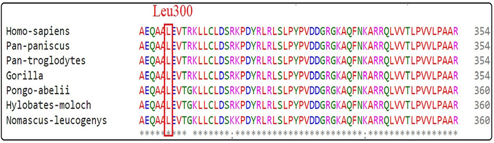
While multiple sequence alignment confirms that substituted amino acid i.e. Leu300 was highly conserved throughout the species as in Figure 6.
Protein–protein docking
The study of wild-type and mutant DNAAF2 with close interactor DNAAF4 protein has revealed remarkable alteration in docking sites due to identified variant (Table II). Wild-type DNAAF2 protein was interacting with DNAAF4 protein by 17 hydrogen and 6 unfavorable bonds though1 12 residues including, Glu378, Asp371, Ala379, Glu368, Glu356, Arg354, Lys305, Ser321, Arg319, Arg317, Gln247, Glu343, Pro241, Ser240, Asp45 and Glu42. While in the case of mutant DNAAF2 protein, it interacts with close interactor DNAAF4 protein by 17 hydrogens and 3 unfavorable bonds through 12 residues, including Lys531, Asp530, Glu636, His607, Ser641, Ser604, Pro576, Ala421, Glu419, Pro418, Glu655, His400, Asp401, Thr402, Gln665, Gln378.
Discussion
In the present genetic study, WES combined with in silico functional analysis identifies homozygous variant DNAAF2; NM_018139.3, c.899T>G, p.(Leu300Arg) in a single Chinese patient. Sanger sequencing confirmed that the variant was segregating in the family in the autosomal recessive pattern. Pathogenicity of the variant was validated by using additional clinical testing, TEM analysis, and in silico functional analysis. The patient was infertile and diagnosed with PCD; moreover, an upright radiograph revealed scoliosis. Semen morphological analysis showed that patient sperm flagella were reduced in size, bent, and irregular in shape, and even absent flagella were also noted in the sperm of the patient, which confirms the finding of a previous study that Sperm flagella length is considerably shortened in patients having DNAAF2 variant .
DNAAF2 gene is present on chromosome no 14, and it encodes 837 amino acids long protein. DNAAF2 protein is highly conserved throughout the species . DNAAF2 is dispersed in cell types having cilia, and it interrelates with some other proteins that are limited to the cilia functioning . A previous study also confirmed the finding that DNAAF2 variants caused combined ODA and IDA flaws that result in infertility in humans. More studies also recognized that pathogenic variants in the DNAAF4 and DNAAF2 genes in individuals have flaws in the ODA and IDA assembly, situs inversus, and PCD ; ; .
It has been documented that DNAAF2 is mainly involved in the preassembly of dynein in the cytoplasm, which is important for the normal functioning of the motile cilia. However pathogenic variants in the DNAAF2 result in damage of both IDAs and ODAs ; .
Previously different DNAAF2 variants i.e., c.564dupG, c.1160A>G, c.31delG, c.C156A, c.822del, c.998C>T, c.1891G>A, c.2027_2028delCT, c.1199_1214dup16, c.177_178insA, c.1555delG, c.1901T>C, c.491T>C, c.472G>T, c.23C>A and c.26C>A, have been documented
Table II. Protein-protein interaction of wild-type and mutant DNAAF2 protein with close interactor DNAAF4 protein.
| Interacting Protein | Interacting residues in DNAAF2 protein | Interacting residues in close interactor DNAAF4 protein | Type of interacting bonds |
| Wild-type DNAAF2 protein + Close interactor DNAAF4 protein | Glu378,Asp371,Ala379,Glu368, Glu356,Arg354,Lys305,Ser321, Arg319,Arg317,Gln247,Glu343, Pro241,Ser240,Asp45, Glu42 | Phe220,Glu217,Asn215, Lys198,Lys194,Ala161, Glu170,Glu145,Asn150, Asp146,Lys93,Arg100, Asn218,Glu116,Ala121, Arg123,Ser131,Lys127, Glu139 | 17 hydrogen bonds and 6 salt bridges |
| Mutant DNAAF2 protein + Close interactor DNAAF4 protein | Lys531,Asp530,Glu636,His607, Ser641,Ser604,Pro576,Ala421, Glu419,Pro418,Glu655,His400, Asp401,Thr402,Gln665,Gln378 | Asp405,Thr374,Lys408, Lys299,Arg371,Lys367, Asp295,Lys336,Lys338, Asn274,Arg271,Ala267, Ile240,Arg245,Ser237, Glu222 | 18 hydrogen bonds and 3 salt bridges |
to cause PCD in different ethnicities of the world including Dutch and Chinese ; ; ; ; ; ; .
As for Chinese children, DNAFF2 gene variants are sporadic. As Guan et al. (2021) reported in 81 Chinese children, the genes with the highest incidence of variants that caused PCD were DNAH11, followed by HYDIN, DNAH5, CCDC39, DNAH1, and CCNO; no DNAAF2 gene variant was detected . In Chinese adult patients, Sun et al. (2020) have identified two compound heterozygous DNAAF2 gene variants i.e. c.156C>A; p.(Tyr52Term) and c.26C>A; p.(Ser9Term) in them, which lead to the defect of ODAs and IDAs resulting in PCD with the manifestation of male infertility similar finding was also found in our patient. Recently, Lu et al. (2021) found two heterozygous variants i.e., c.491T>C; p.(Leu164Pro) and c.822del; p.(Ala275Profs*10) in two females having major phenotypes of PCD, sinusitis bronchiectasis, and infertility, besides, the one with c.491T>C variant also had scoliosis .
PCD is a genetic ciliopathic defect which is caused due abnormality and failure of the motile cilia functioning . Cilia in different organs may include, i.e. sperm, embryonic node, and ependymal cells .
Different PCD-causing genes of the DNAAF family, including DNAAF2 are documented to affect the preassembly of both ODA and IDA . As directed by the TEM scanning in the current study, the ultrastructure of the ODAs and IDAs of the ppatient’s (II:1) sperm flagella appeared disturbed as compared to the normal ultra-structural prearrangement of control’ (I:1) sperm flagella .
These results are also comparable with results documented by Omran et al. (2008) who exhibited that male mutant fish showed abnormality in sperm motility resulting in infertility . The variation in the DNAAF genes that encode the dynein subunits or the components essential for the assembly, transport, or docking of axonemal dyneins, results in different developmental disorders .
Conclusion
A current genetic study identified a homozygous variant NM_018139.3, c.899T>G, p.(Leu300Arg) in the DNAAF2 gene, which results in infertility in a single Chinese male patient. TEM analysis found that semen flagella showed defects in the outer dynamic arm (ODAs) and inner dynamic arms (IDAs). Sperm morphological analysis also showed abnormal, bent, coiled, and short-size flagella. This study will also assist in genetic counselling of Chinese families which are at risk of PCD.
Acknowledgment
We are grateful to the affected family for their volunteer participation in the present study.
Disclosure of interest
The authors declare that they have no conflict of interest.
Funding
The current study was supported by the Medical and Health Science Technology Development program of Shandong Province,China(202204050706).
IRB approval
This study was approved by a committee of the Ethical Review Board of the Reproductive Hospital Affiliated to Shan Dong University.
Ethical approval
Notified consent was obtained from the concerned individuals. This study was approved by a committee of the Ethical Review Board from the Reproductive Hospital Affiliated to Shan Dong University.
Data availability statement
The researchers possess the data.
Consent for publication
The consent for the publication of images and clinical details has been obtained
Statement of conflict of interest
The authors have declared no conflict of interest.
Figures
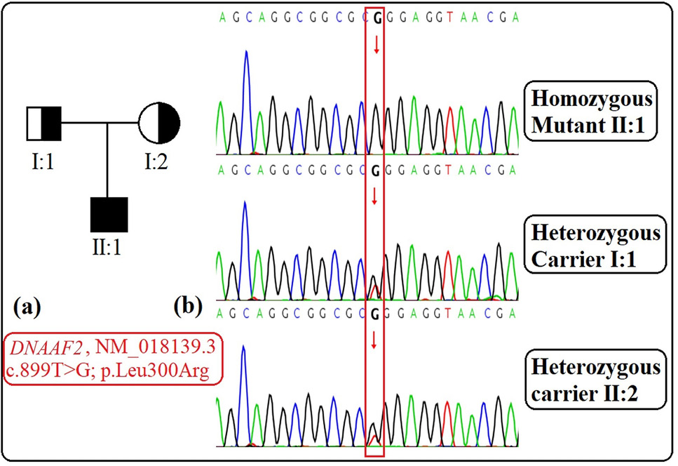
a) Family pedigree showing autosomal recessive mode of inheritance (b) Sangers sequencing chromatograms showing segregation of variant c.899C>G in the family in DNAAF2 gene.
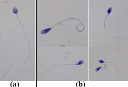
Sperm morphological analysis (a) Normal control sperm morphology (b) Patient sperm showing bent, absent, short and irregular sperm flagella.
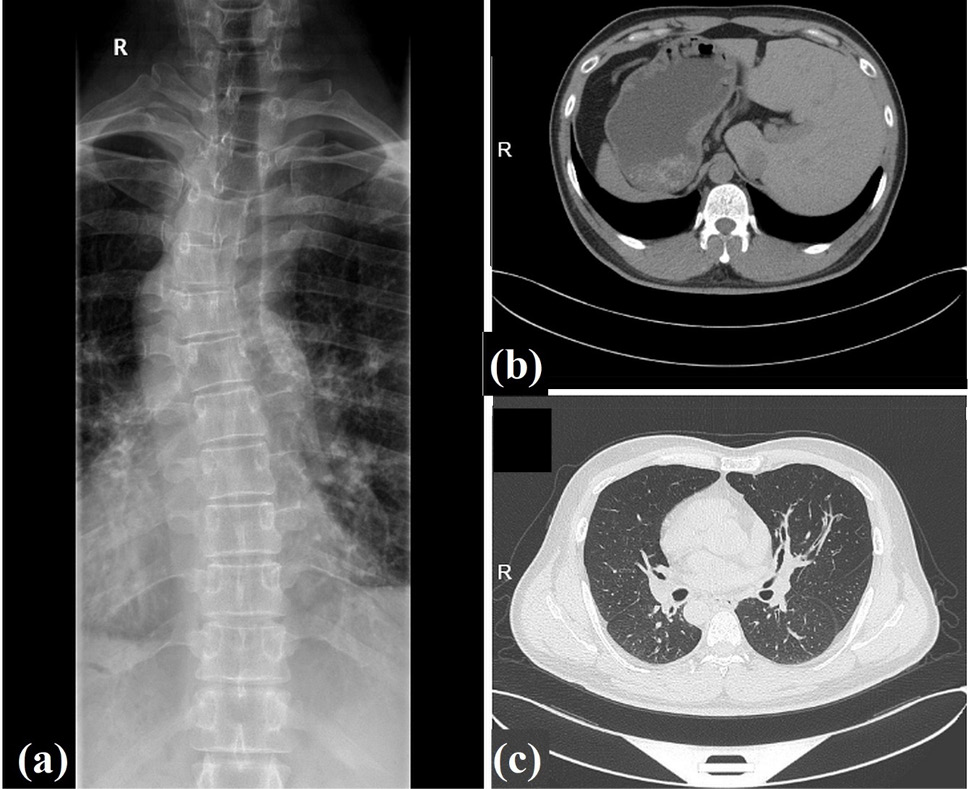
Clinical features of patient. (a) Radiographic image of patient with bronchiectasis showing right left-sided lumbar scoliosis (b and c) The chest high-resolution computed tomography (HRCT) scan of patient showing bronchiectasis and the situs inversus.
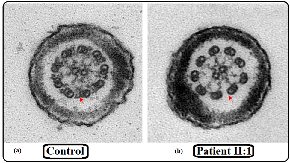
Transmission election microscopic examination revealed that ODAs were not present in the sperm flagella of the patients.
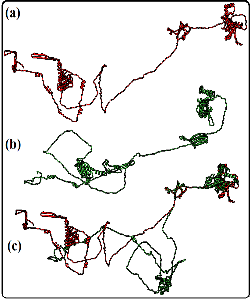
(a) 3D model of wild-type DNAAF2 protein (b) 3D model of wild-type DNAAF2 protein (c) 3D model of superimpose structure of wild-type and mutant DNAAF2 proteins.
References
- Aebi, M., 2005. The adult scoliosis. Eur. Spi J., 14. https://doi.org/10.1007/s00586-005-1053-9[CrossRef]
- Alhathal, N., Maddirevula, S., Coskun, S., Alali, H., Assoum, M., Morris, T., Deek, H.A., Hamed, S.A., Alsuhaibani, S., Mirdawi, A., Ewida, N., Al-Qahtani, M., Ibrahim, N., Abdulwahab, F., Altaweel, W., Dasouki, M.J., Assiri, A., Qabbaj, W. and Alkuraya, F.S., 2020. A genomics approach to male infertility. Genet. Med., 22: 1967-1975.https://doi.org/10.1038/s41436-020-0916-0[CrossRef]
- Aprea, I., Raidt, J., Höben, I.M., Loges, N.T., Nöthe-Menchen, T., Pennekamp, P., Olbrich, H., Kaiser, T., Biebach, L., Tüttelmann, F., Horvath, J., Schubert, M., Krallmann, C., Kliesch, S. and Omran, H., 2021. Defects in the cytoplasmic assembly of axonemal dynein arms cause morphological abnormalities and dysmotility in sperm cells leading to male infertility. PLoS Genet., 17: https://doi.org/10.1371/journal.pgen.1009306[CrossRef]
- Beiras, R., Bellas, J., Cachot, J., Cormier, B., Cousin, X., Engwall, M., Gambardella, C., Garaventa, F., Keiter, S., Le Bihanic, F., López-Ibáñez, S., Piazza, V., Rial, D., Tato, T. and Vidal-Liñán, L., 2018. Ingestion and contact with polyethylene microplastics does not cause acute toxicity on marine zooplankton. J. Hazard. Mater., 360. https://doi.org/10.1016/j.jhazmat.2018.07.101[CrossRef]
- Blanchon, S., Legendre, M., Bottier, M., Tamalet, A., Montantin, G., Collot, N., Faucon, C., Dastot, F., Copin, B., Clement, A., Filoche, M., Coste, A., Amselem, S., Escudier, E., Papon, J.F. and Louis, B., 2020. Deep phenotyping, including quantitative ciliary beating parameters, and extensive genotyping in primary ciliary dyskinesia. J. med. Genet., 57: https://doi.org/10.1136/jmedgenet-2019-106424[CrossRef]
- Brandt, T., Sack, L.M., Arjona, D., Tan, D., Mei, H., Cui, H., Gao, H., Bean, L.J.H., Ankala, A., Del Gaudio, D., Knight, J.A., Vincent, L.M., Reavey, C., Lai, A., Richard, G. and Meck, J.M., 2020. Adapting ACMG/AMP sequence variant classification guidelines for single-gene copy number variants. Genet. Med., 22: 336-344. https://doi.org/10.1038/s41436-019-0655-2[CrossRef]
- Emiralioğlu, N., Taşkıran, E.Z., Koşukcu, C., Bilgiç, E., Atilla, P., Kaya, B., Günaydın, Ö., Yüzbaşıoğlu, A., Tuğcu, G.D., Ademhan, D., Eryılmaz, P.S., Gharibzadeh, H.M., Yalçın, E., Doğru, D., Kiper, N., Alikaşifoğlu, M. and Özçelik, U., 2020. Genotype and phenotype evaluation of patients with primary ciliary dyskinesia: First results from Turkey. Pedi Pulmo, 55: 383-393. https://doi.org/10.1002/ppul.24583[CrossRef]
- Grimes, D.T., Boswell, C.W., Morante, N.F.C., Henkelman, R.M., Burdine, R.D. and Ciruna, B., 2016. Zebrafish models of idiopathic scoliosis link cerebrospinal fluid flow defects to spine curvature. Science, 352: 1341-1344. https://doi.org/10.1126/science.aaf6419[CrossRef]
- Guan, Y., Yang, H., Yao, X., Xu, H., Liu, H., Tang, X., Hao, C., Zhang, X., Zhao, S., Ge, W. and Ni, X., 2021. Clinical and genetic spectrum of children with primary ciliary dyskinesia in China. Chest, 159: https://doi.org/10.1016/j.chest.2020.06.045[CrossRef]
- Kozakov, D., Hall, D.R., Xia, B., Porter, K.A., Padhorny, D., Yueh, C., Beglov, D. and Vajda, S., 2017. The ClusPro web server for protein-protein docking. Nat. Prot., 12: 255–278. https://doi.org/10.1038/nprot.2016.169[CrossRef]
- Lee, L., 2011. Mechanisms of mammalian ciliary motility: Insights from primary ciliary dyskinesia genetics. Gene, 473: 57-66. https://doi.org/10.1016/j.gene.2010.11.006[CrossRef]
- Li, H., and Durbin, R., 2009. Fast and accurate short read alignment with Burrows-Wheeler transform. Bioinformation, 25: 1754–1760. https://doi.org/10.1093/bioinformatics/btp324[CrossRef]
- Lu, C., Yang, D., Lei, C., Wang, R., Guo, T. and Luo, H., 2021. Identification of two novel dnaaf2 variants in two consanguineous families with primary ciliary dyskinesia. Pharm. Pers Med., 14: 1415-1423. https://doi.org/10.2147/PGPM.S338981[CrossRef]
- Lucas, J.S., Davis, S.D., Omran, H. and Shoemark, A., 2020. Primary ciliary dyskinesia in the genomics age. Lancet Resp. Med., 8: 202--216. https://doi.org/10.1016/S2213-2600(19)30374-1[CrossRef]
- Mirra, V., Werner, C. and Santamaria, F., 2017. Primary ciliary dyskinesia: An update on clinical aspects, genetics, diagnosis, and future treatment strategies. Front. Pediatr., 5. https://doi.org/10.3389/fped.2017.00135[CrossRef]
- Mitchison, H.M., Schmidts, M., Loges, N.T., Freshour, J., Dritsoula, A., Hirst, R.A., O’Callaghan, C., Blau, H., Al-Dabbagh, M., Olbrich, H., Beales, P.L., Yagi, T., Mussaffi, H., Chung, E.M.K., Omran, H. and Mitchell, D.R., 2012. Mutations in axonemal dynein assembly factor DNAAF3 cause primary ciliary dyskinesia. Nat. Genet., 44: 381–389. https://doi.org/10.1038/ng.1106[CrossRef]
- Omran, H., Kobayashi, D., Olbrich, H., Tsukahara, T., Loges, N.T., Hagiwara, H., Zhang, Q., Leblond, G., O’Toole, E., Hara, C., Mizuno, H., Kawano, H., Fliegauf, M., Yagi, T., Koshida, S., Miyawaki, A., Zentgraf, H., Seithe, H., Reinhardt, R. and Takeda, H. 2008. Ktu/PF13 is required for cytoplasmic preassembly of axonemal dyneins. Nature, 456: 611–616. https://doi.org/10.1038/nature07471[CrossRef]
- Paff, T., Kooi, I.E., Moutaouakil, Y., Riesebos, E., Sistermans, E.A., Daniels, H.J.M.A., Weiss, J.M.M., Niessen, H.H.W.M., Haarman, E.G., Pals, G. and Micha, D., 2018. Diagnostic yield of a targeted gene panel in primary ciliary dyskinesia patients. Hum. Muta., 39: 653-665. https://doi.org/10.1002/humu.23403[CrossRef]
- Pettersen, E.F., Goddard, T.D., Huang, C.C., Couch, G.S., Greenblatt, D.M., Meng, E.C. and Ferrin, T.E., 2004. UCSF Chimera - A visualization system for exploratory research and analysis. J. Comput. Chem., 25: 1605-1612. https://doi.org/10.1002/jcc.20084[CrossRef]
- Richards, S., Aziz, N., Bale, S., Bick, D., Das, S., Gastier-Foster, J., Grody, W.W., Hegde, M., Lyon, E., Spector, E., Voelkerding, K. and Rehm, H.L., 2015. Standards and guidelines for the interpretation of sequence variants: A joint consensus recommendation of the American College of Medical Genetics and Genomics and the Association for Molecular Pathology. Genet. Med., 17: 405-424. https://doi.org/10.1038/gim.2015.30[CrossRef]
- Sun, M., Zhang, Y., Jiyun, Y., Wang, Y., Tan, H., Wang, H., Lei, T., Li, X., Zhang, X., Xiong, W., Dou, K. and Ma, Y., 2020. Novel compound heterozygous DNAAF2 mutations cause primary ciliary dyskinesia in a Han Chinese family. J. Assist. Reprod. Genet., 37: 2159–2170. https://doi.org/10.1007/s10815-020-01859-7[CrossRef]
- Szklarczyk, D., Gable, A.L., Lyon, D., Junge, A., Wyder, S., Huerta-Cepas, J., Simonovic, M., Doncheva, N. T., Morris, J.H., Bork, P., Jensen, L.J. and Von Mering, C., 2019. STRING v11: Protein-protein association networks with increased coverage, supporting functional discovery in genome-wide experimental datasets. Nucleic Acid Res., 47: D607–D613. https://doi.org/10.1093/nar/gky1131[CrossRef]
- Thorvaldsdóttir, H., Robinson, J.T. and Mesirov, J., P.2013. Integrative genomics viewer (IGV): High-performance genomics data visualization and exploration. Br. Bioinf., 14: 178–192. https://doi.org/10.1093/bib/bbs017[CrossRef]
- Venkatraman, E.S. and Olshen, A.B., 2007. A faster circular binary segmentation algorithm for the analysis of array CGH data. Bioinformatics, 23: 657–663. https://doi.org/10.1093/bioinformatics/btl646[CrossRef]
- Wallace, A.C., Laskowski, R.A. and Thornton, J.M., 1995. Ligplot: A program to generate schematic diagrams of protein-ligand interactions. Prog. Eng. Des. Select, 8: 127–134. https://doi.org/10.1093/protein/8.2.127[CrossRef]
- Wang, K., Li, M. and Hakonarson, H., 2010. Annovar: Functional annotation of genetic variants from high-throughput sequencing data. Nucleic Acid Res., 38: https://doi.org/10.1093/nar/gkq603[CrossRef]
- World Health Organization. 2010. Global recommendations on physical activity for health.
- Yang, J., Yan, R., Roy, A., Xu, D., Poisson, J. and Zhang, Y., 2014. The I-TASSER suite: Protein structure and function prediction. Nat. Methods, 12. https://doi.org/10.1038/nmeth.3213[CrossRef]
- Zhu, N., Welch, C.L., Wang, J., Allen, P.M., Gonzaga-Jauregui, C., Ma, L., King, A.K., Krishnan, U., Rosenzweig, E.B., Ivy, D.D., Austin, E.D., Hamid, R., Pauciulo, M.W., Lutz, K.A., Nichols, W.C., Reid, J.G., Overton, J.D., Baras, A., Dewey, F.E. and Chung, W.K., 2018. Rare variants in SOX17 are associated with pulmonary arterial hypertension with congenital heart disease. Genome Med., 10. https://doi.org/10.1186/s13073-018-0566-x[CrossRef]
Services
Article usage statistics
Loading…