
A consanguineous Pakistani family with three affected siblings was investigated to determine the genetic diagnosis of an inherited contracture disorder. Whole-exome sequencing was performed for four participants. Variants were filtered based on homozygosity in the three patients and heterozygosity in the obligate carrier (mother), predicted effect of variants on the encoded protein, and their frequencies in public databases. Sanger sequencing was performed to explore the segregation of the variant with the phenotype. All patients had congenital limb contractures. These included camptodactyly of hands and feet, ptosis, adducted thumb and clubfoot morphology. A novel homozygous missense variant in ECEL1 c.2051A>G, p.(Tyr684Cys) was identified in all three patients. The variant was absent from the DNA of 500 ethnically matched control samples as well as from all public databases. In conclusion, this study reports a family with clinical features of distal arthrogryposis type 5D and extends the genotype spectrum of the disorder.
Research Article · Volume 55, Issue 1, 9 November 2022 · Open Access
A Novel ECEL1 Variant Associated with a Congenital Contracture Disorder
· · · · · · · ·
Affiliations & Notes ⌄
- 1School of Biological Sciences, University of the Punjab, Quaid-i-Azam Campus, Lahore 54590, Pakistan 2Institute of Neurogenetics, University of Luebeck 23538, Lübeck, Germany 3Department of Neurology, University of Luebeck 23538, Lübeck, Germany 4University of Okara, Okara, Pakistan 5Department of Human Genetics and Molecular Biology, University of Health Sciences, Lahore, Pakistan. * Corresponding author: katja.lohmann@neuro.uni-luebeck.de
- 2naz.sbs@pu.edu.pk
Article Info ⌄
- Journal:
- Pakistan Journal of Zoology
- Published:
- 9 November 2022

From the article
- All online tools predicted the ECEL1 p.(Tyr684Cys) variant to be damaging or deleterious to the protein (Table II).
- The Tyr684 residue is located within a peptidase M13 domain that is important for the activity of the protein as it contains the active site of the enzyme.
- The variant may disturb the domain structure and interaction between the domains, which could affect the function of the protein.
- ECEL1 encodes endothelin converting enzyme like 1 (ECEL1), a member of neprilysin family of endopeptidases (peptidase family M13).
Selected from the article's own text.
Abstract
Introduction
Arthrogryposis multiplex congenita (AMC) is a heterogeneous disorder involving contractures of the distal parts of the limbs which affect joint mobility. More than ten subtypes of DA have been identified on the basis of phenotypic features. Most DA subtypes are inherited as autosomal dominant disorders, and are caused by pathogenic variants in genes encoding contractile proteins of myofibers . Distal arthrogryposis type 5D (DA5D) (OMIM# 615065) is a subtype of DA inherited as an autosomal recessive disorder. It is characterized by severe camptodactyly of the hands, adducted thumbs and wrists, mild camptodactyly of the toes, clubfoot and/or a calcaneovalgus deformity, extension contractures of the knee, unilateral ptosis or ptosis that is more severe on one side. A round-shaped face, arched eyebrows, a bulbous, upturned nose, and micrognathia are also characteristic . Mutations in ECEL1 encoding endothelin converting enzyme like 1 have been identified in DA5D families (Dieterich et al., 2013; . Here, we report a family presenting characteristic features of DA5D with a novel homozygous missense variant in ECEL1.
Materials and methods
Ethics statement
This study was approved by the Institutional Review Board of School of Biological Sciences, University of the Punjab, Lahore, Pakistan. All participants provided written informed consent.
Subjects
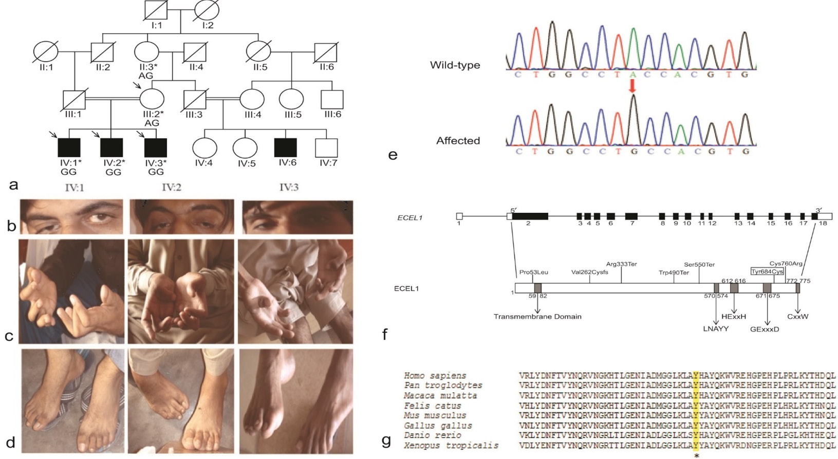
Family RDHR-01 had four affected male individuals in two consanguineous marriages (Fig. 1a). Three individuals (IV:1, IV:2, IV:3) were videotaped according to a standardized video protocol. The videos were evaluated by movement disorder specialists (N.B., T.B., A.M.).
Molecular analyses
Blood samples were collected from all available family members and genomic DNA was extracted by a standard protocol. Exome sequencing was performed for four individuals of the family at Centogene AG (Rostock, Germany). NimblegenSeqCap EZ Human Exome Library v2.0 was used for exome capturing, enrichment and sequencing was performed on an Illumina HiSeq 2000 machine with a medium coverage of 100X. Data were analyzed and filtered to remove those variants with minor allele frequencies equal to or greater than 0.01 in the public databases such as gnomAD and 1000 genomes and with a high number of homozygotes or hemizygotes (>20). All exonic and splice site variants were prioritized if they were present in the data of the three affected siblings in the homozygous state and heterozygous in that of the unaffected parent.
Sanger sequencing was performed on samples for the available family members to confirm the segregation of the candidate variants. DNA from 200 ethnically matched controls was sequenced to determine the frequency of the variant in the Pakistani population together with the exome data from 300 unrelated in-house ethnically matched individuals.
In silico predictions
We performed in silico analysis by using online prediction tools such as PolyPhen2 (http://genetics.bwh.harvard.edu/pph2/), MutationTaster (http://www.mutationtaster.org/), PROVEAN (http://provean.jcvi.org/protein_batch_submit.php?species=human), SIFT (https://sift.bii.a-star.edu.sg/www/SIFT_seq_submit2.html) and FATHMM (http://fathmm.biocompute.org.uk/inherited.html) among others in order to predict the pathogenicity of the identified variants. Moreover, HOPE (https://www3.cmbi.umcn.nl/hope/) was used to predict the effect of mutant amino acid residue on the protein.
Results
Clinical data
The proband, IV:1, now aged 30 years in family RDHR-01 was reported to have upper and lower limbs contractures at birth. The two affected siblings (IV:2, aged 28 years and IV:3, aged 26 years) were reported to have similar clinical features at birth as exibited by the proband (Table I). All affected individuals had ptosis. Individual IV:1 exhibited unilateral ptosis which was more severe on the right side (Fig. 1b). Mild bilateral ptosis were observed in individuals IV:2 and IV:3. Bulbous nose and arched eyebrows were observed in individual IV:2. The posture deformity of the hands associated with curved fingers, and adducted thumbs of both hands were present in all affected individuals. The thumbs of the hands were curved inside medially (Fig. 1c). The lower limbs abnormalities were associated with problems in knee joints; as the joints were not flexible, the right leg could not be bent and remained straight. The feet exhibited pes cavus. The posture deformity of feet in individual IV:2 resembles clubfoot deformity in which the foot rotated towards the other foot (Fig. 1d).
Table I. Clinical features of family RDHR-01.
| Patients ID | IV:1 | IV:2 | IV:3 | Symptoms in all 59 patients* |
| Sex | M | M | M | Not reported |
| Onset | At birth | At birth | At birth | At birth |
| Age at examination (yrs) | 30 | 28 | 26 | Varied |
| Ptosis | + | + | + | 44 (74%) |
| Arched eye brows | ˗ | + | + | 10 (17%) |
| Bulbous nose | + | + | + | 22 (32%) |
| Hands and/or fingers contracture | + | + | + | 59 (100%) |
| Adducted thumbs | + | + | + | 30 (51%) |
| Foot and or toe contracture | ˗ | + | + | 50(85%) |
| Club foot | ˗ | + | + | 16 (27%) |
| Pes cavus | ˗ | + | + | 7 (12%) |
M, male; yrs, years; +, present; -, absent; *; ; ; ; ; ; ; ; ; ; ; .
Molecular data
Only four different missense variants fulfilled our filtering criteria applied to the exome data analysis (Table II). All these variants segregated with the phenotype and were absent from the DNA of the ethnically matched controls (1000 chromosomes). The novel missense variant in ECEL1, c.2051A>G, p.(Tyr684Cys) (NM_004826.2) best explained the disorder in the family based on phenotypic overlap with patients described with other pathogenic variants in this gene, complete conservation of the amino acid among different vertebrates orthologues (Fig. 1g), the highest pathogenicity scores (Table II). In addition, the variant was not present in any public database.
Results of in silico analyses predictions for ECEL1 p.(Tyr684Cys)
All online tools predicted the ECEL1 p.(Tyr684Cys) variant to be damaging or deleterious to the protein (Table II). The wild type amino acid Tyr is aromatic, partially hydrophobic and non-polar. In contrast, the mutant amino Cys is hydrophobic, polar, and sulphur containing. The Tyr684 residue is located within a peptidase M13 domain that is important for the activity of the protein as it contains the active site of the enzyme. The variant may disturb the domain structure and interaction between the domains, which could affect the function of the protein.
Discussion
ECEL1 encodes endothelin converting enzyme like 1 (ECEL1), a member of neprilysin family of endopeptidases (peptidase family M13). Members of this family are zinc containing type II integral membrane proteins with a short cytosolic N-terminal tail and a long C-terminal extra cytosolic domain containing the catalytic site . The ECEL1 protein (O95672, UniProt https://www.uniprot.org) consist of 775 amino acids with an N-terminal cytoplasmic domain (1-59 residues), a single putative membrane-spanning region (60-82 residues) and a large luminal C-terminal domain (83-775 residues) that contains a zinc-binding motif and the active site (Fig. 1f).
ECEL1 and distal arthrogryposis type 5D
Pathogenic variants in ECEL1 were first identified as a cause for distal arthrogryposis type 5D in eleven families by two independent groups ; . To date, 48 variants of ECEL1 (Human Gene Mutation Database accessed March 2022) have been reported from different ethnic groups affected with distal arthrogryposis type 5D. ECEL1 is highly expressed in the central and the peripheral nervous system in humans and rodents . It plays an important role in the development of neuromuscular junctions and intramuscular axonal branching during fetal life both in mice and humans . Mice deficient for the homologous murine gene, Ecel1, die immediately after birth due to respiratory failure. Knock-in mice with pathogenic variants show impaired axonal arborization of spinal motor nerves and axon guidance in abducens nerves . Defects in terminal branching of motor neurons to the end plate of skeletal muscles result in poor formation of neuromuscular junction . This may explain the pathogenesis in individuals with ECEL1 variant and congenital contracture disorders.
The affected individuals in family RDHR-01 showed the characteristic clinical features of distal arthrogryposis type 5D. The 48 variants have been identified in a total of 59 individuals with distal arthrogryposis type 5D in several families ; ; ; ; ; ; ; ; ; ; ; ; ; ; ; ; . Pronounced unilateral ptosis more severe on the right side, adducted thumbs, and the limbs contracture presented by RDHR01 are consistent with features observed in reported patients of distal arthrogryposis, type 5D (Table I). Arched eye brows (17%) and Pes cavus (12%) are the less common phenotypes associated with previously reported ECEL1 variants (Table I). Both of these relatively rare phenotypes were observed in two of the three patients in our family.
Possible effects of missense variant ECEL1 p.(Tyr684Cys)
The missense variant identified in family RDHR-01 was located near the zinc coordination motif of ECEL1 (Fig. 1f) and therefore may affect the zinc binding ability of protein. Moreover, in silico analyses indicated that the identified variant may change protein configuration, thus adversely affecting protein activity. The allelic spectra of ECEL1 variants is wide and no clear genotype-phenotype correlation has been identified as yet. The patients with truncating variants do not show more severe presentation than the patients with missense variants ; . The affected individuals in the family presented here manifest a severe phenotype due to a missense variant.
In conclusion, this report adds to the cases of distal arthrogryposis type 5D and expands the variant spectrum of ECEL1.
Acknowledgments
We express our gratitude to all members of the family RDHR-01. Work reported here was supported by award # LO1555/8-1 German Research Foundation (KL) and award #2877 from Higher Education Commission (SN), Pakistan.
Statement of conflict of interest
The authors have declared no conflict of interest.
Figures

Segregating variants in family RDHR-01.
*, position according to the human genome build GRCh37/hg19; GERP-RS, genomic evolutionary rate profiling-rejected substitutions; CADD, combined annotation dependent depletion; REVEL, rare exome variant ensemble learner; ClinPred, (https://sites.google.com/site/clinpred/); ExAC, exome aggregation consortium; ,**, predictions according to PolyPhen2; MutationTaster, PROVEAN, SIFT and FATHMM, respectively; D, deleterious or damaging, P, pathogenic; N, neutral; T, tolerated; B, benign; NA, not available.
References
- Alesi, V., Sessini, F., Genovese, S., Calvieri, G., Sallicandro, E., Ciocca, L., Mingoia, M., Novelli, A. and Moi, P., 2021. A new intronic variant in ECEL1 in two patients with distal arthrogryposis Type 5D. Int. J. mol. Sci., 22: 2106. https://doi.org/10.3390/ijms22042106[CrossRef]
- Bamshad, M., Van Heest, A.E. and Pleasure, D., 2009. Arthrogryposis: A review and update. J. Bone Joint Surg. Am., 91: 40-46. https://doi.org/10.2106/JBJS.I.00281[CrossRef]
- Barnett, C.P., Todd, E.J., Ong, R., Davis, M.R., Atkinson, V., Allcock, R., Laing, N. and Ravenscroft, G., 2014. Distal arthrogryposis type 5D with novel clinical features and compound heterozygous mutations in ECEL1. Am. J. med. Genet. A., 164: 1846-1849. https://doi.org/10.1002/ajmg.a.36342[CrossRef]
- Bayram, Y., Karaca, E., Coban Akdemir, Z., Yilmaz, E.O., Tayfun, G.A., Aydin, H., Torun, D., Bozdogan, S. T., Gezdirici, A., Isikay, S., Atik, M.M., Gambin, T., Harel, T., El-Hattab, A.W., Charng, W.-L., Pehlivan, D., Jhangiani, S.N., Muzny, D.M., Karaman, A., Celik, T., Yuregir, O.O., Yildirim, T., Bayhan, I.A., Boerwinkle, E., Gibbs, R.A., Elcioglu, N., Tuysuz, B. and Lupski, J.R., 2016. Molecular etiology of arthrogryposis in multiple families of mostly Turkish origin. J. clin. Invest., 126: 762-778. https://doi.org/10.1172/JCI84457[CrossRef]
- Dieterich, K., Quijano-Roy, S., Monnier, N., Zhou, J., Fauré, J., Smirnow, D.A., Carlier, R., Laroche, C., Marcorelles, P., Mercier, S., Mégarbané, A., Odent, S., Romero, N., Sternberg, D., Marty, I., Estournet, B., Jouk, P.-S., Melki, J. and Lunardi, J., 2013. The neuronal endopeptidase ECEL1 is associated with a distinct form of recessive distal arthrogryposis. Hum. mol. Genet., 22: 1483-1492. https://doi.org/10.1093/hmg/dds514[CrossRef]
- Dohrn, N., Le, V.Q., Petersen, A., Skovbo, P., Pedersen, I.S., Ernst, A., Krarup, H. and Petersen, M.B., 2015. ECEL1 mutation causes fetal arthrogryposis multiplex congenita. Am. J. med. Genet. A., 167: 731-743. https://doi.org/10.1002/ajmg.a.37018[CrossRef]
- Gowda, M., Mohan, S., Ramesh, D. and Chinta, N., 2021. Distal arthrogryposis type 5D in a South Indian family caused by novel deletion in ECEL1 gene. Clin. Dysmorphol., 30: 100-103.
- Hall, J.G. and Kiefer, J., 2016. Arthrogryposis as a syndrome: Gene ontology analysis. Mol. Syndromol., 7: 101-109.
- Hamzeh, A.R., Nair, P., Mohamed, M., Saif, F., Tawfiq, N., Khalifa, M., Al-Ali, M.T. and Bastaki, F., 2017. A novel variant in the endothelin-converting enzyme-like 1 (ECEL1) gene in an Emirati child. Med. Princ. Pract., 26: 195-198. https://doi.org/10.1159/000456034[CrossRef]
- Huddar, A., Polavarapu, K. and Preethish-Kumar, V., 2021. Expanding the phenotypic spectrum of ECEL1-associated distal arthrogryposis. Children (Basel), 8: 909. https://doi.org/10.3390/children8100909[CrossRef]
- Jin, J.-Y., Liu, D.-Y., Jiao, Z.-J., Dong, Y., Li, J. and Xiang, R., 2020. The novel compound heterozygous mutations of ECEL1 identified in a family with distal arthrogryposis type 5D. BioMed Res. Int., 2020: 2149342.
- McMillin, M.J., Below, J.E., Shively, K.M., Beck, A.E., Gildersleeve, H.I., Pinner, J., Gogola, G.R., Hecht, J.T., Grange, D.K., Harris, D.J., Earl, D.L., Jagadeesh, S., Mehta, S.G., Robertson, S.P., Swanson, J.M., Faustman, E.M., Mefford, H.C., Shendure, J., Nickerson, D.A. and Bamshad, M.J., 2013. Mutations in ECEL1 cause distal arthrogryposis type 5D. Am. J. Hum. Genet., 92: 150-156. https://doi.org/10.1016/j.ajhg.2012.11.014[CrossRef]
- Nagata, K., Kiryu-Seo, S., Tamada, H., Okuyama-Uchimura, F., Kiyama, H. and Saido, T.C., 2016. ECEL1 mutation implicates impaired axonal arborization of motor nerves in the pathogenesis of distal arthrogryposis. Acta Neuropathol., 132: 111-126. https://doi.org/10.1007/s00401-016-1554-0[CrossRef]
- Nagata, K., Takahashi, M., Kiryu-Seo, S., Kiyama, H. and Saido, T.C., 2017. Distinct functional consequences of ECEL1/DINE missense mutations in the pathogenesis of congenital contracture disorders. Acta Neuropathol. Commun., 5: 83. https://doi.org/10.1186/s40478-017-0486-9[CrossRef]
- Patil, S.J., Rai, G.K., Bhat, V., Ramesh, V.A., Nagarajaram, H.A., Matalia, J. and Phadke, S.R., 2014. Distal arthrogryposis type 5D with a novel ECEL1 gene mutation. Am. J. med. Genet. A., 164: 2857-2862. https://doi.org/10.1002/ajmg.a.36702[CrossRef]
- Rai, A., Puri, R.D. and Phadke, S.R., 2018. Extending the phenotype and an ECEL1 gene mutation in distal arthrogryposis type 5D. Clin. Dysmorphol., 27: 130-134. https://doi.org/10.1097/MCD.0000000000000236[CrossRef]
- Shaaban, S., Duzcan, F., Yildirim, C., Chan, W.-M., Andrews, C., Akarsu, N.A. and Engle, E.C., 2014. Expanding the phenotypic spectrum of ECEL1-related congenital contracture syndromes. Clin. Genet., 85: 562-567. https://doi.org/10.1111/cge.12224[CrossRef]
- Shaheen, R., Al-Owain, M., Khan, A.O., Zaki, M.S., Hossni, H.A.A., Al-Tassan, R., Eyaid, W., Alkuraya, F.S., 2014. Identification of three novel ECEL1 mutations in three families with distal arthrogryposis type 5D. Clin. Genet., 85: 568-572. https://doi.org/10.1111/cge.12226[CrossRef]
- Stattin, E.-L., Johansson, J., Gudmundsson, S., Ameur, A., Lundberg, S., Bondeson, M.-L. and Wilbe, M., 2018. A novel ECEL1 mutation expands the phenotype of distal arthrogryposis multiplex congenita type 5D to include pretibial vertical skin creases. Am. J. med. Genet. A., 176: 1405-1410.
- Ullmann, U., D’Argenzio, L., Mathur, S., Whyte, T., Quinlivan, R., Longman, C., Farrugia, M.E., Manzur, A., Willis, T., Jungbluth, H., Pitt, M., Cirak, S., Feng, L., Stewart, W., Mein, R., Phadke, R., Sewry, C., Sarkozy, A. and Muntoni, F., 2018. ECEL1 gene related contractural syndrome: Long-term follow-up and update on clinical and pathological aspects. Neuromuscul. Disord., 28: 741-749. https://doi.org/10.1016/j.nmd.2018.05.012[CrossRef]
- Umair, M., Khan, A., Hayat, A., Abbas, S., Asiri, A., Younus, M., Amin, W., Nawaz, S., Khan, S., Malik, E., Alfadhel, M. and Ahmad, F., 2019. Biallelic Missense Mutation in the ECEL1 underlies distal arthrogryposis type 5 (DA5D). Front. Pediatr., 7: 343-343. https://doi.org/10.3389/fped.2019.00343[CrossRef]
Services
Article usage statistics
Loading…