
Leber Congenital Amaurosis (LCA) (OMIM: 204000,604537) is a severe inherited retinal dystrophy characterized by early-onset vision loss and photoreceptor dysfunction. Mutations in the LCA5 gene (HGNC ID: 31923), encoding the ciliary protein Lebercilin, have been associated with disrupted photoreceptor function. This study reports the segregation and in silico analysis of two pathogenic LCA5 variants NM_001122769.3, NP_001116241.1 p.(Pro384Glnfs*18) and p.(Gln479Profs*16)) identified in two independent Pakistani families, LUPCG18 and LUVI-31, affected by LCA. Pedigree analysis confirmed autosomal recessive pattern of inheritance, and variant pathogenicity was supported by bioinformatic tools. Structural modeling revealed loss of three hydrogen bonds for p.(Gln479Profs*16) followed by early protein truncation, suggesting structural destabilization and loss of function for Lebercilin. Hence, our findings expand the mutational spectrum of LCA5 and highlight its role in the pathogenesis of LCA within the Pakistani population.
Research Article · Volume 58, Issue 6, 2 October 2026 · Open Access
Identification and Segregation Analysis of LCA5 Variants in Two Pakistani Families with Leber Congenital Amaurosis
· · ·
Affiliations & Notes ⌄
- 1Center of Excellence in Molecular Biology, University of the Punjab, Lahore, Pakistan 2Liaquat University of Medical and Health Sciences, Jamshoro, Pakistan 3Centre for Applied Molecular Biology, University of the Punjab, Lahore, Pakistan * Corresponding author: asima.saman@cemb.edu.pk, asif.naeem@cemb.edu.pk
Article Info ⌄
- Journal:
- Pakistan Journal of Zoology
- Published:
- 2 October 2026

From the article
- This study expands the mutational landscape of LCA5 by providing evidence of two pathogenic frameshift variants NM_001122769.3 c.1151delC (p.Pro384Glnfs*18) and c.1435dupC (p.Gln479Profs*16), segregating in two unrelated Pakistani families LUPCG18 and LUVI-31 diagnosed with LCA.
- The identification of a novel variant (c.1435dupC) highlights the need for further population-based screening to help identify more mutations in LCA5 causative for LCA.
- Our Protein modeling and in silico analyses predicted that LCA5 protein variants p.(Pro384Glnfs*18) and p.(Gln479Profs*16) disrupt protein stability and folding, either due to absence structurally functional.
- Interaction network analyses using STRING revealed extensive protein-protein interaction clusters, with LCA5 associated with other essential retinal dystrophy proteins such as RDH12, RPGRIP1, AIPL1, and USH2A (Fig. 4C, D).
Selected from the article's own text.
Abstract
Introduction
Leber Congenital Amaurosis (LCA) is a severe, early-onset retinal dystrophy characterized by profound visual impairment from birth along with nystagmus and abnormal electroretinogram (ERG) responses . LCA primarily affects photoreceptors, has a prevalence ranging from 1 in 372 in certain rural South Indian populations to 1 in 9000 in Korea . Though this prevalence varies globally, LCA remains a significant cause of inherited childhood blindness worldwide . Approximately, 5% of all inherited retinal dystrophies (IRDs) are caused by LCA and is considered to be the most severe form . The genetic etiology of LCA is highly heterogeneous, with mutations in over 28 genes reported till now to be associated with the LCA phenotype, showing the complex genetic landscape underlying this condition . Among these genes, LCA5, encoding the centrosomal protein lebercilin, is associated with the severe form of LCA associated with its mutations . Lebercilin, a ciliary protein, is widely expressed in ciliated epithelia throughout the body however, mutations in LCA5 typically result in a retinal dysfunction in humans . In the mouse retina, Lca5 mRNA expression increases during development within photoreceptor cells, indicating specialized early stage function of Lebercilin in these cells .
The LCA5, encoding the lebercilin protein is essential for outer segment maintenance and photoreceptor survival , 2008). Mutations in LCA5 disrupt ciliary transport mechanisms hence, causing photoreceptor dysfunction and degeneration ; . Although LCA5 mutations are a rare cause of LCA globally, their incidence and mutation spectrum in the Pakistani population remain understudied.
This study describes the identification and detailed segregation analysis of two LCA5 frameshift variants NM_001122769.3 c.1435dupC p.(Gln479Profs*16) and c.1151delC p.(Pro384Glnfs*18) segregating in two unrelated Pakistani families, LUVI-31 and LUPCG18 respectively. Using genetic, bioinformatic, and protein structural analyses, we provide evidence supporting the pathogenicity of these variants and their role in LCA related disease etiology.
Materials and Methods
Study participants and clinical evaluation
This study was approved by the ethical committee of National Centre of Excellence in Molecular Biology, University of the Punjab, Lahore. Two unrelated Pakistani families, LUPCG18 and LUVI-31, were recruited from the Sindh province of Pakistan. Both families presented with vision impairment (HP: 0001133), nystagmus (HP:0000639), and photophobia (HP:0000613). Comprehensive clinical evaluations were conducted including detailed ophthalmological examinations and systemic phenotypic assessments including skeletal, behavioral and neurological features. Blood samples were taken after the written informed consents were obtained from all participants in accordance with institutional ethical guidelines.
Genetic analysis
Genomic DNA was extracted from peripheral blood samples using the organic extraction method. Whole exome sequencing (WES) was subsequently performed, and variants were filtered based on parameters as outlined previously . Prioritized candidate variants were then validated and segregated in family members through Sanger sequencing.
In silico analysis and expression study
The UCSC Genome Browser and MetaDome protein prediction tool were utilized to generate bar diagrams illustrating gene and protein features, respectively. MetaDome was further used to assess the mutation tolerance ratio at specific amino acid positions. PolyPhen-2 was used to evaluate the conservation scores of both variants. Transcriptomic expression levels in human retinal organoids were analyzed using the UCSC Cell Browser to identify impacted cell types. Protein-protein interaction analyses, including STRING and binary interaction networks , were performed to identify LCA5 protein-associated interacting proteins involved in disease pathogenesis. For structural insights, 3D protein modeling was conducted by extracting Protein Data Bank (PDB) files from AlphaFold (Jumper et al.,2014), followed by analysis with PyMOL to assess effects on hydrogen bonding and overall protein conformation.
Results
LCA5 variants segregation
In family LUPCG18, a rare frameshift deletion (NM_001122769.3) c.1151delC, previously reported , was identified (Fig. 1B). This variant occurring in exon 8 of LCA5 leads to premature truncation of Lebercilin in non-structural domain region (Fig. 2A).
Similarly for family LUVI-31, a novel variant (NM_001122769.3) c.1435dupC was segregated with the disease in an autosomal recessive pattern (Fig. 1A). The variant occurs in exon9 resulting into a frameshift mutation causing a premature protein truncation in a non-structural domain of the protein (Fig. 2A). Hence, both variants were homozygous in affected individuals and heterozygous in unaffected carriers, supporting recessive inheritance (Fig. 1A).
Bioinformatics analysis for variant pathogenicity
Table I. LCA5 variants details in family LUVI-31 and LUPCG18.
|
Family |
LUPCG18 |
LUVI-31 |
|
Gene |
LCA5 |
|
|
Accession IDs |
||
|
cDNA change |
c.1151delC |
c.1435dupC |
|
Genomic change |
||
|
Protein change |
p.(Pro384Glnfs*18) |
p.(Gln479Profs*16) |
|
Variant Known |
Known |
Novel |
|
Exon no. |
8 |
9 |
|
Allele frequency (gnomAD) |
0.00000399 |
0 |
|
PloyP100 |
1.197 |
4.219 |
3D protein modeling
Protein modeling for p.(Pro384Glnfs*18) does not show any hydrogen bond impact however, as proline is a ringed structure it provides more stability and rigidity to protein site and produces sharp curves for protein folding whereas glutamine has a linear structure and is more flexible amino acid as compared to proline. Hence, this mutation occurs in the loop region of the protein followed by early truncation resulting in structural instability and loss of function (Fig. 3A, B). p.(Gln479Profs*16) on the other hand results in into loss of three hydrogen bonds when Gln479 changes to Pro479. Wild type Gln479 makes two bonds with Asp475, one with each Arg476 and Arg482, respectively, and two with Asn483. When mutation was created from Gln479 to Pro479 a single hydrogen bond of 3.0 Å with Arg476 and two bonds with the length of 3.3 Å and 3.4 Å with Asp475 was lost (Fig. 3 A, C). Also, as described above proline is a more rigid amino acid causing kinks in proteins which can impact protein folding and stability.
Expression analysis of LCA5 in retinal organoids
Expression analysis was done on dataset of 285,441 single cells from retinal organoids with fully function retinal layers and synapse activation among cells, available at UCSC cell browser. Data shows overall higher expression of LCA5 in rods, cones, amacrine, retinal pigmented epithelium (RPE) cells, astrocytes and Muller cells (Fig. 4A, B).
Protein-protein interactions
Discussion
This study expands the mutational landscape of LCA5 by providing evidence of two pathogenic frameshift variants NM_001122769.3 c.1151delC (p.Pro384Glnfs*18) and c.1435dupC (p.Gln479Profs*16), segregating in two unrelated Pakistani families LUPCG18 and LUVI-31 diagnosed with LCA. Both variants were found to be causative for the disease in an autosomal recessive pattern of inheritance as shown through pedigree analysis (Fig. 1).
Our findings align with previously reported mutations and their phenotype are consistent with previous reported individuals carrying mutations in LCA5 showing phenotype of vision impairment (HP:0001103), nystagmus (HP:0000639), and photophobia (HP:0000613) ; ; ; ; . The identification of a novel variant (c.1435dupC) highlights the need for further population-based screening to help identify more mutations in LCA5 causative for LCA.
Frameshift mutations, similar to those identified in our study, usually result in premature truncation of the Lebercilin protein and loss of normal function . Our Protein modeling and in silico analyses predicted that LCA5 protein variants p.(Pro384Glnfs*18) and p.(Gln479Profs*16) disrupt protein stability and folding, either due to absence structurally functional
regions or by altering amino acids responsible for hydrogen bond formation like for variation p.(Gln479Profs*16) three essential hydrogen bonds were lost in protein helical structure, while proline substitutions leads to cause local rigidity and kinks within the mutant LCA5 protein structure.
Mutation tolerance scores from MetaDome show mutant amino acid regions to be tolerant to amino acid changes (Fig. 2) however, the conservation analyses with PolyPhen-2 supported the likely deleterious nature of these variants (Table I), predicting their evolutionary constraint to LCA5 loci and supporting their pathogenicity to cause disease.
These structural disruptions impair Lebercilin’s function at the photoreceptor connecting cilium, disrupting protein trafficking essential for outer segment maintenance and phototransduction . Loss of Lebercilin thus leads to photoreceptor degeneration characteristic of LCA5-related LCA. Accurate variant identification supports patient stratification for emerging gene augmentation therapies, such as AAV-delivered LCA5 replacement vectors in preclinical development, and antisense oligonucleotide approaches to mitigate nonsense-mediated decay ; ; .
Single-cell transcriptomic profiling of human retinal organoids shows significant high RNA expression of LCA5 across all major retinal cell types, including rods, cones, RPE, astrocytes, and Müller cells (Fig. 4A, B). These results align with the previous studies highlighting Lebercilin’s predominant role in photoreceptor maintenance and ciliary function . Most people with Lebercilin mutations only show symptoms in the eyes, even though the protein is found throughout the body, this is likely because Lebercilin is especially critical for developing and maintaining ciliary function in retina .
Interaction network analyses using STRING revealed extensive protein-protein interaction clusters, with LCA5 associated with other essential retinal dystrophy proteins such as RDH12, RPGRIP1, AIPL1, and USH2A (Fig. 4C, D). Disruption of these networks due to mutations could affect retinal structure and function.
According to Leiden Open Variation Database (LOVD) 100 different variants are reported till date, predominantly frameshifts and nonsense mutations causing loss of function for LCA5. The identification of a LCA5 novel variant p.(Gln479Profs*16) identified in this study signifies the need and importance of screening such genes with time to identify new genetic mutations, especially in underrepresented populations.
However, this study lacks functional characterization of the identified variants, and future studies are required to prove their actual pathogenicity.
Summary
This study identified two pathogenic frameshift variants in LCA5 in Pakistani families affected by LCA. Both variants, c.1151delC and c.1435dupC (NM_001122769.3), co-segregate with autosomal recessive inheritance and cause early-onset vision impairment (HP:0001103), nystagmus (HP:0000639), and photophobia (HP:0000613). Both the variants were predicted to be causing functional loss of protein through in silico analysis. Overall, this study not only expands the known LCA5 mutational spectrum but also demonstrates the value of combined clinical, genetic, and in silico approaches for variant interpretation. Hence, these findings support future genotype-driven diagnostic and therapeutic strategies in IRDs.
Declarations
Acknowledgements
We would like to thank family members of family LUPCG18 and LUVI-31 for participating in this study.
Funding
This study was not supported by any funding grant.
IRB statement
This study received approval from the Institutional Review Boards at the National Centre of Excellence in Molecular Biology, University of the Punjab, Lahore, Pakistan and Liaquat University of Medical and Health Sciences, Jamshoro, Pakistan.
Ethical statement
This study was approved by the ethics committee of National Centre of Excellence in Molecular Biology, University of the Punjab Lahore (No: IRB-ID/2023), and the informed consent was obtained from guardian of the patients and all participants.
Informed consent statement
Informed consent was obtained from all the individuals involved in the study.
Generative AI and AI assisted technology statement
The authors declare that no generative AI and AI assisted technology was used in the creation of this manuscript.
Statement of conflict of interest
The authors have declared no conflict of interest.
Figures
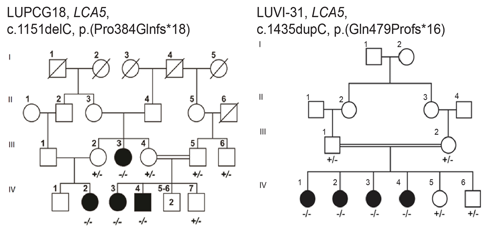
Homozygous frameshift mutations segregate in two Pakistani families in autosomal recessive pattern. (A) Pedigree of the LUPCG18 family showing segregation for the variant NM_001122769.3 c.1151delC (p.Pro384Glnfs*18). (B) Pedigree of the LUVI-31 showing segregating of the variant c.1435dupC (p.Gln479Profs*16). Black Filled symbols indicate affected individuals, while white symbols represent unaffected family member. The squares represent males while the circles represent females of each family, the diagonal line on any of these shows deceased individuals. Double line between male and female represent consanguineous marriage. Genotypes below symbols show the LCA5 variant status (+/+; homozygous wild type, +/-; heterozygous carrier, -/-; homozygous mutant).
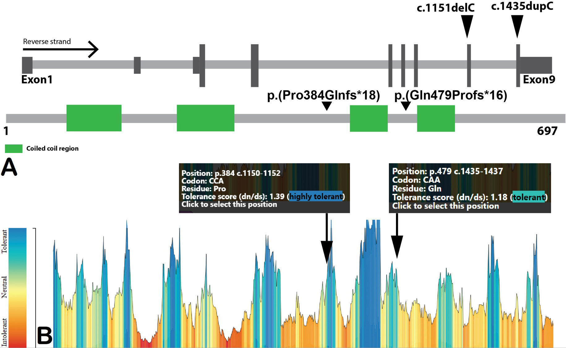
Genomic structure and mutation tolerance of the LCA5 gene. (A) Schematic representation of the LCA5 gene (NM_001122769.3) showing 9 exons (left to right exon1 to 9) organization and position of organization and position of identified variants c.1151delC and c.1435dupC in exons 8 and 9 respectively marked by black arrowheads. The lower bar shows LCA5 linear protein structure having 697 amino acids, green boxes show coiled coil regions however gray bar area represent non-structural domain regions. (B) MetaDome-based bar graph showing mutation tolerance ratios across the LCA5 protein. Peaks indicate regions with higher tolerance to no tolerance region for amino acid variation (colored blue meaning tolerant, yellow= slightly intolerant and red/orange = highly intolerant). The positions of Pro384 and Gln479 are highlighted, showing both variants reside within relatively tolerant regions, as indicated by their tolerance scores.
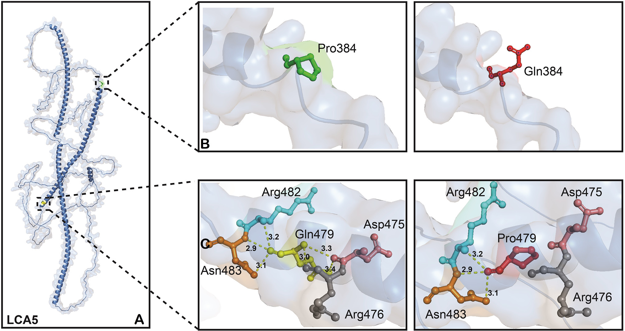
Structural modeling of LCA5 variants p.(Pro384Glnfs*18) and p.(Gln479Profs*16). (A) The overall structure of the wild type LCA5 protein with highlighted mutation sites in dashed boxes. (B) Comparative modeling of Pro384 (left box) and Gln384 (right box) reveals that the ringed structure of proline while glutamine confers shows linear structure. Both amino acids form no neighboring hydrogen bond and occur in loop region of the protein. (C) Wild-type Gln479 (left box) forms six hydrogen bonds with Asp475, Arg476, Arg482, and Asn483 respectively. Substitution to Pro479 (right box) leads to the loss of three key hydrogen bonds; one with Arg476 and two with Asp475. Yellow dashed lines show hydrogen bonding between amino acids and values represent bond length of these bonds in Armstrong (Å).
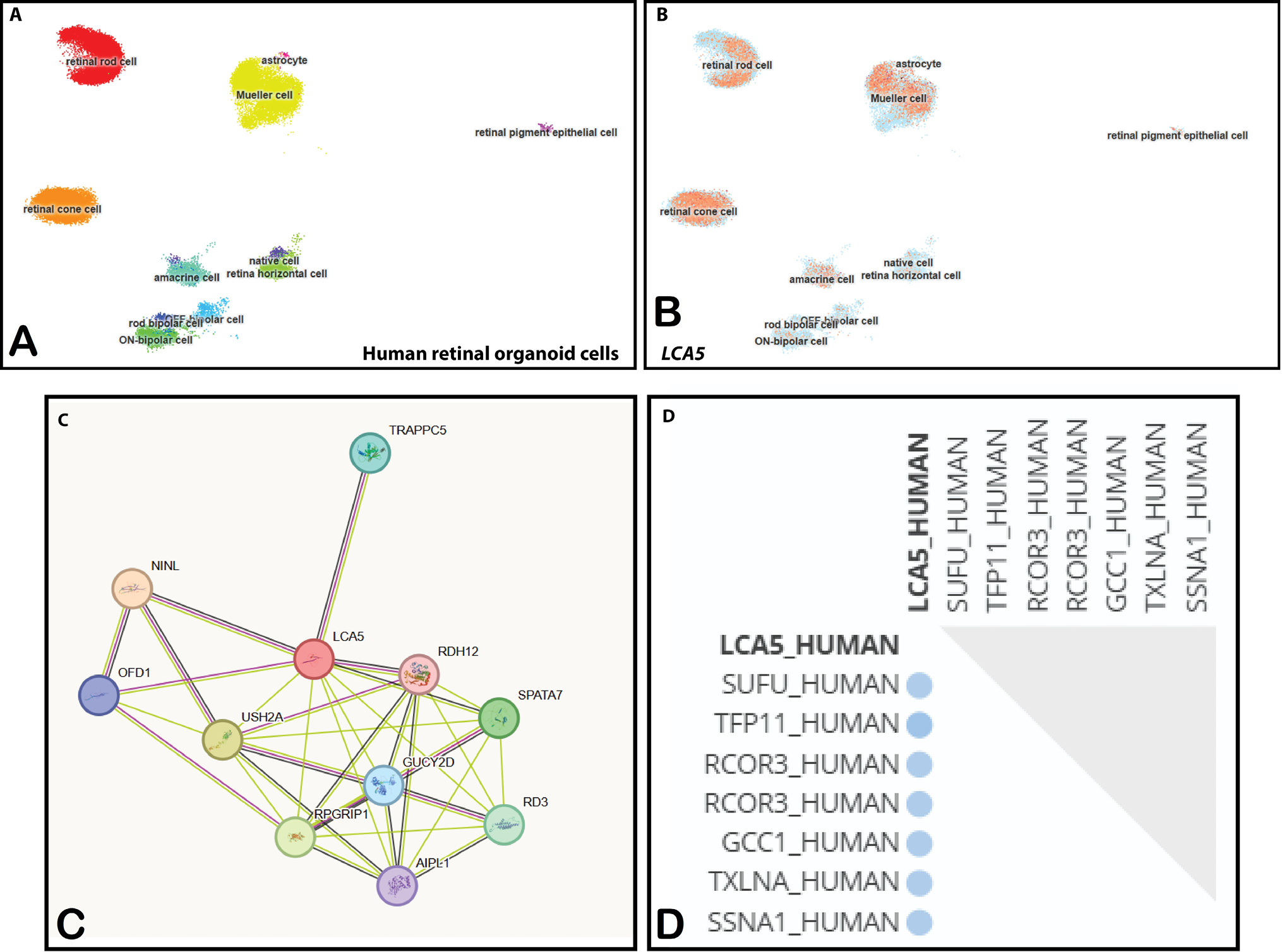
Expression profile and interaction networks of LCA5 in the retina. (A) Clusters of annotated human retinal organoid cell types (n=285,441) visualized using single cell transcriptomic data. (B) Expression of LCA5 across major retinal cell populations of human retinal organoids, with high signal (orange color) detected in retinal rod and cone photoreceptors, Müller glia, astrocytes, bipolar, amacrine, horizontal, and retinal pigment epithelial (RPE) cells. Blue color represent absence of LCA5 expression in cells. (C) STRING-based protein-protein interaction network showing LCA5 and its interactions with other IRDs proteins. (D) Binary interaction analysis identifying direct protein partners of LCA5 using multiple annotation databases; core interactors are listed alongside LCA.
References
- Adzhubei, I., Jordan, D.M. and Sunyaev, S.R., 2013. Predicting functional effect of human missense mutations using PolyPhen‐2. Curr. Protoc. Hum. Genet., 76: 7-20. https://doi.org/10.1002/0471142905.hg0720s76[CrossRef]
- Collin, R.W.J., van Minkelen, R., den Hollander, A.I., Vooistra, R.F., Gijsen, M., Marco, F.L., Willemsen, R., Rehman Khan, M., Riazuddin, S., Cremers, F.P.M. and Riazuddin, S., 2012. Antisense oligonucleotide (AON)-based therapy for Leber congenital amaurosis. Investigative Ophthalmol. Visual Sci., 53: 8842. https://doi.org/10.1038/mtna.2012.3[CrossRef]
- Corton, M., Avila-Fernandez, A., Vallespín, E., López-Molina, M.I., Almoguera, B., Martín-Garrido, E., Tatu, S.D., Khan, M.I., Blanco-Kelly, F. and Riveiro-Alvarez, R., 2014. Involvement of LCA5 in Leber congenital amaurosis and retinitis pigmentosa in the Spanish population. Ophthalmology, 121: 399–407. https://doi.org/10.1016/j.ophtha.2013.08.028[CrossRef]
- Den Hollander, A.I., Koenekoop, R.K., Mohamed, M.D., Arts, H.H., Boldt, K., Towns, K.V, Sedmak, T., Beer, M., Nagel-Wolfrum, K. and McKibbin, M., 2007. Mutations in LCA5, encoding the ciliary protein lebercilin, cause Leber congenital amaurosis. Nat. Genet., 39: 889–895. https://doi.org/10.1038/ng2066[CrossRef]
- Den Hollander, A.I., Roepman, R., Koenekoop, R.K. and Cremers, F.P.M., 2008. Leber congenital amaurosis: genes, proteins and disease mechanisms. Progr. Retinal Eye Res., 27: 391–419. https://doi.org/10.1016/j.preteyeres.2008.05.003[CrossRef]
- Faber, S., Mercey, O., Junger, K., Garanto, A., May-Simera, H., Ueffing, M., Collin, R.W.J., Boldt, K., Guichard, P. and Hamel, V., 2023. Gene augmentation of LCA5-associated Leber congenital amaurosis ameliorates bulge region defects of the photoreceptor ciliary axoneme. JCI Insight, 8: e169162. https://doi.org/10.1172/jci.insight.169162[CrossRef]
- Ghaffar, A., Akhter, T., Strømme, P., Misceo, D., Khan, A., Frengen, E., Umair, M., Isidor, B., Cogné, B. and Khan, A.A., 2024. Variants of NAV3, a neuronal morphogenesis protein, cause intellectual disability, developmental delay, and microcephaly. Commun. Biol., 7: 831. https://doi.org/10.1038/s42003-024-06466-1[CrossRef]
- Jacobson, S.G., Aleman, T.S., Cideciyan, A.V., Sumaroka, A., Schwartz, S.B., Windsor, E.A.M., Swider, M., Herrera, W. and Stone, E.M., 2009. Leber congenital amaurosis caused by Lebercilin (LCA5) mutation: retained photoreceptors adjacent to retinal disorganization. Mol. Vision, 15: 1098.
- Jumper, J., Evans, R., Pritzel, A., Green, T., Figurnov, M., Ronneberger, O., Tunyasuvunakool, K., Bates, R., Žídek, A., Potapenko, A., Bridgland, A., Meyer, C., Kohl, S.A.A., Ballard, A.J., Cowie, A., Romera-Paredes, B., Nikolov, S., Jain, R., Adler, J., Back, T., Petersen, S., Reiman, D., Clancy, E., Zielinski, M., Steinegger, M., Pacholska, M., Berghammer, T., Bodenstein, S., Silver, D., Vinyals, O., Senior, A.W., Kavukcuoglu, K., Kohli, P. and Hassabis, D., 2021. Highly accurate protein structure prediction with AlphaFold. Nature, 596: 583–589. https://doi.org/10.1038/s41586-021-03819-2[CrossRef]
- Li, L., Zhou, J. and Ran, J., 2025. Retinal photoreceptor cilia and ciliopathies: Molecular mechanisms and therapeutic strategies. Semin. Cell Dev. Biol., 174: 103635. https://doi.org/10.1016/j.semcdb.2025.103635[CrossRef]
- Li, Y., Wang, H., Peng, J., Gibbs, R.A., Lewis, R.A., Lupski, J.R., Mardon, G. and Chen, R., 2009. Mutation survey of known LCA genes and loci in the Saudi Arabian population. Invest. Ophthalmol. Visual Sci., 50: 1336–1343. https://doi.org/10.1167/iovs.08-2589[CrossRef]
- Mackay, D.S., Borman, A.D., Sui, R., van den Born, L.I., Berson, E.L., Ocaka, L.A., Davidson, A.E., Heckenlively, J.R., Branham, K. and Ren, H., 2013. Screening of a large cohort of leber congenital amaurosis and retinitis pigmentosa patients identifies novel LCA 5 mutations and new genotype–phenotype correlations. Hum. Mutat., 34: 1537–1546. https://doi.org/10.1002/humu.22398[CrossRef]
- Munir, A., Afsar, S. and Rehman, A.U., 2024. A systematic review of inherited retinal dystrophies in Pakistan: Updates from 1999 to April 2023. BMC Ophthalmol., 24: 55. https://doi.org/10.1186/s12886-024-03319-7[CrossRef]
- Orchard, S., Ammari, M., Aranda, B., Breuza, L., Briganti, L., Broackes-Carter, F. and Hermjakob, H., 2014. The MIntAct project IntAct as a common curation platform for 11 molecular interaction databases. Nucl. Acids Res., 42: D358–D363. https://doi.org/10.1093/nar/gkt1115[CrossRef]
- Perez, G., Barber, G.P., Benet-Pages, A., Casper, J., Clawson, H., Diekhans, M., Fischer, C., Navarro, G.J., Hinrichs, A.S., Lee, C.M., Nassar, L.R., Raney, B.J., Speir, M.L., van Baren, M.J., Vaske, C.J., Haussler, D., Kent, W.J. and Haeussler, M., 2025. The UCSC genome browser database: 2025 update. Nucl. Acids Res., 53: D1243–D1249. https://doi.org/10.1093/nar/gkae974[CrossRef]
- Ramprasad, V.L., Soumittra, N., Nancarrow, D., Sen, P., McKibbin, M., Williams, G.A., Arokiasamy, T., Lakshmipathy, P., Inglehearn, C.F. and Kumaramanickavel, G., 2008. Identification of a novel splice-site mutation in the Lebercilin (LCA5) gene causing Leber congenital amaurosis. Mol. Vision, 14: 481.
- Russell, S.R., Drack, A.V., Cideciyan, A.V., Jacobson, S.G., Leroy, B.P., Van Cauwenbergh, C., Wu, Z., Schwartz, F.T., Bejjani, B.A. and Girach, A., 2022. Intravitreal antisense oligonucleotide sepofarsen in Leber congenital amaurosis type 10: A phase 1b/2 trial. Nat. Med., 28: 1014–1021. https://doi.org/10.1038/s41591-022-01755-w[CrossRef]
- Sato, S., Morimoto, T., Tanaka, S., Hotta, K., Fujikado, T., Tsujikawa, M. and Nishida, K., 2020. Novel mutation identified in Leber congenital amaurosis: A case report. BMC Ophthalmol., 20: 313. https://doi.org/10.1186/s12886-020-01577-9[CrossRef]
- Skorczyk-Werner, A., Sowińska-Seidler, A., Wawrocka, A., Walczak-Sztulpa, J. and Krawczyński, M.R., 2023. Molecular background of Leber congenital amaurosis in a Polish cohort of patients—novel variants discovered by NGS. J. appl. Genet., 64: 89–104. https://doi.org/10.1007/s13353-022-00733-9[CrossRef]
- Szklarczyk, D., Kirsch, R., Koutrouli, M., Nastou, K., Mehryary, F., Hachilif, R. and Von Mering, C., 2023. The STRING database in 2023: Protein–protein association networks and functional enrichment analyses for any sequenced genome of interest. Nucl. Acids Res., 51: D638-D646. https://doi.org/10.1093/nar/gkac1000[CrossRef]
- Tareen, J.K., Khan, H., Ghani, S., Khan, S., Khan, B., Wu, Y., Khan, M.A., Shah, S.S.U.D., Hussain, A. and Kiyani, M.M., 2025. Whole exome sequencing reveals pathogenic variants in CNGA3, CACNA1F, and RPGRIP1 in consanguineous Pakistani families with diverse retinal phenotypes. PLoS One, 20: e0327176. https://doi.org/10.1371/journal.pone.0327176[CrossRef]
- Tehreem, R., Chen, I., Shah, M.R., Li, Y., Khan, M.A., Afshan, K., Chen, R. and Firasat, S., 2022. Exome sequencing identified molecular determinants of retinal dystrophies in nine consanguineous Pakistani families. Genes, 13: 1630. https://doi.org/10.3390/genes13091630[CrossRef]
- Wiel, L., Baakman, C., Gilissen, D., Veltman, J.A., Vriend, G. and Gilissen, C., 2019. MetaDome: Pathogenicity analysis of genetic variants through aggregation of homologous human protein domains. Hum. Mutat., 40: 1030–1038. https://doi.org/10.1101/509935[CrossRef]
- Zobor, D., Brühwiler, B., Zrenner, E., Weisschuh, N. and Kohl, S., 2023. Genetic and clinical profile of retinopathies due to disease-causing variants in Leber congenital amaurosis (LCA)-associated genes in a large German cohort. Int. J. mol. Sci., 24: 8915. https://doi.org/10.3390/ijms24108915[CrossRef]
Services
Article usage statistics
Loading…