Research Article · Volume 13, Issue 5, 19 April 2025 · Open Access
Exposure to Environmental Perfluorooctanoic Acid (PFOA) Induced Hepatocellular Apoptosis and Alteration in Serum Biomarkers in Diabetic Guinea Pigs
·
Affiliations & Notes ⌄
1Department of Pathology and Poultry Disease, College of Veterinary Medicine, University of Karbala, Iraq. *Correspondence | Zahraa K. Shakir, Department of Pathology and Poultry Disease, College of Veterinary Medicine, University of Karbala, Iraq
2Email: zahraa.shakir@uokerbala.edu.iq
3Department of Pathology and Poultry Disease, College of Veterinary Medicine, University of Baghdad, Iraq
Our results showed significant increasing of serum alkaline phosphatase (ALP) concentrations after 4 weeks of PFOA exposure.
In conclusion, PFOA exposure induced apoptosis in the liver of diabetic and non- diabetic guinea pigs by activation of caspase-3.
Moreover, PFOA induces liver toxicity and promotes the production of reactive oxygen species via increasing ALP enzyme and decreasing glutathione reductase activity.
Our results suggested that PFOA was associated with liver damage and induce apoptosis in diabetic and non-diabetic animals since PFOA itself can contribute to hyperglycemia.
Selected from the article's own text.
Abstract
We aimed to study the association of serum perfluorooctanoic acid (PFOA) with incident of diabetes and hepatotoxicity in guinea pigs. With increasing rates of diabetes following PFOA exposure, remarkable increase in mortality among PFOA producing workers was occurred from liver disease. The association between diabetes and liver disease are well-known but not following PFOA exposure. In this research, biomarkers of hepatocytes apoptosis, serum alkaline phosphatase (ALP) and serum glutathione reductase (GR) were evaluated in healthy and diabetic male guinea pigs following PFOA exposure. The results showed that PFOA-induced higher expression of liver caspase 3 activity with high levels of serum glucose and non-diabetic guinea pigs exposed to PFOA. Further, significant increased (p <0.05) in serum ALP in both diabetic and non-diabetic animals, respectively after PFOA exposure. At the end of experimental period of 4 weeks, both diabetic and non-diabetic guinea pigs showed significant decrease in GR levels following PFOA treatment. In conclusion, evaluation of liver caspase 3 activity, serum ALP and GR following PFOA treatment was significantly affected by diabetes. PFOA induce higher mitochondrial-mediated apoptosis with increased liver enzyme ALP and oxidative stress in diabetic and non-diabetic animals, respectively. Our findings were constant with the previous studies that indicated exposure to PFOA was linked with increasing blood glucose concentrations and liver toxicity.
Per- and polyfluoroalkyl substances (PFAS) are earning worldwide attention due to their bio accumulative, toxicity and resistance to processes of biological degradation in the environment . Per- and polyfluoroalkyl substances including PFOA have been manufactured by using electrochemical flourination and telomerization techniques . The major exposure source of PFOA is drinking water in contaminated communities . Moreover, PFOA was detected in food particularly in meat, fish, dairy products, vegetables and also in human breast milk . Perfluorooctanoic acid has been widely used since 1940s due to their unique chemical properties, making them very stable and repellents for water, stains and grease ; ; . Perfluorooctanoic acid is used in many industrial applications, such as carpeting, floor wax, upholstery, apparel, textiles, sealants and firefighting foam. Perfluorooctanoic acid also used for the production of another perfluoroalkyl-substituted compounds and polymeric materials .
Perfluorooctanoic acid represent a health and risk concern in spite of its manufacturing has been reduced in recent years . Perfluorooctanoic acid was highly persistent chemical that cannot degrade environmentally or break down by enzymes in the biological systems . Systemic absorption of PFOA and other PFAS compounds occur by oral, inhalation and dermal routes. After absorbed, PFOA is widely distributed in the body and accumulating mainly in the liver, kidney and blood . All animal and human data showed that PFOA not metabolized after absorption due to the high stability and low reactivity of carbon-fluorine bonds in PFOA chain .
Perfluorooctanoic acid have toxic effects in both animals and humans, like hepatotoxicity, immunotoxicity, genotoxicity and neurotoxicity (DeWitt et al., 2008). Perfluorooctanoic acid has also been related with hepatic damage resulting in fatty degeneration due to alteration in the antioxidant enzyme activity and induction of oxidative stress in hepatocytes . Perfluorooctanoic acid exposure association with pancreatic damage and incidence of diabetic . Moreover, PFOA can accumulate in mitochondria and change fatty acid (precursor of cholesterol) and mitochondrial transport process-related gene expression, leading to inhibit production of cholesterol .
The aim of this research was to evaluate the relation between PFOA exposure and incidence of liver damage in diabetic and non-diabetic animals.
MATERIALS AND METHODS
Ethics and Animals
All procedures and experimental design used in this study were reviewed and approved by the Scientific Committee of the Department of Pathology, College of Veterinary Medicine, University of Baghdad, Iraq in compliance with the ethical principles of animal welfare .
Forty (40) male guinea pigs (Cavia porcellus) (aged 16-19 weeks and weighing 500-550g) were selected for this study. The animals apparently healthy and kept under hygienic environment at 22±4°C in air-conditioned room, a relative humidity of 50±10%, and the light system was 12/12 hrs. light/dark cycle. The air of the room was continuously changed by ventilation vacuum along period of experiment (28 days).
Experimental Design
Experimental animals (40 male guinea pigs) were divided randomly and equally into 4 groups. Group 1 (G1) represented as a control. Group 2 (G2) induced diabetic male guinea pigs using alloxan monohydrate. Animals in Group 3 (G3) were received 100 mg/kg BW of PFOA. Group 4 (G4) diabetic male guinea pigs were received 100 mg/kg BW. dose of PFOA orally by using stomach gavage. The duration of the experiment was 28 days.
Induction of Diabetes in Experimental Animals
Diabetes was induced by injection of alloxan monohydrate intraperitoneally (200 mg/kg/BW.) in 24 hours intervals, respectively. Blood glucose test was performed before and after the injection as described in Akunneh and Aduema (2018) with modification.
Diabetes induced in two groups of animals, G2 and G4, respectively and animals with blood glucose levels higher than 200 mg/dL were supposed to be diabetic and included in the study ; ; .
Sampling and Laboratory Tests
Blood samples (2.5ml) were collected from each animal weekly by cardiac puncture (Arrak, 2012). Serum was extracted by centrifugation at 3000 rpm for 15 min and stored in deep freezer in polyethylene Eppendorf tubes at (-20ºC) for later tests .
Biochemical tests were performed by using commercially kits. Caspase 3 antibody kit from biorbyt company (British). Alkaline phosphatase kit from (SPINRACT, Spain) and guinea glutathione reductase kit from (SunLong Biotech, China).
Caspase-3 Activity in Liver
Rabbit polyclonal caspase-3 antibody was used for detection caspase-3 activity in liver . The paraffin sections of the selected specimen were heated in microwave oven (30 min at 90˚C) to retrieval the antigen then Hydrogen peroxide was applied with incubation for 20 min. After that, the slides rinsed with DW. 100 μl of Reagent 1 was added and incubate for 20 min then slides were drained and blotted without washing. 100 μl of primary antibody was placed into the tissue section and incubated for 1 hour at 37˚C. 100 μl of the antibody amplifier applied into the sections with incubated at 37ºC for 60 min. 100 μl of HRP polymer conjugate was placed into the tissue section and incubated for 60 min at 37˚C. 50 μl of the DAB-substrate chromogen (20 microliter of DAB mixed with 1ml of substrate diluent) was placed into the tissue section and incubated for 5 min at 37˚C. The slides immersed in a bath of Mayer’s Hematoxylin for 1 minute and then washed three times in D.W, 1 min each. Slides were dehydrated by soaked in ethanol (50, 70, 90, absolute) and then in xylene for 5 min. Evaluation of IHC results was performed by high powered light microscope ; .
Statistical Analysis
The data of the experiment were analyzed by using the Graph pad prism Statistical (version 8.0.2). Two-way ANOVA and Tukey’s multiple comparisons test were performed to evaluate significant differences among means of the groups. The results were expressed as mean ± stander errors and P < 0.05 was considered statistically significant.
RESULTS
Serum Alkaline Phosphatase Concentrations
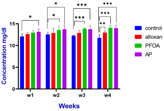
Figure 1 showed that serum alkaline phosphatase (ALP) concentrations were significantly increase (13.1±0.5) in diabetic group that exposed to PFOA (G4) compared with control group (12.06±0.5) at the 1st week of the experiment. Likewise, ALP concentrations were significantly increasing in G3 (13.53±0.8, 13. 8±0.1, 14 ±0.5) and in G4 (13.7±0.7, 13.7±0.7, 13.97±0.6) at weeks 2,3 and 4, respectively compared with control group. Moreover, ALP concentration increased significantly (12.9±0.3) in G2 (diabetic group) at 4th week compared with control group (11.7±0.7).
Guinea Glutathione Reductase Concentrations
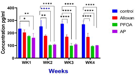
Figure 2 illustrated that glutathione reductase concentrations were significantly reduced (p <0.05) in G3 (127.54±12.1) and G4 (114.9±9.4) compared with the control group (234.08±33) during the 1st week of the experiment. In the same way, GR activity was significantly reduced (p <0.05) in G2 (177.37±6.7, 169.7±13.7, 167.9±24.6), G3 (101.97±4, 98.33±15.3, 94.62 ±4.05) and G4 (107.7±3.6, 108.19±6.5, 100.8±1.3), respectively compared with control (G1) at 2nd, 3rd and 4th weeks of the experiment.
Caspase 3 Activity in Liver
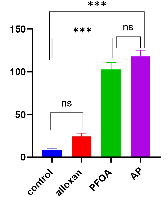
Figure 3 illustrate caspase 3 activity in control (G1) and treated guinea pigs with alloxan (G2), PFOA (G3) and PFOA + alloxan (G4). The results showed that caspase 3 activity was significantly rise (p<0.05) in G2, G3 and G4 (24.3 ±3.2, 103.3±6.1, 109.6±8.5) compared with the control group (8±2.1), respectively at the end of the experiment. Further, there were no significant differences (p≥0.05) between G3 that received PFOA and G4 diabetic animals dosed with PFOA.
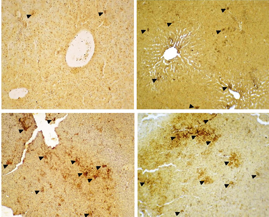
A photomicrograph of liver tissue sections stained immunohistochemically with caspase-3 antibody represented the normal caspase-3 activity in control group (G1) 3.26% (Figure 4A). Diabetic guinea pig (G2) shows mild increase 9.92% of caspase-3 activity when compared with control group 3.26% (Figure 4B). Additionally, guinea pig in G3 that dosed with PFOA illustrations increasing caspase-3 activity 42.10% compared with G1as shown in (Figure 4C). Diabetic male guinea pig that receives PFOA (G4) displays elevation caspase-3 activity 44.60% compared with control group (Figure 4D).
DISCUSSION
Our results showed significant increasing of serum alkaline phosphatase (ALP) concentrations after 4 weeks of PFOA exposure. These results are in agreement with Qazi et al. (2010) who reported that that dietary exposure of male mice to 0.002% PFOA for 10 days lead to elevated the activity of alkaline phosphatase (ALP). Furthermore, PFOA administration at dose 1.5, 5 and 10 mg/kg/day for 17,14 and 18 day in mice induced a significantly increase in serum ALP level ; .
Additionally, PFOA can disrupt hepatic metabolism and may be associated with increasing ALP enzyme in a representative sample of Canadian adults .
Yang et al. (2014) revealed that exposure to PFOA cause structural damage in the liver with degeneration leading to leakage of large quantities of ALP into the blood stream.
The results of our study displayed that the serum glutathione reductase concentrations were significantly decreased (p <0.05) in G3 that dosed with PFOA and in diabetic animals that dosed with PFOA (G4) compared with control group (G1) and diabetic guinea pigs (G2), respectively.
Previous reports suggested that PFOA lead to decrease the activity of glutathione reductase (GR) Zhang et al. (2019). PFOA treatmenet also resulting in increased in oxidative stress and inhibition activity of glutathione reductase . Moreover, alloxan treated group (G2) in this study decrease GR activity and this agree with West (2000) who reported that diabetic animals have decreased the activity of GR.
PFOA induce liver toxicity cause degeneration, necrosis, and accumulation of fat droplets inside nucleus of hepatocytes due to disturbance in lipid metabolic pathways ; ; .
The CD36 protein which also called fatty acid translocase (FAT), is member of cell surface proteins that located on differentiated adipocytes and other cells. This protein has important in regulation of uptake and distribution of fatty acids inside the cells ; . PFOA exposure in mice cause expressions of CD36 protein in hepatocytes leading to lipid accumulation inside hepatocytes .
Our study reported diabetic and non-diabetic guinea pig that receives PFOA showed high caspase-3 activity when compare with control group. PFOA induced mitochondrial membrane potential collapse and reduced adenosine triphosphate levels, cardiolipin peroxidation and cytochrome c release . In addition, PFOA exposure results in β cell apoptosis in pancreatic tissue and glucose-enhance insulin secretion . PFOA induced mitochondrial apoptosis presumably by the effect on the expression of genes related apoptosis such as p53, Bcl-2, Bax and Caspase-3 ; .
According to the results in this study (as seen in Figure 1, 3 and 4), the alloxan treated group (G2) showed significant increase in caspase-3 activity in liver tissues, serum ALP and reduction in GR activity. However, these changes were significantly differences in animals that dosed with PFOA (G3 and G4).
Moreover, the non-significant result of caspase-3 activity in liver, serum ALP and GR between non-diabetic group (G3) and diabetic animals (G4) toxicated with PFOA, suggested that the exposure to PFOA directly induced diabetic like action in guinea pigs.
Emerging the toxicity data from G3 and G4 and compared with G2 (induced diabetes in guinea pigs) have been shown constant results related that diabetes (DM) one of the health conditions which can be caused by perfluorooctanoic acid (PFOA). These findings indicated the toxic effect of PFOA lead to damaging of hepatocytes in diabetic and non-diabetic animals since PFOA itself can contribute to hyperglycemia.
CONCLUSIONS AND RECOMMENDATIONS
In conclusion, PFOA exposure induced apoptosis in the liver of diabetic and non- diabetic guinea pigs by activation of caspase-3. Moreover, PFOA induces liver toxicity and promotes the production of reactive oxygen species via increasing ALP enzyme and decreasing glutathione reductase activity. Our results suggested that PFOA was associated with liver damage and induce apoptosis in diabetic and non-diabetic animals since PFOA itself can contribute to hyperglycemia.
ACKNOWLEDGEMENTS
I would like to thank the staff of Department of pathology at the College of Veterinary Medicine/University of Baghdad, for their assistance in completing the research requirements.
NOVELTY STATEMENTS
evaluate the relation between PFOA exposure and incidence of liver damage in diabetic and non-diabetic animals. We found PFOA is associated with liver damage and induces apoptosis in diabetic and non-diabetic animals since PFOA itself can induce hyperglycemia.
AUTHOR’S CONTRIBUTIONS
The first auther work the experimental methods and writing, the second auther discuss the results with data analysis.
Conflict of Interest
The authors have declared no conflict of interest.
Figures
Figure 1:.
Effect of perfluorooctanoic acid, on serum alkaline phosphatase concentrations (mg/dL) in the control, alloxan induced diabetic, treatment animals with PFOA at 100 mg/kg BW and diabetic guinea pigs exposed to PFOA at 100 mg/kg BW during the experimental period of 4 weeks. Mean ± SE, n = 10.
Figure 2:.
Effect of perfluorooctanoic acid, on serum glutathione reductase activities (mg/dL) in the control, alloxan induced diabetic, treatment animals with PFOA at 100 mg/kg BW and diabetic guinea pigs exposed to PFOA at 100 mg/kg BW during the experimental period of 4 weeks. Mean ± SE, n = 10.
Figure 3:.
Effect of perfluorooctanoic acid, on apoptosis activities in the control, alloxan induced diabetic, treatment animals with PFOA at 100 mg/kg BW and diabetic guinea pigs exposed to PFOA at 100 mg/kg BW during the experimental period of 4 weeks. Mean ± SE, n = 10.
Figure 4:.
Immunohistochemical staining in guinea pigs liver. Localization of caspase 3 appears as dark brown color staining. (A) control group (G1). (B) diabetic group (G2). (C) Represented immunohistochemical staining of caspase-3 in liver of guinea pig in PFOA treated group (G3) at 100 mg/kg bw shows increasing expression of caspase-3 activity. (D) Represent diabetic male guinea pigs exposed to PFOA 100mg/kg bw (G4) reveled high expression of caspase-3 activity in liver. (X10).
References
Abduljalel ME , Al-Saadi RN (2022). Toxicopathological effect of benzene, toluene, ethylbenzene, and xylenes (btex) as a mixture and the protective effect of citicoline in male rats followings 90-day oral exposure. Red Vet., 23(3): 399–414.
Akunneh W, Aduema W (2018). The effects of the leaf extracts of vernonia amygdalina, ocimum gratissimum and phyllanthus amarus on blood glucose level of alloxan-induced diabetic guinea pigs. Loj Med. Sci., 2 (1)-2018. 127. https://doi.org/10.32474/LOJMS.2018.02.000127[CrossRef]
Al-Aaraje ZK, Al-Saadi RN (2023). Toxico-pathological effects of perfluorooctanoic acid (PFOA) in normal and diabetic male guinea pigs following oral exposure. J. Surv. Fish. Sci., 10(3S): 2906-2920.
Al-Mzaien KA (2012). Assessment the antioxidant and hypolipidmic effect of black cumin (nigella sativa l.) flavonoids in induced oxidative stressed male rabbits. Iraqi J. Vet. Med., 36(2): 163-173. https://doi.org/10.30539/iraqijvm.v36i2.459[CrossRef]
Andersen ME, Butenhoff JL, Chang SC, Farrar DG, Kennedy Jr GL, Lau C, Olsen GW, Seed J, Wallace KB (2008). Perfluoroalkyl acids and related chemistries—toxicokinetics and modes of action. Toxicol. Sci., 102(1): 3-14. https://doi.org/10.1093/toxsci/kfm270[CrossRef]
Araak J K (2012). The Protective role of date palm pollen (phoenix dactylifera l.) on liver function in adult male rats treated with carbon tetrachloride. Iraqi J. Vet. Med., 36(0E): 132-142. https://doi.org/10.30539/iraqijvm.v36i0E.409[CrossRef]
Aslan, Ozcan F, Kucuksayan E (2013). Increased small dense ldl and decreased paraoxonase enzyme activity reveals formation of an atherogenic risk in streptozotocin-induced diabetic guinea pigs. J. Diabetes Res., 860190. https://doi.org/10.1155/2013/860190[CrossRef]
ATSDR (2021). Toxicological profile for perfluoroalkyls. Agency for Toxic Substances and Disease Registry (ATSDR). Atlanta, GA: U.S. Department of Health and Human Services, Public Health Service.
Borghese MM, Liang C , Owen J, Fisher MJ (2022). Individual and mixture associations of perfluoroalkyl substances on liver function biomarkers in the canadian health measures survey. 21(1): 1-11. https://doi.org/10.1186/s12940-022-00892-6[CrossRef]
Cui Y, Liu W, Xie W, Yu W, Wang C, Chen H (2015). Investigation of the effects of perfluorooctanoic acid (pfoa) and perfluorooctane sulfonate (pfos) on apoptosis and cell cycle in a zebrafish (danio rerio) liver cell line. Int. J. Environ. Res. Public Health., 12(12): 15673-15682. https://doi.org/10.3390/ijerph121215012[CrossRef]
De Silva AO, Armitage JM, Bruton TA, Dassuncao C, Heiger‐Bernays W, Hu XC, Kärrman A, Kelly B, Ng C, Robuck AJ (2021). PFAS Exposure Pathways for Humans and Wildlife: A Synthesis of Current Knowledge and Key Gaps in Understanding. Environ. Toxicol. Chem., 40 (3): 631–657 https://doi.org/10.1002/etc.4935.[CrossRef]
DeWit JC, Copeland CB, Strynar MJ, Luebke RW (2008). Perfluorooctanoic acid–induced immunomodulation in adult c57bl/6j or c57bl/6n female mice. Environ. Health Perspect., 116(5): 644-650. https://doi.org/10.1289/ehp.10896[CrossRef]
Dietz C, Infanger M, Romswinkel A, Strube F , Kraus A (2019). Apoptosis induction and alteration of cell adherence in human lung cancer cells under simulated microgravity. Int. J. Mol. Sci., 20(14): 3601. https://doi.org/10.3390/ijms20143601[CrossRef]
European Food Safety Authority (2012). Perfluoroalkylated substances in food: occurrence and dietary exposure. EFSA J., 10(6): 2743. https://doi.org/10.2903/j.efsa.2012.2743[CrossRef]
Everds NE, Kennedy GL (2015). Serum perfluorooctanoic acid (pfoa) concentrations in normal and hyperlipidemic female hamsters dosed orally with ammonium perfluorooctanoate (apfo) for up to 30 days. Toxicol. Rep., 2: 70-77. https://doi.org/10.1016/j.toxrep.2015.01.013[CrossRef]
Glatz JF, Luiken JJ (2017). From fat to fat (cd36/sr-b2): Understanding the regulation of cellular fatty acid uptake. Biochimie, 136: 21-26. https://doi.org/10.1016/j.biochi.2016.12.007[CrossRef]
Glatz JF, Nabben M, Luiken JJ (2022). Cd36 (sr-b2) as master regulator of cellular fatty acid homeostasis. Curr. Opin. Lipidol., 33(2): 103. https://doi.org/10.1097/MOL.0000000000000819[CrossRef]
Glüge J, Scheringer M, Cousins IT, DeWitt JC, Goldenman G, Herzke D, Lohmann R, Ng CA, Trier X, Wang ZJ (2020). An overview of the uses of per-and polyfluoroalkyl substances (pfas). Environ. Sci. Processes Impacts, 22(12): 2345-2373. https://doi.org/10.1039/D0EM00291G[CrossRef]
He X, Wu D, Xu Y, Zhang Y, Sun Y, Chang X, Zhu Y, Tang W (2022). Perfluorooctanoic acid promotes pancreatic β cell dysfunction and apoptosis through er stress and the atf4/chop/trib3 pathway. Environ Sci Pollut Res, 29(56): 84532-84545. https://doi.org/10.1007/s11356-022-21188-9[CrossRef]
Kannan K, Corsolini S, Falandysz J, Fillmann G, Kumar KS, Loganathan BG, Mohd MA, Olivero J, Wouwe NV, Yang J (2004). Perfluorooctanesulfonate and related fluorochemicals in human blood from several countries. Environ. Sci. Technol., 38(17): 4489-4495. https://doi.org/10.1021/es0493446[CrossRef]
Li K, Gao P, Xiang P, Zhang X, Cui X , Ma LQ (2017). Molecular mechanisms of PFOA-induced toxicity in animals and humans: Implications for health risks. Environ. int, 99: 43-54. https://doi.org/10.1016/j.envint.2016.11.014[CrossRef]
Lindstrom AB, Strynar MJ, Libelo E (2011). Polyfluorinated compounds: Past, present, and future. Environ. Sci. Technol., 45(19): 7954-7961. https://doi.org/10.1021/es2011622[CrossRef]
Margolis R , Sant KE (2021). Associations between exposures to perfluoroalkyl substances and diabetes, hyperglycemia, or insulin resistance: A scoping review. J Xenobiot, 11(3): 115-129. https://doi.org/10.3390/jox11030008[CrossRef]
McCutcheon KR (2022). Pfas compounds pfoa and gen x are teratogenic to sea urchin embryos. (Master’s thesis, Boston University).
Obaid QA, Khudair KK , Al-Shammari AM (2021). 2-Deoxyglucose glycolysis inhibitor augment oncolytic virotherapy to induce oxidative stress and apoptosis in breast cancer (Part Ⅲ). Iraqi J. Vet. Med., 45(2): 26-32. https://doi.org/10.30539/ijvm.v45i2.1257[CrossRef]
Paul AG, Jones KC, Sweetman AJ (2009). A first global production, emission, and environmental inventory for perfluorooctane sulfonate. Environ. Sci. Technol., 43(2): 386-392. es802216n_si_001.pdf (620.75 kb). https://doi.org/10.1021/es802216n[CrossRef]
Qazi MR, Abedi MR, Nelson BD, DePierre JW, Abedi-Valugerdi MJ (2010). Dietary exposure to perfluorooctanoate or perfluorooctane sulfonate induces hypertrophy in centrilobular hepatocytes and alters the hepatic immune status in mice. Int. Immunopharmacol. 10(11): 1420-1427. https://doi.org/10.1016/j.intimp.2010.08.009[CrossRef]
Razak MR, Aris AZ, Zainuddin AH, Yusoff FM, Yusof ZNB, Kim SD, Kim KW (2023). Acute toxicity and risk assessment of perfluorooctanoic acid (pfoa) and perfluorooctanesulfonate (pfos) in tropical cladocerans moina micrura. Chemosphere. 313: 137377. https://doi.org/10.1016/j.chemosphere.2022.137377[CrossRef]
Serrano L, Iribarne-Durán LM, Suárez B, Artacho-Cordón F, Vela-Soria F, Peña-Caballero M, Hurtado JA, Olea N, Fernández MF, Freire C (2021). Concentrations of perfluoroalkyl substances in donor breast milk in southern spain and their potential determinants. Int. J. Hyg. Environ., 236: 113796. https://doi.org/10.1016/j.ijheh.2021.113796[CrossRef]
Shakir ZK, Muhsain SN, Al-Saadi RN (2023). Effect of Perfluorooctanoic Acid on Kidney Function in Diabetic and Non-Diabetic Male Guinea Pigs. Iraqi J. Vet. Med., 47(2): 73-80. https://doi.org/10.30539/ijvm.v47i2.1515[CrossRef]
Sheng N, Li J, Liu H, Zhang A, Dai J (2016). Interaction of perfluoroalkyl acids with human liver fatty acid-binding protein. Arch. Toxicol., 90: 217–227 (2016). https://doi.org/10.1007/s00204-014-1391-7[CrossRef]
Shnawa ZK, Abass DA (2022). Effect of P-glycoprotein inhibitor (carvedilol) on developmental outcome methotrexate are given alone and in combination of pregnant rats. Iraqi J. Vet. Med., 46(2): 36-42. https://doi.org/10.30539/ijvm.v46i2.1410[CrossRef]
Suh KS, Choi E M, Kim Y J, Hong SM, Park SY, Rhee SY, Oh S, Kim SW, Pak YK, Choe W (2017). Perfluorooctanoic acid induces oxidative damage and mitochondrial dysfunction in pancreatic β-cells. Mol. Med. Rep., 15(6): 3871-3878. https://doi.org/10.3892/mmr.2017.6452[CrossRef]
Taher IA (2016). The effect of melatonin on adrenal gland and pancreas function in alloxan–induced diabetes in adult female rabbits. Iraqi J. Vet. Med., 40(1): 38-46. https://doi.org/10.30539/iraqijvm.v40i1.136[CrossRef]
Tian M, Huang Q, Wang H, Martin FL, Liu L, Zhang J, Shen H (2019). Biphasic effects of perfluorooctanoic acid on steroidogenesis in mouse leydig tumour cells. Reprod. Toxicol., 83: 54-62. https://doi.org/10.1016/j.reprotox.2018.11.006[CrossRef]
Verma P (2022). Alkaline Phosphatase: A Review Article. Int. J. Biochem. Biomol., 8(1): 23-28.
Wang Q, Chen W, Zhang B, Gao Z, Zhang Q, Deng H,Han L, Shen XL (2022). Perfluorooctanoic acid induces hepatocellular endoplasmic reticulum stress and mitochondrial-mediated apoptosis in vitro via endoplasmic reticulum-mitochondria communication. Chem. Biol. Interact., 354: 109844. https://doi.org/10.1016/j.cbi.2022.109844[CrossRef]
Wen Y, Chen J, Li J, Arif W, Kalsotra A, Irudayaraj J (2020). Effect of pfoa on DNA methylation and alternative splicing in mouse liver. Toxicol. Lett., 329, 38-46. https://doi.org/10.1016/j.toxlet.2020.04.012[CrossRef]
West IC (2000). Radicals and oxidative stress in diabetes. Diabet. Med., 17(3): 171-180. https://doi.org/10.1046/j.1464-5491.2000.00259.x[CrossRef]
Wu X, Liang M, Yang Z, Su M, Yang B (2017). Effect of acute exposure to pfoa on mouse liver cells in vivo and in vitro. Environ. Sci. Pollut. Res. Int., 24(31): 24201-24206. https://doi.org/10.1007/s11356-017-0072-5[CrossRef]
Xu M, Wan J, Niu Q, Liu R (2019). Pfoa and pfos interact with superoxide dismutase and induce cytotoxicity in mouse primary hepatocytes: A combined cellular and molecular methods. Environ. Res., 175: 63-70. https://doi.org/10.1016/j.envres.2019.05.008[CrossRef]
Yahia D, Abd El-Nasser M, Abedel-Latif M, Tsukuba C, Yoshida M, Sato I, Tsuda S (2010). Effects of perfluorooctanoic acid (pfoa) exposure to pregnant mice on reproduction. J. Toxicol. Sci., 35(4): 527-533. https://doi.org/10.2131/jts.35.527[CrossRef]
Yan S, Wang J, Dai J (2015). Activation of sterol regulatory element-binding proteins in mice exposed to perfluorooctanoic acid for 28 days. Arch. Toxicol., 89(9): 1569-1578. https://doi.org/10.1007/s00204-014-1322-7[CrossRef]
Yang B, Zou W, Hu Z, Liu F, Zhou L, Yang S, Kuang H, Wu L, Wei J, Wang J, Zou T, Zhang D (2014). Involvement of oxidative stress and inflammation in liver injury caused by perfluorooctanoic acid exposure in mice. Biomed. Res. Int., 409837. https://doi.org/10.1155/2014/409837[CrossRef]
Zhang J, Sun N, Sun J, Wang B, Chen X, Lv Z, Liu J, Zhao B, Yuan Z (2021). The combined effects of wood vinegar and perfluorooctanoic acid on enzymatic activities, DNA integrity and gene transcription in dugesia japonica. https://doi.org/10.21203/rs.3.rs-906219/v1[CrossRef]
Zhang J, Wang B, Zhao B, Li Y, Zhao X, Yuan Z (2019). Blueberry anthocyanin alleviate perfluorooctanoic acid-induced toxicity in planarian (dugesia japonica) by regulating oxidative stress biomarkers, atp contents, DNA methylation and mrna expression. Environ. Pollut., 245: 957-964. https://doi.org/10.1016/j.envpol.2018.11.094[CrossRef]