Research Article · Volume 13, Issue 5, 20 April 2025 · Open Access
Cold Stress Modulates the Phenotype of Lymphocyte Subsets and the Function of Blood Phagocytes in Goats
· · ·
Affiliations & Notes ⌄
1Department of Microbiology, College of Veterinary Medicine, King Faisal University, Al-Ahsa, Saudi Arabia. *Correspondence | Jamal Hussen, Department of Microbiology, College of Veterinary Medicine, King Faisal University, Al-Ahsa, Saudi Arabia
This indicates that cold stress negatively affects T helper-cell-mediated immunity in goats.
The results indicate a compromising effect of ambient temperature drop on adaptive and innate immune cells in goats.
Whether these effects are related to the young age of the goat kids used in this study and whether older goats may show higher resistance to cold stress is still to be investigated in future studies.
Additionally, as the current study only focused on the short-time effects of temperature change on selected parameters of goat immune cells, it would be interesting to see how chronic cold stress for a longer period or how the exposure to extremely colder temperatures would modulate the goat immune system.
Selected from the article's own text.
Abstract
The accelerated climate change, resulting in sudden fluctuations in ambient temperatures, is one of the most important challenges for animal health and production worldwide. Few studies focused on the impact of cold stress on the immune response of goats. The aim of the present study was to investigate the impact of changes in ambient temperature (normal versus cold temperatures) on the distribution of lymphocyte subsets in goat blood and the antimicrobial activity of blood phagocytes. Composition of lymphocyte was analyzed by using flow cytometry and antibody staining. In addition, the ability of neutrophils and monocytes to engulf S. aureus was investigated ex vivo. The drop in ambient temperature was associated with a decrease in the percentage and absolute count of T helper cells with increased frequency of cytotoxic T cells, indicating a negative impact of cold stress on the cell-mediated immunity in goats. This was also supported by the cold-stress-induced decrease in the CD4/CD8 ratio as well as the observed decrease in CD44 expression on CD8+ T cells. In addition, the reduced capacity of both phagocytic cells after ambient temperature drop indicates reduced antimicrobial functionality of these innate immune cells. Collectively, the results indicate a compromising effect of ambient temperature drop on the adaptive and innate immune cells in goats. Further studies are required to identify the mechanisms mediating the observed changes in immune cell phenotype and function in goats.
The impact of thermal stress on the immune system has been extensively investigated in several veterinary species ; . Most studies, however, focused on the adaptation of the immune system to heat stress conditions ; . Although climate change-induced colder winter is currently observed in several desert areas, only few studies investigated the impact of cold stress on the immune system of livestock animals in these area ; ; ; ; ; ; ; . In pigs, cold stress was associated with increased basal levels of the pro-inflammatory cytokines IL-1β, and IL-6 with enhanced upregulation of tissue cytokines in response to LPS stimulation . Studies in rodents showed that housing at sub-optimal temperatures is associated with reduced energy availability for driving the immune responses resulting in significant changes in the functionality of the immune system . In rats, short-term cold stress resulted in decreased plasma levels of the T cell cytokines IFN-γ, IL-2, IL-4, and the percentage of regulatory CD4+CD25+Foxp3+ T cells .
CD4+ helper T cells and CD8+ cytotoxic T cells are two lymphocyte subsets with key roles in the adaptive immune response to intracellular and extracellular pathogens, respectively . On the other hand, neutrophils and monocytes are innate immune cells of the first line of defense with the capacity to phagocytose and kill pathogens .
Limited data are available on the impact of low ambient temperatures on the immune system of goat kids. The current study aimed, therefore, to investigate the impact of ambient temperature drop on some phenotypic and functional parameters related to innate and adaptive immune response in goat kids.
MATERIALS AND METHODS
Study Design
The study was conducted between October and December 2024 in the eastern province of Saudi Arabia. For this arid region (Al-Ahsa region; N 25°17′8.0844″, E 49°29′11.3316″), the climate is characterized by a long hot and dry summer from April to November (day temperatures range between 40°C and 50°C) and a short moderate winter from December (day temperatures range between 15°C and 29°C). Environmental temperatures (Table 1) were measured using a digital thermometer (ThermoPro TP50 Digital Hygrometer) with the measurement range from -40 to + 80ºC. Blood samples were collected at 07: 00 am on the following dates: 01.10.2024, 07.10.2024, 14.10.2024, 28.10.2024, 04.11.2024, 20.11.2024, and 03.12.2024.
Table 1: Time-related changes in ambient temperature (ºC) and humidity level.
Date
Temp. Max.
Temp.min.
Temp. Average
Humidity level
01.10.2024
41.3
33.6
37.5
60.8
07.10.2024
40.2
31.6
35.9
61.2
14.10.2024
39.3
28.5
33.9
68.2
28.10.2024
37.7
25.3
31.5
42.7
04.11.2024
35.2
24.8
30.0
59.3
20.11.2024
33.6
14.6
24.1
53.5
03.12.2024
17.3
9.4
13.4
50.8
Animals and Sampling
Blood samples were collected from five goat kids aged between 4 and 6 months including two males and 3 females of the Jabali goat breed. The goats were kept on the farm of the Scientific Research Station at King Faisal University in Al-Ahsa region (Saudi Arabia). Blood samples (5 mL) were collected from the jugular vein into vacutainer heparin tubes (Guangzhou Improve Medical Instruments Co., Ltd; Guangzhou, China) and transported to the laboratory for further processing.
Cell Separation
Separation of white blood cells (WBC) by hypotonic lysis of red blood cells was performed according to a previously published protocol for camel leukocytes . For this, 6 mL of distilled water was added to 2 mL blood for 20 seconds in a 15 mL sterile falcon tube followed by the addition of 6 mL 2x PBS to restore tonicity. After centrifugation at 1000 xg for 15 min at 4ºC, the lysis step was repeated twice (two rounds of centrifugation at 500 and 250 xg for 10 min each round) for complete removal of erythrocytes. Finally, the pellet was resuspended in cold PBS (2 x 10⁶ cell / mL).
Flow Cytometric Analysis of Leukocyte Subsets
Staining of separated cells with monoclonal antibodies (mAbs) was performed as previously described for bovine leukocytes . Briefly, isolated WBCs (1 x 10⁵ cell/well) were incubated in a 96-well plate for 15 min at 4°C with monoclonal antibodies to the lymphocyte markers CD4, CD8, CD335 (NKp46), BAQ44A (B cell), WC1 (Ƴδ T cell), and CD44. After washing in PBS-containing bovine serum albumin (BSA; 0.5%), the cells were subsequently incubated with secondary antibodies to mouse immunoglobulin isotypes (Table 2) for 15 min at 4°C. Labeled cells were finally washed two times and analyzed by flow cytometry (Becton Dickinson Accuri C6 flow cytometer; Becton Dickinson Biosciences, San Jose, California, USA).
Table 2: Monoclonal antibodies.
Antigen
Antibody clone
Labeling
Source
Isotype
CD4
GC50A1
-
Kingfisher
Mouse IgM
CD8
CC63
-
Biorad
Mouse IgG2a
WC1
CC15
FITC
Biorad
Mouse IgG2a
CD335
EC1.1
-
Kingfisher
Mouse IgG1
CD44
BAT31A
-
Kingfisher
Mouse IgG1
Mouse IgM
poly
APC
Thermofisher
Goat IgG
Mouse IgG1
poly
FITC
Thermofisher
Goat IgG
Mouse IgG2a
poly
PE
Thermofisher
Goat IgG
Phagocytosis
The phagocytosis activity of neutrophils and monocytes was analysed according to a protocol previously described for bovine phagocytes . Briefly, separated WBC (1 x 10⁵ cell/well) were incubated in vitro in a 96-well plate with 50 µL of FITC-conjugated inactivated Staphylococcus aureus (S. aureus) bacteria (2 × 10⁸ cells/mL in RPMI medium; Institute of Immunology, Hannover University of Veterinary Medicine, Germany). After incubation for 45 min at 37°C, flow cytometric analysis of phagocytosis was performed on the Accuri C6 flow cytometer.
Statistical Analyses
Statistical analysis was performed using Prism software (GraphPad). The comparison between means at different timepoints of the study was performed using the repeated measure One way analysis of variance (ANOVA) test in combination with Bonferroni test for normally distributed data and the Friedman test with Kruskal-Wallis test for not normally distributed data. P values less than 0.05 indicate significant effects. The Common Language Effect Size (CLES) and the confidence intervals were calculated based on Cohen’s d using an online calculation tool (https://www.psychometrica.de/effect_size.html) and presented it alongside the p-values.
RESULTS
Relative Composition of Blood Lymphocytes
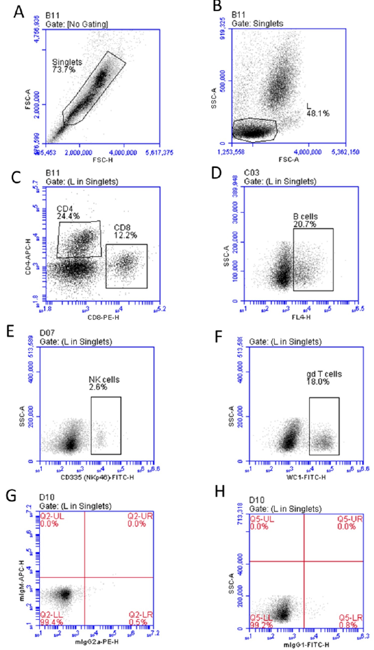
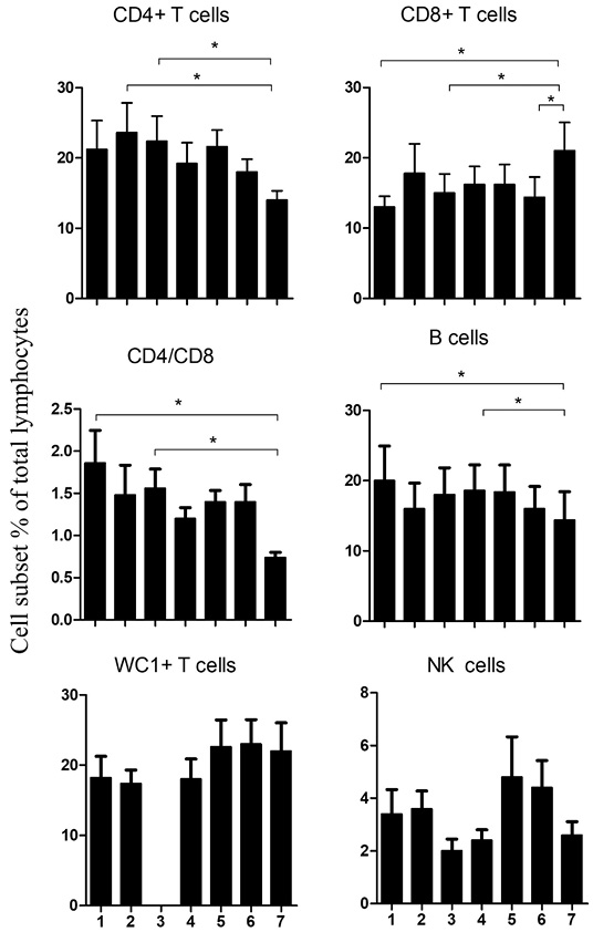
The relative frequency of lymphocyte subsets was analyzed using flow cytometry (Figure 1A, 1B, 1C,1D, 1E, 1F, 1G and 1H). The fraction of CD4+ T cells showed a gradual decrease with decreased ambient temperatures. The percentage of CD4+ T cells reached the lowest value on time point 7. The difference was, however, only significant (p < 0.05) in comparison to values of time points 2 (CLES = 0.96; CI = 0.82 - 4.1) and 3 (CLES = 0.97; CI = 0.90 - 4.25) (Figure 2). In contrast to this, the percentage of CD8+ T cells was significantly (p < 0.05) higher on time point 7 than at time point 1 (CLES = 0.97; CI = -4.35 - -0.95), 3 (CLES = 0.893; CI = -3.2 - -0.29), and 6 (CLES = 0.725; CI = -0.45 - 2.1). This change in the percentages of CD4+ and CD8+ T cells resulted in significantly lower CD4 to CD8 ratio (CD4/CD8) on time point 7 than time points 1 (CLES = 1; CI = 2.5 - 7.6) and 3 (CLES = 0.99; CI = 1.92 – 6.3) (p < 0.05). The lowest percentage of B cells was found on time point 7, which was significantly lower than time points 1 (CLES = 0.66; CI = -1.86 – 0.67) and 4 (CLES = 0.65; CI = -1.80 – 0.73). No significant changes were observed in the percentages of WC1+ T cells or natural killer (NK) cells at any time point (p > 0.05) (Figure 2). No data was available for WC1+ T cells on time point 3 due to low cell staining quality. Descriptive statistics of the analyzed parameters are presented in Table 3. In addition, the raw data for each time point and each animal and each analyzed parameter is shown in supplementary Table S1.
Count of Blood Lymphocyte Subsets in Goat Blood
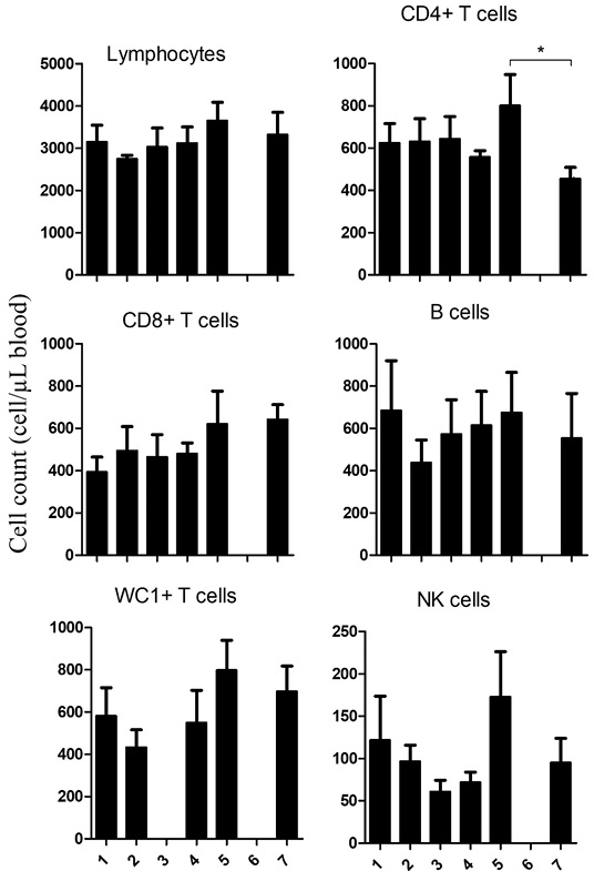
The absolute number of total lymphocytes was calculated by counting leukocytes under microscope after the addition of Turk solution, to lyse the red blood cells, followed by multiplication of lymphocyte percentage with total white blood cell count (Table 3). The total lymphocyte count did not show any significant changes at any time point (p > 0.05) (Figure 3). Only CD4+ T cells were significantly reduced on point 7 compared to time point 5 (CLES = 0.98; CI = 1.3 - 5.0; p < 0.05) (Figure 3). Although some tendential differences were observed over the study period, the numbers of CD8+ T cells, B cells, Ƴδ T cells (WC1), or NK cells did not show significant changes on any time point (p > 0.05) (Figure 3).
Changes in the Expression Level of the Activation Marker CD44 on Goat Lymphocytes
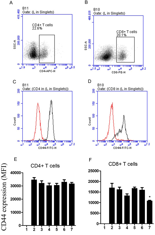
The expression density of CD44 was analysed by flow cytometry (Figure 4A, 4B, 4C and 4D). The abundance of the activation marker CD44 showed significant time-related changes only for CD8+ T cells (Table 3). The mean fluorescence intensity (MFI) of CD44 on CD8+ T cells was lowest on time point 7. The difference was significant between time point 7 (effect sizes f and d = 0.85 and 2.55; calculated based on group means from ANOVA) and all other time points (p < 0.05) (Figure 4E and 4F).
Impact of Ambient Temperature Change on Phagocytic Activity of Goat Neutrophils and Monocytes
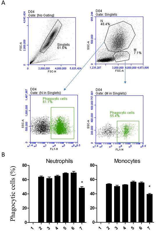
The phagocytosis function of neutrophils and monocytes were analyzed by using flow cytometry after incubating the cells with FITC-labeled and inactivated staphylococcus aureus bacteria (Figure 5A). For both neutrophils and monocytes, the percentage of phagocytosis-positive cells was significantly lower on time point 7 (effect sizes f and d = 1.9 and 5.9; calculated based on group means from ANOVA) than on all other time points (p < 0.05) (Figure 5B).
DISCUSSION
The evaluation of changes in the distribution of lymphocyte subsets in blood is one of the most popular methodologies for evaluating the status of the immune system. The present study investigated the impact of changes in ambient temperature on the distribution of lymphocyte cell subsets in goat blood and the phagocytic function of blood neutrophils and monocytes.
Although the total number of lymphocytes did not show significant changes during the study period, lymphocyte composition was significantly affected by the change in ambient temperature. The drop in ambient temperature was associated with a decrease in the percentage and absolute count of CD4+ T cells (T helper cells) with increased frequency of CD8+ T cells (cytotoxic T cells). Due to their essential role in all immune cell activities including the activation of B cells for antibody production and cytotoxic T cells for killing virus-infected cells, reduced numbers of T helper cells usually reflect compromised adaptive immune responses
Table 3: Lymphocyte populations, marker expression, and phagocytosis activity.
Time 1
Time 2
Time 3
Time 4
Time 5
Time 6
Time 7
Mean± SEM
Mean± SEM
Mean± SEM
Mean± SEM
Mean± SEM
Mean± SEM
Mean±SEM
Total lymphocyte count1
3146 ± 397
2747 ± 82
3030 ± 445
3123 ± 380
3651 ± 435
-
3324 ± 525
CD4+ T cells %2
21 ± 4 ab
24 ± 4 a
22 ± 4 a
19 ± 3 ab
22 ± 2 ab
18 ± 2 ab
14 ± 1 b
CD8+ T cells %2
12 ± 2 a
18 ± 4 ab
15 ± 3 a
16 ± 3 ab
16 ± 3 ab
14 ± 3 a
21 ± 4 b
B cells %2
20 ± 5 a
16 ± 4 ab
18 ± 4 ab
19 ± 4 a
18 ± 4 ab
16 ± 3 ab
14 ± 4 b
Ƴδ T cells %2
18 ± 3 a
16 ± 3 a
-
17 ± 3 a
23 ± 4 a
24 ± 3 a
22 ± 4 a
NK cells %2
3 ± 0.9 a
4 ± 0.7 a
2 ± 0.4 a
2 ± 0.4 a
5 ± 2 a
4 ± 1 a
3 ± 0.5 a
CD4/CD8 ratio
2 ± 0.4 a
1 ± 0.4 ab
2 ± 0.2 a
1 ± 0.1 ab
1 ± 0.1 ab
1 ± 0.2 ab
0.7 ± 0.06 b
CD4+ T cells count1
625 ± 91 ab
632 ± 108 ab
645 ± 105 ab
558 ± 29 ab
802 ± 147 a
-
455 ± 53 b
CD8+ T cells count1
393 ± 71 a
495 ± 114 a
465 ± 105 a
481 ± 50 a
622 ± 155 a
-
643 ± 70 a
B cells count1
685 ± 235 a
439 ± 107 a
574 ± 162 a
615 ± 161 a
675 ± 190 a
-
554 ± 211 a
Ƴδ T cells count1
582 ± 133 a
433 ± 83 a
-
551 ± 152 a
799 ± 141 a
-
699 ± 119 a
NK cells count1
122 ± 52 a
97 ± 19 a
61 ± 13 a
72 ± 12 a
173 ± 53 a
-
95 ± 29 a
CD44 MFI on CD4 T cells
-
36566± 2449a
32024± 1818a
30207± 1881a
30409± 1509a
32853± 1809a
31535± 1145a
CD44 MFI on CD8 T cells
-
16946± 2191a
16152± 1176a
13214± 929 a
16693± 667 a
15926± 1186 a
10801± 313 b
Phagocytic neutrophils (%)3
-
64 ± 1 a
62 ± 2 a
65 ± 1 a
69 ± 0.9 a
70 ± 2 a
48 ± 2 b
Phagocytic monocytes (%)4
-
54 ± 1 a
50 ± 2 a
53 ± 1 a
57 ± 1 a
55 ± 1 a
39 ± 1 b
1: cell/µl blood; 2:percent of total lymphocytes; MFI: mean fluorescence intensity; 3: percent of total neutrophils; 4: percent of total monocytes. Different small letters indicate significant differences between the means (p < 0.05).
. This indicates that cold stress negatively affects T helper-cell-mediated immunity in goats. The cold-stress-induced decrease in the CD4/CD8 ratio, which represents a marker of immune competence , is in support of this. The frequency of immune cells in blood is regulated by the balance between their production in the bone marrow and cell migration and mobilization to tissues. For the identification of the mechanisms responsible for the observed change in the frequency of lymphocyte subsets one may discuss the role of altered thymic output , changes in peripheral survival of lymphocytes, or changed migration patterns of these cells under cold stress. As the exposure of broiler chicken to cold stress was associated with improved development and function of the thymic gland reflected in an altered expression of several TLRs and cytokines , a role of altered thymic output in the observed changes in CD4 and CD8 frequencies seems unlikely. Although short-term cold exposure in humans was associated with decreased numbers of CD4+ cells without changing the abundance of CD8+ cells , the study did not identify the exact mechanism behind this effect. Furthermore, cold-stress induced cell apoptosis has been reported in the published literature ; . Identified mechanisms include the inhibition of the Nrf2-Keap1 signaling pathway and modulation of the AMPK/PI3K/AKT/mTOR pathways. However, it is still unknown whether exposure of goats to cold stress may differently impact cell apoptosis in T cell subsets leading to selective decrease in CD4+ T cells. As the mobilization of T cell subsets between their compartments in the bone marrow, blood, and lymphatic tissues is regulated by several cytokines and chemokines as well as the expression of homing receptors, gene expression analysis of distinct T cell chemokines, colony stimulating factors, and homing receptors would support a better understanding of the mechanisms behind the observed alterations in the distribution of T cell subsets. In addition, the analysis of lymphocyte functions such as proliferation and cytotoxicity would uncover the impact of cold stress on immune cell function in goats. Furthermore, cytokine production measurement or gene expression analysis of T helper type 1 and type 2 cytokines after in vitro stimulation may provide insights into T helper cell polarization in goats under cold stress.
In the present study, no temperature-induced changes were observed in the abundance of NK cells in goat blood. NK cells represent only a minor fraction of lymphocytes in blood. Therefore, the observed minimal changes in their frequency under cold stress could be due to small sample size. In addition, the reported enhancement of NK cell function in humans after exposure to cold temperatures suggests possible positive effect of cold stress on NK cell function . Therefore, future studies with larger animal size are important to answer the question whether goat NK cells are affected by or might possess higher resistance to cold stress.
Caprine Ƴδ T cells can be divided into different subsets ; . Therefore, although the total number of or Ƴδ T cells was not affected by cold stress, the detailed enumeration of all subsets in goat blood may uncover possible changes in distinct Ƴδ T cell subsets.
The hyaluronan receptor CD44 is a glycoprotein with wide range cellular expression and involvement in several cellular activities, including cell adhesion, migration, and activation ; . Although not proven through in-vitro analysis in this study, the observed decrease in CD44 expression on CD8+ T cells suggests reduced activation status of these cytotoxic cells under cold conditions. Given their role in the immune response to viruses , these results suggest impaired immune response to viral infection in goats under cold stress. The identification of exact mechanisms through which cold stress impairs T cell functions and the involvement of CD44 in these effects represent interesting areas for future in vitro or in vivo studies. It is especially important to see whether cold stress directly modulates distinct pathways such as mTOR leading to modulated expression of CD44 on CD8+ T cells or whether the effect is indirectly mediated by changed cytokines expression such as the proinflammatory cytokine IL-1beta .
Neutrophils and monocytes represent cells of the first defense barrier contributing to effective innate immunity against microbes through their ability to engulf and kill pathogens . In the present study, the reduced phagocytosis percentages of both phagocytic cells in goats after ambient temperature drop indicate reduced antimicrobial functionality of these innate immune cells. However, additional functional analysis on phagocytes by analyzing oxidative burst, bacterial killing, Netosis, and degranulation is required to confirm the observed cold stress-induced reduction in antimicrobial activity. Bacterial phagocytosis by neutrophils and monocytes is mediated by opsonin receptors, IgG receptors FcγRII and FcγRIII and the C3b receptors CR1 and CR3, and phagocytosis priming receptors. As the bacteria used for the phagocytosis assay in the present study was non-opsonized bacteria, the role of altered expression of FC and complement receptors in the observed changes in phagocytosis function can be excluded. It is most likely that cold stress affects the expression of other receptors involved in the direct priming of phagocytosis. Such receptors include scavenger receptors, TLRs, the complement receptors C3aR and C5aR, IL-8R, and TNFR . Additionally, it is also possible that cold stress-induced changes in the cytoskeletal dynamics in neutrophils and monocytes are responsible for the observed decrease in their phagocytosis function . Furthermore, the impact of energy availability and nutrient status on the immune system has been intensively investigated in the literature ; . Whether the observed decrease in phagocytic activity could be linked to changes in metabolic pathways or energy availability in immune cells under cold stress, is still to be investigated.
The results of the current study were generated based on samples collected from only five goat kids, which is a limitation of the present study. Therefore, future studies should consider increasing the number of animals and including animals of different age groups and breeds in order to provide conclusions that are more robust.
CONCLUSIONS AND RECOMMENDATIONS
The present study evaluated the impact of cold stress on the immunophenotype of blood lymphocytes and the antimicrobial function of neutrophils and monocytes. The results indicate a compromising effect of ambient temperature drop on adaptive and innate immune cells in goats. Whether these effects are related to the young age of the goat kids used in this study and whether older goats may show higher resistance to cold stress is still to be investigated in future studies. Additionally, as the current study only focused on the short-time effects of temperature change on selected parameters of goat immune cells, it would be interesting to see how chronic cold stress for a longer period or how the exposure to extremely colder temperatures would modulate the goat immune system. Especially longitudinal studies would help in answering the question whether chronic and or extreme cold stress will more profoundly impact the goat immune system or whether the animals will adapt to the chronic cold stress after a specify time of exposure.
ACKNOWLEDGEMENTS
The authors thank the Scientific Research Station at King Faisal University for providing the experimental animals.
NOVELTY STATEMENTS
The present study employed membrane immunofluorescence and flow cytometry to investigate the impact of cold stress on the immune system of goat kids. Specifically, the study performed a time course analysis of cold-stress-induced changes in the distribution of lymphocyte cell subsets in goat blood and the phagocytic function of blood neutrophils and monocytes.
AUTHOR’S CONTRIBUTIONS
Jamal Hussen designed the study, Mohammed Ali Al Hejji, Mohammed Ali Al-Sukruwah, and Baraa Falemban collected the samples. Jamal Hussen did the flow cytometry and wrote the first draft of the manuscript. All authors revised and approved the manuscript.
Funding
This work was supported by the Deanship of Scientific Research, Vice Presidency for Graduate Studies and Scientific Research, King Faisal University, Saudi Arabia [KFU250139].
Data Availability
The datasets generated during the current study are available from the corresponding author on reasonable request
Ethics Approval
This study was performed in line with the principles of the Declaration of Helsinki. Approval was granted by the Ethics Committee of King Faisal University, Saudi Arabia (KFU-REC-2024-JUN-ETHICS1843). All measures were taken to minimize stress and discomfort to the animals during blood sampling.
The authors have no relevant financial or non-financial interests to disclose.
Figures
Figure 1:.
Gating strategy for the flow cytometric analysis of lymphocyte subsets. Leukocytes were separated from goat blood, labeled with antibodies to cell surface markers, and analyzed by flow cytometry. After gating on signle cells (for the exclusion of cell doublets) in a forward scatter aria (FSC-A) against forward scatter height (FSC-H) density plot (A), lymphocytes (L) were gated based on their FSC and side scatter (SSC) characteristics (B). The lymphocyte subsets CD4+ ab T cells, CD8+ ab T cells (C), BAQ44A+ B cells (D), NKp46+ (CD335) natural killer (NK) cells (E), and WC1+ gd T cells (F) were identified based on positive staining with the corresponding mAb. Isotype control staining with secondary anti-mouse IgG2, anti-mouse IgM (G), and anti-mouse IgG1 (H) was performed in parallel.
Figure 2:.
Relative percentages of lymphocyte subsets in goat blood. Leukocytes were separated from goat blood, labeled with fluorescent antibodies, and analyzed by flow cytometry. The percentage of CD4+ T cells, CD8+ T cells, natural killer (NK) cells, B cells, and WC1+ Ƴδ T cells in addition to the CD4 to CD8 ratio (CD4/CD8) were calculated and presented in graph (n = 5 goats; * indicates significant differences ((One-Way Anova; p < 0.05).
Figure 3:.
Cell counts of goat lymphocytes subsets in blood. Leukocytes were separated from goat blood, counted under microscope, labeled with fluorescent antibodies, and analyzed by flow cytometry. The absolute numbers of CD4+ ab T cells, CD8+ ab T cells, the NKp46+ natural killer (NK) cells, B cells, and WC1+ Ƴδ T cells were calculated by multiplication of their percentages with the absolute number of lymphocytes. As the absolute leukocyte count was missing for the time point 2, no absolute count of lymphocyte subsets could be calculated for this time point. (n = 5 goats; * indicates significant differences (One-Way Anova; p < 0.05)).
Figure 4:.
Flow cytometric analysis of the abundance of CD44 on goat CD4+ and CD8+ T cells. Leukocytes were separated from goat blood and labeled with fluorescent antibodies to CD4, CD8, and CD44 and analyzed by flow cytometry. After the identification of CD4+ (A) and CD8+ (B) cell subsets within the lymphocyte population, overlapping histrograms were prepared to show the abundance of CD44 on CD4+ (C) and CD8+ T cells (D). Staining with anti-CD44 antibody was shown as black line, while control antibody staining was shown as red line. The expression levels of CD44 were calculated as mean fluorescence intensity (MFI) for CD4+ T cells (E) and CD8+ T cells (F). (n = 5 goats; * indicates significant differences (One-Way Anova; p < 0.05)).
Figure 5:.
Phagocytosis activity. Blood leukocytes were incubated in vitro with FITC-labeled and inactivated Staphylococcus aureus bacteria and analyzed by flow cytometry. A) Cell duplets were excluded based on forward scatter (FSC) and side scatter (SSC) properties. After gating on neutrophils (N) and monocytes (M), phagocytic cells were identified based on the increased fluorescence in the FITC channel. B) the percentage of phagocytosis-positive cells within neutrophils and monocytes was calculated and presented. (n = 5 goats; * indicates significant differences (One-Way Anova; p < 0.05)).
References
Alhussien MN, Hussen J, De Matteis G (2024). Editorial: Heat stress and immune responses in livestock: Current challenges and intervention strategies. Front. Vet. Sci., 11: 1366274. https://doi.org/10.3389/fvets.2024.1366274[CrossRef]
Becker M, Serr I, Salb VK, Ott VB, Mengel L, Bluher M, Weigmann B, Hauner H, Tschop MH, Daniel C (2019). Short-term cold exposure supports human treg induction in vivo. Mol. Metab., 28: 73-82. https://doi.org/10.1016/j.molmet.2019.08.002[CrossRef]
Brenner IK, Castellani JW, Gabaree C, Young AJ, Zamecnik J, Shephard RJ, Shek PN (1999). Immune changes in humans during cold exposure: Effects of prior heating and exercise. J. Appl. Physiol., 87(2): 699-710. https://doi.org/10.1152/jappl.1999.87.2.699[CrossRef]
Brooks DG, Tishon A, Oldstone MBA, McGavern DB (2021). Prevention of cd8 t cell deletion during chronic viral infection. Viruses, 13(7). https://doi.org/10.3390/v13071189[CrossRef]
Cong P, Liu Y, Liu N, Zhang Y, Tong C, Shi L, Liu X, Shi X, Liu Y, Tong Z (2018). Cold exposure induced oxidative stress and apoptosis in the myocardium by inhibiting the nrf2-keap1 signaling pathway. BMC Cardiovasc Disord., 18(1): 36. https://doi.org/10.1186/s12872-018-0748-x[CrossRef]
Dale DC, Boxer L, Liles WC (2008). The phagocytes: Neutrophils and monocytes. Blood, 112(4): 935-945. https://doi.org/10.1182/blood-2007-12-077917[CrossRef]
Foster LC, Arkonac BM, Sibinga NE, Shi C, Perrella MA, Haber E (1998). Regulation of cd44 gene expression by the proinflammatory cytokine interleukin-1beta in vascular smooth muscle cells. J. Biol. Chem., 273(32): 20341-20346. https://doi.org/10.1074/jbc.273.32.20341[CrossRef]
Frank JW, Carroll JA, Allee GL, Zannelli ME (2003). The effects of thermal environment and spray-dried plasma on the acute-phase response of pigs challenged with lipopolysaccharide. J. Anim. Sci.,81(5): 1166-1176. https://doi.org/10.2527/2003.8151166x[CrossRef]
Fu Y, Zhang S, Zhao N, Xing L, Li T, Liu X, Bao J, Li J (2022). Effect of mild intermittent cold stimulation on thymus immune function in broilers. Poult. Sci., 101(10): 102073. https://doi.org/10.1016/j.psj.2022.102073[CrossRef]
Guan H, Nagarkatti PS, Nagarkatti M (2009). Role of cd44 in the differentiation of th1 and th2 cells: Cd44-deficiency enhances the development of th2 effectors in response to sheep rbc and chicken ovalbumin. J. Immunol., 183(1): 172-180. https://doi.org/10.4049/jimmunol.0802325[CrossRef]
Hangalapura BN, Kaiser MG, Poel JJ, Parmentier HK, Lamont SJ (2006). Cold stress equally enhances in vivo pro-inflammatory cytokine gene expression in chicken lines divergently selected for antibody responses. Dev. Comp. Immunol., 30(5): 503-511. https://doi.org/10.1016/j.dci.2005.07.001[CrossRef]
Hennig J, Laschefski U, Becker H, Rammsayer T, Netter P (1993). Immune cell and cortisol responses to physically and pharmacologically induced lowering of body core temperature. Neuropsychobiology, 28(1-2): 82-86. https://doi.org/10.1159/000119005[CrossRef]
Hoter A, Rizk S and Naim HY (2019). Cellular and molecular adaptation of arabian camel to heat stress. Front. Genet., 10: 588. https://doi.org/10.3389/fgene.2019.00588[CrossRef]
Hu GZ, Yang SJ, Hu WX, Wen Z, He D, Zeng LF, Xiang Q, Wu XM, Zhou WY, Zhu QX (2016). Effect of cold stress on immunity in rats. Exp. Ther. Med., 11(1): 33-42. https://doi.org/10.3892/etm.2015.2854[CrossRef]
Hu L, Brito LF, Abbas Z, Sammad A, Kang L, Wang D, Wu H, Liu A, Qi G, Zhao M (2021). Investigating the short-term effects of cold stress on metabolite responses and metabolic pathways in inner-mongolia sanhe cattle. Animals, (Basel) 11(9). https://doi.org/10.3390/ani11092493[CrossRef]
Hussen J (2021). Bacterial species-specific modulatory effects on phenotype and function of camel blood leukocytes. BMC Vet. Res., 17(1): 241. https://doi.org/10.1186/s12917-021-02939-1[CrossRef]
Hussen J, Duvel A, Sandra O, Smith D, Sheldon IM, Zieger P, Schuberth HJ (2013). Phenotypic and functional heterogeneity of bovine blood monocytes. PLoS One, 8(8): e71502. https://doi.org/10.1371/journal.pone.0071502[CrossRef]
Hussen J, Koy M, Petzl W, Schuberth HJ (2016). Neutrophil degranulation differentially modulates phenotype and function of bovine monocyte subsets. Innate Immunol., 22(2): 124-137. https://doi.org/10.1177/1753425915620911[CrossRef]
Iddir M, Brito A, Dingeo G, Fernandez Del Campo SS, Samouda H, La Frano MR, Bohn T (2020). Strengthening the immune system and reducing inflammation and oxidative stress through diet and nutrition: Considerations during the covid-19 crisis. Nutrients 12(6). https://doi.org/10.3390/nu12061562[CrossRef]
Kang HJ, Lee IK, Piao MY, Gu MJ, Yun CH, Kim HJ, Kim KH, Baik M (2016). Effects of ambient temperature on growth performance, blood metabolites, and immune cell populations in korean cattle steers. Asian-Australas. J. Anim. Sci., 29(3): 436-443. https://doi.org/10.5713/ajas.15.0937[CrossRef]
Kim WS, Ghassemi Nejad J, Lee HG (2023). Impact of cold stress on physiological, endocrinological, immunological, metabolic, and behavioral changes of beef cattle at different stages of growth. Animals (Basel), 13(6). https://doi.org/10.3390/ani13061073[CrossRef]
Kizaki T, Suzuki K, Hitomi Y, Iwabuchi K, Onoe K, Ishida H, Izawa T, Ji LL and Ohno H (2001). Activation and apoptosis of murine peritoneal macrophages by acute cold stress. Biochem. Biophys. Res. Commun., 283(3): 700-706. https://doi.org/10.1006/bbrc.2001.4843[CrossRef]
Luckheeram RV, Zhou R, Verma AD, Xia B (2012). Cd4(+)t cells: Differentiation and functions. Clin. Dev Immunol 2012: 925135. https://doi.org/10.1155/2012/925135[CrossRef]
Morgado JN, Lamonaca E, Santeramo FG, Caroprese M, Albenzio M and Ciliberti MG (2023). Effects of management strategies on animal welfare and productivity under heat stress: A synthesis. Front. Vet. Sci., 10: 1145610. https://doi.org/10.3389/fvets.2023.1145610[CrossRef]
Mylvaganam S, Freeman SA, Grinstein S (2021). The cytoskeleton in phagocytosis and macropinocytosis. Curr. Biol., 31(10): R619-R632. https://doi.org/10.1016/j.cub.2021.01.036[CrossRef]
Pikula J, Heger T, Bandouchova H, Kovacova V, Nemcova M, Papezikova I, Piacek V, Zajickova R, Zukal J (2020). Phagocyte activity reflects mammalian homeo- and hetero-thermic physiological states. BMC Vet. Res., 16(1): 232. https://doi.org/10.1186/s12917-020-02450-z[CrossRef]
Ponta H, Sherman L, Herrlich PA (2003). Cd44: From adhesion molecules to signalling regulators. Nat. Rev. Mol. Cell. Biol., 4(1): 33-45. https://doi.org/10.1038/nrm1004[CrossRef]
Ron R, Moreno E, Martinez-Sanz J, Branas F, Sainz T, Moreno S, Serrano-Villar S (2023). Cd4/cd8 ratio during human immunodeficiency virus treatment: Time for routine monitoring? Clin. Infect. Dis., 76(9): 1688-1696. https://doi.org/10.1093/cid/ciad136[CrossRef]
Soehnlein O, Lindbom L (2010). Phagocyte partnership during the onset and resolution of inflammation. Nat. Rev. Immunol., 10(6): 427-439. https://doi.org/10.1038/nri2779[CrossRef]
Thapa P, Farber DL (2019). The role of the thymus in the immune response. Thorac. Surg. Clin., 29(2): 123-131. https://doi.org/10.1016/j.thorsurg.2018.12.001[CrossRef]
van Kessel KP, Bestebroer J, van Strijp JA (2014). Neutrophil-mediated phagocytosis of staphylococcus aureus. Front. Immunol., 5: 467. https://doi.org/10.3389/fimmu.2014.00467[CrossRef]
Vialard F, Olivier M (2020). Thermoneutrality and immunity: How does cold stress affect disease? Front. Immunol., 11: 588387. https://doi.org/10.3389/fimmu.2020.588387[CrossRef]
Worku D, Hussen J, De Matteis G, Schusser B, Alhussien MN (2023). Candidate genes associated with heat stress and breeding strategies to relieve its effects in dairy cattle: A deeper insight into the genetic architecture and immune response to heat stress. Front. Vet. Sci., 10: 1151241. https://doi.org/10.3389/fvets.2023.1151241[CrossRef]
Xu J, Strasburg GM, Reed KM, Bello NM, Velleman SG (2023). Differential effects of temperature and mtor and wnt-planar cell polarity pathways on syndecan-4 and cd44 expression in growth-selected turkey satellite cell populations. PLoS One, 18(2): e0281350. https://doi.org/10.1371/journal.pone.0281350[CrossRef]
Xu Q, Wang YC, Liu R, Brito LF, Kang L, Yu Y, Wang DS, Wu HJ, Liu A (2017). Differential gene expression in the peripheral blood of chinese sanhe cattle exposed to severe cold stress. Genet. Mol. Res., 16(2). https://doi.org/10.4238/gmr16029593[CrossRef]
Yirsaw A, Baldwin CL (2021). Goat gammadelta t cells. Dev. Comp. Immunol., 114: 103809. https://doi.org/10.1016/j.dci.2020.103809[CrossRef]
Yirsaw AW, Gillespie A, Britton E, Doerle A, Johnson L, Marston S, Telfer J, Baldwin CL (2021). Goat gammadelta t cell subpopulations defined by wc1 expression, responses to pathogens and cytokine production. Dev. Comp. Immunol., 118: 103984. https://doi.org/10.1016/j.dci.2020.103984[CrossRef]