
The aim of our present study was to assess the effects of Kanglaite injection on apoptosis and expression of apoptosis-related factors Bcl-2 and Bax protein and the activities of Caspase-3 and Caspase-8 in colon cancer SW480 cells were studied. Compared with the control group without medication, the proliferation inhibition rate of cells processed with Kanglaite injection (10, 20 and 40 μL/mL) significantly increased (p <0.05), the apoptosis rate increased (p <0.05), the expression of Bcl-2 protein decreased (p <0.05), the expression of Bax protein significantly increased (p <0.05), and the activity of Caspase-3 and Caspase-8 increased (p <0.05) in a dose-dependent manner. Hence, this can be concluded that Kanglaite injection can effectively inhibit the proliferation and induce apoptosis of SW480 cells, which may be correlated with the inhibition of Bcl-2 expression, the promotion of Bax expression and the enhancement of caspase activity.
Short Communication · Volume 53, Issue 1, 30 December 2020 · Open Access
The Effect of Kanglaite Injection on Apoptosis of Colon Cancer SW480 Cells and Expression of Apoptosis-Related Factors
·
Affiliations & Notes ⌄
- 1Gastrointestinal Surgery, Quzhou People’s Hospital, Quzhou324000, Zhejiang, China , * Corresponding author: qyjs8zt@163.com
Article Info ⌄
- Journal:
- Pakistan Journal of Zoology
- Published:
- 30 December 2020

Abstract
Materials and methods
Human colon cancer SW480 cells were cultured in a culture medium containing 10% fetal bovine serum, 100 U/mL penicillin and 100 μg/mL streptomycin RPMI-1640 on the condition of 37°C and 5% CO2, and digested and passaged in a conventional manner. Experiments were performed when about 80% cells fused. All experiments were performed in the logarithmic growth phase of the cells. The cells were divided into control group (0 μL/mL Kanglaite injection) and Kanglaite injection treatment group (10, 20 and 40 μL/mL).
The proliferation of cells in each treatment group was examined with MTT assay. Human colon cancer SW480 cells in the logarithmic growth phase were trypsinized, and inoculated into 96-well plates at 1×10⁴ cells/well, and cultured for 24 h. After the supernatant was discarded, 200 μL Kanglaite injection (10, 20 and 40 μL/mL) was taken to process the cells for 48 h, and each group was provided with 3 parallel wells. Another control group was set up. 20 μL MTT (5 mg·mL-1 PBS, 4°C, and protected from light) was added at each time point after cell dosing. After 4 h of incubation, the medium in the well was aspirated, and 150 μL DMSO was added to each well. After shaking for 10 min in microplate reader, the crystals were completely melted, and the OD value was measured by a microplate at a wavelength of 490 nm. Cell proliferation inhibition rate (%) = (1 - experimental group OD value / control group OD value) × 100%.
Cell apoptosis in each treatment group was detected by flow cytometry. SW480 cells in the logarithmic growth phase were inoculated in a 6-well plate at a concentration of 4×10 5 cells/mL, 1 mL per well, and cultured for 24 h, processed with 0 (control), 10, 20 and 40 μL/mL Kanglaite injection and cultured for 48 h. After that, the solution was washed twice with PBS (centrifugation at 2,000 rpm for 5 min) to collect the cells, and 500 μL binding buffer was added for cell suspension. The 5 μL annexin V- FITC was added and mixed, and then 5 μL propidium iodide (PI) was added and mixed to react at room temperature in the dark for 10 min. Cell apoptosis was detected by flow cytometry within 1 h.
The expression of Bax and Bcl-2 protein in each experiment group was detected with Western blot. SW480 cells in the logarithmic growth phase were inoculated into 6-well plate with the method above, and processed with 0 (control), 10, 20 and 40 μL/mL Kanglaite injection. After 48 h, the total protein was extracted, the protein concentration was measured with BCA, and the protein loading was determined to be 30 μg. After SDS-PAGE gel electrophoresis, the protein was transferred to PVDF membrane, and was blocked with 5% skim milk for 1 h. Then, primary anti-Bax, Bcl-2 and β-actin were added and incubated overnight at 4°C shaker. After the membrane was washed for 3 times, 10 min per time, the fluorescently labeled secondary antibody was added, and then incubated at 4°C for 1 h, and washed for 3 times with TBST for 10 min each time. The Odyssey 9120 two-color infrared laser imaging system instrument was used to scan and analyze the protein bands to detect changes in the expression of the target protein.
The SW480 cells in the logarithmic growth phase were inoculated into a 6-well plate as described above, and administered at three concentrations of 10, 20 and 40 μL/mL, each of which was subjected to 3 replicate wells, and a negative control was also set. The cells were collected after been processed for 12 h, and the absorbance values of Caspase-3 and Caspase-8 were measured by enzyme-labeled immunoassay at 405 nm with reference to the instructions of Caspase-3 and Caspase-8 activity test kit. Then, the activity value was speculated according to the standard curve.
Data were analyzed using SPSS version 25.0 (SPSS Inc., Chicago, IL, USA). One way analysis of variance (ANOVA) was used to determine the significance of each parameter among different treatments. If the outcome was significant, the ANOVA was followed by LSD-t test. The probability (p) value less than 0.05 were considered significant.
Results and discussion
The results of MTT assay showed that the inhibition rate of cells treated with different concentrations (10, 20 and 40 μL/mL) of Kanglaite injection was significantly higher than that of the control group (p <0.05, Table I), which suggested that Kanglaite injection can inhibit the proliferation of colon cancer SW480 cells, and the inhibition rate increases with the increase of concentration in a dose-dependent manner.
The results of flow cytometry showed that Kanglaite injection in all concentrations could induce apoptosis of SW480 cells, and the apoptosis rate of cells was significantly different from that of the control group (p <0.05, Table I), and the higher the concentration increased, the stronger the ability to induce apoptosis, which shows a dose-dependent manner.
Table I.- Comparison of proliferation and apoptosis of colon cancer SW480 cells in each group (Mean±SD, n=3).
| Parameter | Kanglaite | |||
| 0 μL/mL | 10 μL/mL | 20 μL/mL | 40 μL/mL | |
| Inhibition rate of cells (%) | 0 | 25.73± 2.15* | 38.16± 3.41* | 57.05± 4.38* |
| Apoptosis rate of cells (%) | 3.13 ± 0.84 | 11.76± 1.85* | 22.53± 2.79* | 29.14± 3.22* |
*Significant difference p < 0.05, compared with the control group.
The results of Western blot showed that the expression of Bcl-2 protein significantly decreased with the increase of the concentration of Kanglaite injection. The difference of the expression of Bcl-2 protein between cells treated with 10, 20 and 40 μL/mL Kanglaite injection and cells in the control group was statistical significant (p <0.05, Fig. 1); and the expression of Bax protein increased with the increase of concentration of Kanglaite injection, and the expression of Bax protein treated with Kanglaite injection at 10, 20 and 40 μL/mL was significantly different from that of the control group (p <0.05, Fig. 1), which suggested that Kanglaite injection can significantly inhibit Bcl-2, promote the level of Bax, and reduce the anti-apoptotic effect of Bcl-2, thus promoting cell apoptosis.
Table II.- Comparison of activity of Caspase-3 and Caspase-8 in SW480 cells of different groups of colon cancer (Mean±SD, n=3).
| Parameter | Kanglaite | |||
| 0 μL/mL | 10 μL/mL | 20 μL/mL | 40 μL/mL | |
| Activity of Caspase 3 (×106 U·mg-1 prot.) | 1.27± 0.33 | 7.68± 0.51* | 9.44± 0.47* | 11.69± 0.63* |
| Activity of Caspase 8 (×106 U·mg-1 prot.) | 1.09± 0.26 | 6.35± 0.42* | 8.61± 0.34* | 10.72± 0.75* |
*Significant difference p < 0.05, compared with the control group.
Table II showed that the activity of Caspase-3 and Caspase-8 increased with the action of 10, 20 and 40 μL/mL Kanglaite injection, and the difference was statistically significant compared with the control group (p <0.05, Table II), and the activity of both significantly improved with the increase of concentration.
Previous studies have confirmed that Kanglaite injection can induce apoptosis of gastric cancer cells, down-regulate apoptosis-inhibiting genes, and up-regulate apoptosis-inducing genes, thereby playing a role in tumor therapy. This study found that Kanglaite injection can inhibit the proliferation and induce apoptosis of colon cancer SW480 cells in a dose-dependent manner, which may inhibit the expression of Bcl-2, promote the expression of Bax, and enhance the activity of Caspase-3 and Caspase-8.
The limitation of this study is that only one kind of colon cancer cell was used in this experiment, and the effect of Kanglaite injection on other types and sources of colon cancer cells and whether Kanglaite injection can regulate other factors in the apoptosis pathway needed to be further explored.
Conclusions
It is concluded that Kanglaite injection can effectively inhibit the proliferation and induce apoptosis of SW480 cells, which may be correlated with the inhibition of Bcl-2 expression, the promotion of Bax expression and the enhancement of caspase activity.
Statement of conflict of interest
Authors have declared no conflict of interest.
Figures
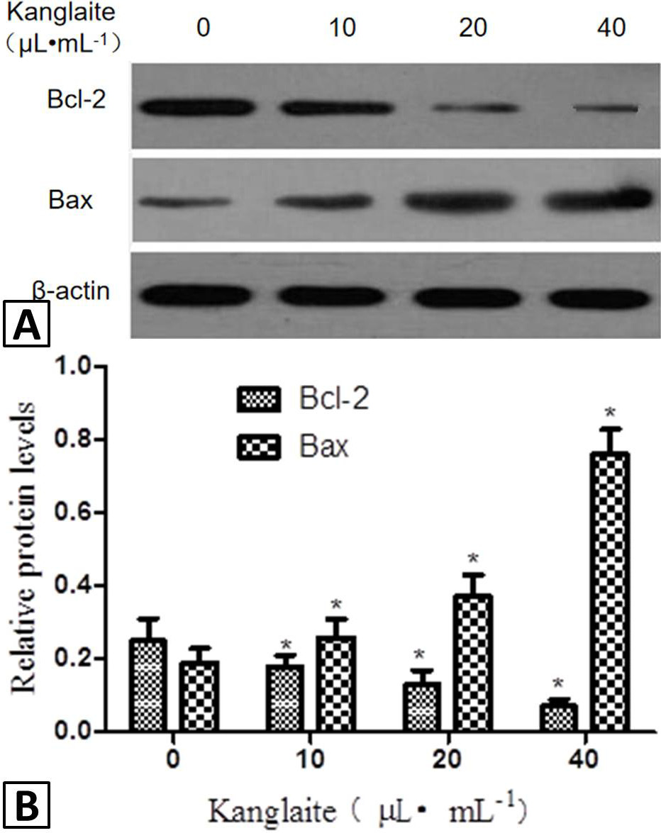
Effect of Kanglaite injection on the expression of Bcl-2 and Bax protein in colon cancer SW480 cells in Western blot (A) and semi quantitative analysis (B). *Significant difference p < 0.05, compared with the control group. Data are expressed as the mean ± SD (n=3).
References
- Andressakis, D., Lazaris, A.C., Tsiambas, E., Kavantzas, N., Rapidis, A. and Patsouris, E., 2008. J. Laryngol. Otol., 122: 1213-1218. https://doi.org/10.1017/S0022215108002636[CrossRef]
- Bratton, S.B., MacFarlane, M., Cain, K. and Cohen, G.M., 2000. Exp. Cell Res., 256: 27-33.
- Huo, X., Zhang, L. and Li, T., 2018. Oncol. Lett., 15: 3056-3060. https://doi.org/10.1006/excr.2000.4835[CrossRef]
- Koohpeyma, H., Goudarzi, I., Elahdadi, Salmani, M., Lashkarbolouki, T. and Shabani, M., 2019. Neurotox. Res., 35: 724-738. https://doi.org/10.1007/s12640-018-9979-y[CrossRef]
- Liu, J., Yu, L. and Ding, W., 2019. Medicine (Baltimore), 98: e16656. https://doi.org/10.1097/MD.0000000000016656[CrossRef]
- Matsuda-Minehata, F., Inoue, N., Goto, Y. and Manabe, N., 2006. J. Reprod. Develop., 52: 695-705. https://doi.org/10.1262/jrd.18069[CrossRef]
- Mineev, V.N., Nesterovich, I.I., Trofimov, V.I., Kashintseva, T.V., Rybakova, M.G. and Grozov, R.V., 2011. Arkh. Patol., 73: 11-14.
- Shan, S.J., Xiao, T., Chen, J., Geng ,S.L., Li, C.P., Xu, X., Hong, Y., Ji, C., Guo, Y., Wei, H., Liu, W., Li, D. and Chen, H.D., 2012. Int. J. mol. Med., 29: 625-629. https://doi.org/10.3892/ijmm.2011.873[CrossRef]
- Twiddy, D., Brown, D.G., Adrain, C., Jukes, R., Martin, S.J., Cohen, G.M., MacFarlane, M. and Cain, K., 2004. J. biol. Chem., 279: 19665-19682. https://doi.org/10.1074/jbc.M311388200[CrossRef]
- Wu, Y., Zhang, J., Hong, Y. and Wang, X., 2018. Med. Sci. Monit., 24: 2901-2906. https://doi.org/10.12659/MSM.909719[CrossRef]
- Zhan, Y.P., Huang, X.E., Cao, J., Lu, Y.Y., Wu, X.Y., Liu, J., Xu, X., Xiang, J. and Ye, L.H., 2012. Asian-Pac. J. Cancer Prev., 13: 5319-5321. https://doi.org/10.7314/APJCP.2012.13.10.5319[CrossRef]
- Zhang, X.W., Liu, L., Zhang, X.Z. and Bo, P., 2017. Exp. Ther. Med., 14: 1789-1794. https://doi.org/10.3892/etm.2017.4650[CrossRef]
Services
Article usage statistics
Loading…