Special Issue: Emerging and Re-Emerging Animal Health Challenges in Low and Middle-Income Countries · Volume 12, Issue 1, 5 December 2024 · Open Access
Evaluation of Proinflammatory Cytokines in Arsenic Induced Hepatotoxicity In Vivo
· ·
Affiliations & Notes ⌄
1Department of Pharmacy, Medical Technical Institute, Northern Technical University, Iraq
2Department of Microbiology, Faculty of Veterinary Medicine, Urmia University, Urmia, Iran. *Correspondence | Ozdan Akram Ghareeb, Department of Pharmacy, Medical Technical Institute, Northern Technical University, Iraq
Arsenic (As), an environmental pollutant, induces hepatic oxidative damage that leads to liver disease. Also, it may be associated with immune-related disorders at high doses. This study aimed to evaluate the As-induced acute hepatotoxicity and the role of immune response through inflammatory cytokines in laboratory rats. Eighteen adult male rats were employed in present study, and they were set into three groups (n = 6/group). First group acted as control (CON) without any treatment, while other two groups were exposed to sodium arsenite orally at a single dose (13 mg/kg) Animals were sacrificed at 1 day and 7 days after exposure of As, and referred as AS-1 and AS-7 groups, respectively. Following that, each rat’s serum was taken for the required biochemical assays, and its liver was removed to assess oxidative stress markers. Comparing treated versus untreated rats, the results showed that AS exposure significantly raised malondialdehyde levels in liver tissue while clearly inhibiting the level of total antioxidants. Furthermore, a time-dependent significant increase in serological concentrations of hepatic enzymes and pro-inflammatory cytokines was noted in comparison to control rats. It was concluded that arsenic-induced hepatotoxicity and pro-inflammatory cytokines response are associated with a time-dependent increasing.
In general, the liver is a critical organ that performs a huge range of functions in the body and maintaining homeostasis , contributing to almost all biochemical pathways to fight diseases ; . So, maintaining liver integrity is essential for the continued health of the body, and guarding against liver damage induction by exposure to various environmental toxicants ; ; . Since long time with the development of technology and the increase of consumer factories and their waste, liver damage caused by chemical pollutants has become overwhelming due to continuous exposure, thus increasing the risk of toxicity ; . Increasingly alarming are the escalating concentrations of environmental pollutants, particularly heavy metals like arsenic . They have raised environmental levels in many regions of the world, and numerous natural, industrial, and human factors have been linked to this . Both people and animals are known to be at risk for major health problems from low concentrations of these heavy metals . Arsenic (As) and many of its compounds are potent toxins and increasing within the ecosystem due to urbanization and industrialization, and its forms could be either organic or inorganic . In vivo, as undergoes several complex metabolic transformations, interacting with macro-molecules inside and outside cells, particularly ones that contained vicinal thiols, although the exact method of action differs based on the arsenic’s chemical structure . The link between exposure to arsenic and several adverse health effects, including hepatotoxic effects, has been shown in many investigations . It is considered a carcinogenic heavy metal that can disrupt DNA synthesis and repair. Its toxic mechanism functions in established pathways through the generation of reactive oxygen species, inactivation of enzymes, and suppression of antioxidant defense . In this regard, some experimental studies have confirmed that development of liver toxicity caused by arsenic is related to generate of cytokine regulatory networks composed of some pro-inflammatory cytokines . Although previous studies have suggested that arsenic induces oxidative stress as well as enhances immune responses, few studies have been conducted to evaluate the possible association between cytokines in the acute phase of toxicity . Management of environment pollutants among affected individuals is a formidable challenge . This study was designed to evaluate pro-inflammatory cytokines in AS-induced hepatotoxicity in rat model.
Animals and treatments
In order to carry out this experimental investigation, eighteen mature male Wistar rats weighed between 180–220 g and aged about eight weeks old were acquired from the Animal Resources Centre of Urmia University (ARCUU). The NIH Institute’s officially approved 1978 criteria for the ethical handling of animals were followed. Before beginning the experiment, they were acclimatized for one week to the proper conditions, which included food and water at all times, A period of twelve hours of light and dark, an identical temperature of 25 °C, and constant humidity. Three groups of rats (n = 6/group) were set up as follows: The first group served as control (CON), which was not given any treatment, while the other two groups received arsenic (Sodium Arsenite NaAsO2) at single oral dose (13 mg/kg), this dosage was chosen in accordance with the Ghosh et al. (2010). Then laboratory animals were euthanized and sacrificed on 1 day and 7 days after exposure (referred as AS-1 and AS-7, respectively). Cardiac blood was collected from each rat by designated tubes, followed ten-minute centrifugation at 3000 rpm, and then kept at -80°C pending the analysis. Also, the livers were separated and homogenized.
Assessment of oxidative stress indicators
A total of 0.3 g of liver tissue was dissected and washed with chilled normal saline, following the preparation of the tissue homogenate and a 10,000×g centrifugation, the supernatant was collected. Then, using commercial ELISA kits, the amounts of glutathione-reducing (GSH), total antioxidant capacity (TAC), and malondialdehyde (MDA) in hepatic tissue were measured.
Assessment of serum hepatic enzymes and cytokines levels
The following liver enzymes were tested in serum: ALT, AST, ALP, and GGT using commercial bio-diagnostic kits according to respective manufacture’s protocol. Applying a typical ELISA protocol, the levels of TNF-α, IL-1β, and IL-6 were determined in serum samples. Samples of serum were gathered, processed, and diluted as needed. As directed by the kit, standards were made by serial dilution. At 450 nm, the absorbance was measured with a micro-plate reader. Concentrations of these cytokines were determined in samples by constructing standard curves from known standards and comparing the absorbance values.
Data analysis
Using the SPSS program (USA), data were statistically processed by one-way test (ANOVA). Tukey post hoc test was subsequently carried out to determine the variations between group means. Data were presented as mean ± standard deviation. The graphs were made with GraphPad Prism. At the 5% level, a result of p<0.05 was set as significant variance.
Results and Discussion
Toxicity of arsenic on hepatic oxidative markers
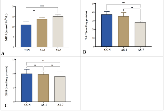
Results showed a significant reduction in TAC level versus a clear raise in MDA content in the hepatic tissues of AS-1 and AS-7 animals when compared to CON rats according to a time-based manner. However, rats exposed to arsenic led to an increase in antioxidant GSH activity compared to control animals but without significant difference as illustrated in Figure 1.
Toxicity of arsenic on serum enzymes levels
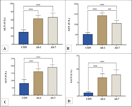
The effect of arsenic on serological level of liver enzymes of intoxicated rats (AS-1 and AS-7) and their comparison with control rats is displayed in Figure 2. The findings demonstrated that, when compared with the control (CON) rats, arsenic exposure considerably raised (p<0.0001) the serological levels of AST, ALT, ALP, and GGT in arsenic treated rats. On the other hand, it was observed that the rats inebriated hepatic enzymes (ALT, ALP, and GGT) increased gradually over time. Otherwise, AST levels in the AS-1 and AS-7 animals showed a significant difference.
Toxicity of arsenic on serum cytokine levels
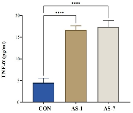
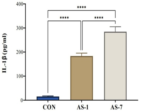
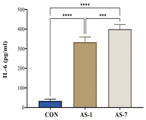
According to results, AS-treated groups showed significantly increased levels of serum cytokines TNF-α, IL-1β, and IL-6 (p<0.0001) in time-related manner on the 7th and 1st day after exposure (respectively) in comparison with the control animals, as presented in Figures 3, 4, 5.
Arsenic is an environmental toxicant that negatively impacts several organ systems in the body, particularly the liver and immune system. In general, arsenic absorption from the body is followed by metabolized to toxic forms, converting to mono-, di-, and methylated pentavalent products. Moreover, it has the property of accumulating in some organs and tissues including the liver
; . The liver is the principal organ for heavy metal intoxication, because the majority of the absorbed exogenous toxicant is trapped in the liver and eventually transferred into other organs ; . In general, accumulation of any toxicant in body depends on route, dose and duration of exposure . To evaluate the hepatotoxic effect of arsenic in exposed rats, hepatic oxidative stress indicators were determined. Many pathological conditions are intrinsically linked to oxidative stress and its consequences, including lipid peroxidation, which is determined by malondialdehyde ; . This is supported by the findings of the current experiment, which show an important rise in its level in liver tissue. It is a reactive aldehyde formed during the peroxidation of polyunsaturated fatty acids . One of the severe effects of oxidative stress is cell death by necrosis or apoptosis, including hepatocytes . Total antioxidant capacity, was significantly reduced in the livers of animals exposed to arsenic, which represents the total non-enzymatic antioxidant capacity and includes the synergistic effects of all antioxidants in liver tissue . Antioxidants neutralize reactive oxygen species, which upset the cellular redox balance, thereby protecting them from oxidative damage. Therefore, when antioxidants level decreases, it causes damage at different molecular levels, increasing the risk of pathological damage such as inflammation ; .
Glutathione is the most abundant intracellular thiol and a critical antioxidant in hepatocytes. It detoxifies ROS and free radicals, protecting the liver from oxidative damage . It conjugates with toxic compounds through the action of glutathione S-transferase (GST), facilitating their excretion. This is particularly important in detoxifying drugs and other xenobiotics . Rats administered sodium arsenite had significantly higher serum concentrations of hepatic enzymes than control rats. This demonstrated its capacity to increase cell membrane permeability and promote liver cell destruction, resulting in liver dysfunction and the release of liver enzymes into the bloodstream ; . In addition, results confirmed the ability of sodium arsenite to cause a rapid inflammatory response in rats after 1 and 7 days of exposure by disturbing the levels of serum cytokines in comparison to non-exposed animals. An important rise in cytokine serum levels suggested an inflammatory response to arsenic exposure. The pro-inflammatory cytokines IL-1 and TNF induce inflammation by increasing the expression of adhesive molecules on endothelial cells, consequently causing leukocytes in circulation to bind to the endothelial . Increased levels of IL-6 and TNF-α disrupt the internal and external homeostasis of these molecules, leading probable cellular death, restricting enzymes and mitochondrial activities, and thus accelerating the induction of various stress genes ; . This experiment’s results are in line with previous studies that has proven that AS is an immunosuppressive agent and exposure to it at non-permissible doses has resulted in a variety of cellular and molecular immune-related disorders ; . It is a potent immunotoxicant that affects the release of cytokines in time and dose-dependent manners . Lipid peroxidation products and pro-inflammatory cytokines were found to positively correlate in arsenic-exposed mice, according to a prior study by Afolabi et al. (2013). Other investigations by Lasram et al. (2014) and Abu-Zeid et al. (2021) found that decrease of antioxidant defense enzymes was linked to apoptosis, immunotoxicity, and liver damage. The ability to activate inflammatory cytokines has been linked to immune-related disorders in humans and rodents exposed to arsenic .
Conclusions
This experimental study confirmed the hepatotoxicity and immunotoxicity of arsenic in a rat model through acute inflammatory response and induction of serum pro-inflammatory cytokines in a time-dependent progression.
Acknowledgements
We express my appreciation to our colleagues who provided us with a stimulating academic environment.
NOVELTY STATEMENT
It represents a new topic that aims to evaluate the levels of serum pro-inflammatory cytokines in acute and subacute hepatotoxicity in vivo.
AUTHOR’S CONTRIBUTION
The research was equally involved in the efforts to complete the experiment.
Conflict of interest
The authors have declared no conflict of interest.
Figures
Figure 1:.
Toxicity of arsenic on oxidative stress indicators in experiment groups (n = 6): A) MDA, B) TAC, and C) GSH. ns: indicates not significant, while symbol* indicate significant as follows: **: at (p < 0.01), ***: at (p < 0.001) ****: at (p<0.0001).
Figure 2:.
Toxicity of arsenic on hepatic enzymes levels: A) ALT, B) AST, C) ALP, and D) GGT in experiment groups (n= 6). symbol* indicate significant as follows: ***: at (p < 0.001) ****: at (p<0.0001).
Figure 3:.
Toxicity of arsenic on serum TNF-α level in experiment rats (n=6/group). ****: significant at (p<0.0001).
Figure 4:.
Toxicity of arsenic on serum IL-1β in experiment rats (n =6 /group). ****: significant at (p<0.0001).
Figure 5:.
Toxicity of arsenic on serum IL-6 in experiment rats (n=6 /group). ***: significant at (p < 0.001) ****: significant at (p<0.0001).
References
Abou-Zeid SM, Aljuaydi SH, AbuBakr HO, Tahoun EA, Di Cerbo A, Alagawany, Farag MR (2021). Astaxanthin mitigates thiacloprid-induced liver injury and immunotoxicity in male rats. Mar. Drugs, 19(9): 525. https://doi.org/10.3390/md19090525[CrossRef]
Afolabi OK, Oyewo EB, Adekunle AS (2013). Assessment of lipid peroxidation markers and proinflammatory cytokines in arsenite-exposed rats. Asian J. Natl. Appl. Sci., 2(2): 8-18.
Ali MM, Hossain D, Al-Imran A, Khan MS, Begum M, Osman MH (2021). Environmental pollution with heavy metals: A public health concern. Heavy metals their environmental impacts and mitigation, pp. 771-783.
Allameh A, Niayesh-Mehr R, Aliarab A, Sebastiani G, Pantopoulos K (2023). Oxidative stress in liver pathophysiology and disease. Antioxidants, 12(9): 1653. https://doi.org/10.3390/antiox12091653[CrossRef]
Barouki R, Samson M, Blanc EB, Colombo M, Zucman-Rossi J, Lazaridis KN, Coumoul X (2023). The exposome and liver disease-how environmental factors affect liver health. J. Hepatol., 79(2): 492-505. https://doi.org/10.1016/j.jhep.2023.02.034[CrossRef]
Bellamri N, Morzadec C, Fardel O, Vernhet L (2018). Arsenic and the immune system. Curr. Opin. Toxicol., 10: 60-68. https://doi.org/10.1016/j.cotox.2018.01.003[CrossRef]
Chen XL, Wang Y, Peng WW, Zheng YJ, Zhang TN, Wang, Zeng QY (2018). Effects of interleukin-6 and IL-6/AMPK signaling pathway on mitochondrial biogenesis and astrocytes viability under experimental septic condition. Int. Immunopharmacol., 59: 287-294. https://doi.org/10.1016/j.intimp.2018.04.020[CrossRef]
Cordiano R, Di Gioacchino M, Mangifesta R, Panzera C, Gangemi S, Minciullo PL (2023). Malondialdehyde as a potential oxidative stress marker for allergy-oriented diseases: An update. Molecules, 28(16): 5979. https://doi.org/10.3390/molecules28165979[CrossRef]
Dangleben NL, Skibola CF, Smith MT (2013). Arsenic immunotoxicity: A review. Environ. Hlth., 12: 1-15. https://doi.org/10.1186/1476-069X-12-73[CrossRef]
Dong L, Liu Y, Wang D, Zhu K, Zou Z, Zhang A (2021). Imbalanced inflammatory response in subchronic arsenic-induced liver injury and the protective effects of Ginkgo biloba extract in rats: Potential role of cytokines mediated cell–cell interactions. Environ. Toxicol., 36(10): 2073-2092. https://doi.org/10.1002/tox.23324[CrossRef]
Donne R, Saroul-Aïnama M, Cordier P, Celton-Morizur S, Desdouets C (2020). Polyploidy in liver development, homeostasis and disease. Nature reviews Gastroenterol. Hepatol., 17(7): 391-405. https://doi.org/10.1038/s41575-020-0284-x[CrossRef]
Duvigneau JC, Luís A, Gorman AM, Samali A, Kaltenecker D, Moriggl R, Kozlov AV (2019). Crosstalk between inflammatory mediators and endoplasmic reticulum stress in liver diseases. Cytokine, 124: 154577. https://doi.org/10.1016/j.cyto.2018.10.018[CrossRef]
Garkal A, Sarode L, Bangar P, Mehta T, Singh DP, Rawal R (2023). Understanding arsenic toxicity: Implications for environmental exposure and human health. J. Hazard. Mater. Lett., 2023: 100090. https://doi.org/10.1016/j.hazl.2023.100090[CrossRef]
Georgiou-Siafis SK, Tsiftsoglou AS (2023). The key role of GSH in keeping the redox balance in mammalian cells: mechanisms and significance of GSH in detoxification via formation of conjugates. Antioxidants, 12(11): 1953. https://doi.org/10.3390/antiox12111953[CrossRef]
Ghareeb OA (2021). Toxicopathological effects of zinc oxide nanoparticles on the liver function and preventive role of silymarin in vivo. Indian J. Forensic Med. Toxicol., 15(2): 3212-3217. https://doi.org/10.37506/ijfmt.v15i2.14863[CrossRef]
Ghareeb OA (2023). Adverse impact of titanium dioxide nanoparticles on hepato-renal functions and improved role of Rosmarinus officinalis. J. Natl. Sci. Biol. Med., 14(1): 33-38.
Ghareeb OA, Ali QA (2023). Pathotoxic impact of zinc oxide nanoparticles on liver function and protective role of Silymarin. Curr. Innov. Dis. Health Res., 3: 153-161. https://doi.org/10.9734/bpi/cidhr/v3/10703F[CrossRef]
Ghareeb OA, Ali QA (2024). Hepatotoxicity induced by some metal nanoparticles in vivo. Glob. Sci. Rev., 23: 25-33.
Ghareeb OA, Ali QA (2024). Pathological disorders caused by atmospheric nanoparticles. Peerian J., 26: 44-51.
Ghareeb OA, Ali QA, Ramadhan SA, Sultan AI (2024). Concepts of one health approach and achieving sustainable development. Eur. J. Pharma. Med. Res., 11(4): 383-389.
Ghosh D, Ghosh S, Sarkar S, Ghosh A, Das N, Saha KD, Mandal AK (2010). Quercetin in vesicular delivery systems: Evaluation in combating arsenic-induced acute liver toxicity associated gene expression in rat model. Chem. Biol. Interact., 186(1): 61-71. https://doi.org/10.1016/j.cbi.2010.03.048[CrossRef]
Giles BH, Mann KK (2022). Arsenic as an immunotoxicant. Toxicol. Appl. Pharmacol., 454: 116248. https://doi.org/10.1016/j.taap.2022.116248[CrossRef]
Gripshover TC, Wahlang B, Head KZ, Young JL, Luo J, Mustafa MT, Cave MC (2023). The environmental pollutant, polychlorinated biphenyl 126, alters liver function in a rodent model of alcohol-associated liver disease. Alcohol Clin. Exp. Res., 47(1): 60-75. https://doi.org/10.1111/acer.14976[CrossRef]
Gupta PK, Gupta PK (2020). Target organ toxicity. Problem solving questions in toxicology: A study guide for the board and other examinations, pp. 83-117. https://doi.org/10.1007/978-3-030-50409-0_7[CrossRef]
Islam LN (2023). An update on the immunotoxic effects of arsenic exposure. In: Handbook of arsenic toxicology. pp. 551-592. https://doi.org/10.1016/B978-0-323-89847-8.00034-1[CrossRef]
Kalas MA, Chavez L, Leon M, Taweesedt PT, Surani S (2021). Abnormal liver enzymes: A review for clinicians. World J. Hepatol., 13(11): 1688. https://doi.org/10.4254/wjh.v13.i11.1688[CrossRef]
Karak P (2022). Arsenic contamination and its impact on the human. Curr. World Environ., 17(1): 58. https://doi.org/10.12944/CWE.17.1.6[CrossRef]
Lasram MM, Lamine AJ, Dhouib IB, Bouzid K, Annabi A, Belhadjhmida N, Gharbi N (2014). Antioxidant and anti-inflammatory effects of N-acetylcysteine against malathion-induced liver damages and immunotoxicity in rats. Life Sci., 107(1-2): 50-58. https://doi.org/10.1016/j.lfs.2014.04.033[CrossRef]
Li W, Jiang X, Qian H, Li X, Su J, Zhang G, Li X (2023). Associations of arsenic exposure with liver injury in US adults: NHANES 2003–2018. Environ. Sci. Pollut. Res., 30(16): 48260-48269. https://doi.org/10.1007/s11356-023-25540-5[CrossRef]
Malik MS, Azam M, Basra MAR (2020). Impact of natural antioxidants on biological systems. Life Sci., 4: 139-162. https://doi.org/10.54692/lgujls.2019.0402105[CrossRef]
Mas-Bargues C, Escriva C, Dromant M, Borras C, Vina J (2021). Lipid peroxidation as measured by chromatographic determination of malondialdehyde. Human plasma reference values in health and disease. Arch. Biochem. Biophys., 709: 108941. https://doi.org/10.1016/j.abb.2021.108941[CrossRef]
Muzamil A, Tahir HM, Ali S, Liaqat I, Ali A, Summer M (2021). Inflammatory process and role of cytokines in inflammation: An overview. Punjab Univ. J. Zool., 36(10): 17582. https://doi.org/10.17582/journal.pujz/2021.36.2.237.252[Full text][CrossRef]
Pánico P, Velasco M, Salazar AM, Picones A, Ortiz-Huidobro RI, Guerrero-Palomo G, Hiriart M (2022). Is arsenic exposure a risk factor for metabolic syndrome? A review of the potential mechanisms. Front. Endocrinol., 13: 878280. https://doi.org/10.3389/fendo.2022.878280[CrossRef]
Pisoschi AM, Pop A, Iordache F, Stanca L, Predoi G, Serban AI (2021). Oxidative stress mitigation by antioxidants. An overview on their chemistry and influences on health status. Eur. J. Med. Chem., 209: 112891. https://doi.org/10.1016/j.ejmech.2020.112891[CrossRef]
Prakash S, Verma AK (2021). Arsenic: It’s toxicity and impact on human health. Int. J. Biol. Innov., 3(1): 38-47. https://doi.org/10.46505/IJBI.2021.3102[CrossRef]
Raju NJ (2022). Arsenic in the geo-environment: A review of sources, geochemical processes, toxicity and removal technologies. Environ. Res., 203: 111782. https://doi.org/10.1016/j.envres.2021.111782[CrossRef]
Ramadhan SA, Ghareeb OA (2021). Toxicity of AgNPs upon liver function and positive role of Tinospora cordifolia: In vivo. Pak. J. Med. Health Sci., 15(6): 2164-2166. https://doi.org/10.53350/pjmhs211562164[CrossRef]
Ramadhan SA, Ghareeb OA (2022). Efficiency of cichorium intybus in reducing hepatotoxicity induced by zinc oxide nanoparticles. Ann. Med. Health Sci. Res., 12(3): 93-96.
Renu K, Mukherjee AG, Gopalakrishnan AV, Wanjari UR, Kannampuzha S, Murali R, Madhyastha H (2023). Protective effects of macromolecular polyphenols, metal (zinc, selenium, and copper)-Polyphenol complexes, and pectin in different organs with an emphasis on arsenic poisoning: A review. Int. J. Biol. Macromol., pp. 126715. https://doi.org/10.1016/j.ijbiomac.2023.126715[CrossRef]
Souza-Arroyo V, Fabián JJ, Bucio-Ortiz L, Miranda-Labra RU, Gomez-Quiroz LE, Gutiérrez-Ruiz MC (2022). The mechanism of the cadmium-induced toxicity and cellular response in the liver. Toxicology, 480: 153339. https://doi.org/10.1016/j.tox.2022.153339[CrossRef]
Sutaria SR, Gori SS, Morris JD, Xie Z, Fu XA, Nantz MH (2022). Lipid peroxidation produces a diverse mixture of saturated and unsaturated aldehydes in exhaled breath that can serve as biomarkers of lung cancer. A review. Metabolites, 12(6): 561. https://doi.org/10.3390/metabo12060561[CrossRef]
Taher GN, Ghareeb OA (2022). Assessment of hepatic biochemical indices in patients with acute cholecystitis. Eur. Med. Res. Period., 13: 94-100.
Taher GN, Abdulla GM, Ghareeb OA (2024). Evaluation of some biochemical indicators in patients with gallstones. World J. Pharma. Med. Res., 10(7): 21-25.
Vašková J, Kočan L, Vaško L, Perjési P (2023). Glutathione-related enzymes and proteins: A review. Molecules, 28(3): 1447. https://doi.org/10.3390/molecules28031447[CrossRef]