Research Article · Volume 10, Issue 8, 14 July 2022 · Open Access
Dermatophytosis in Clinically Infected Cats: Diagnoses and Efficacy Therapy
· · ·
Affiliations & Notes ⌄
1Doctoral Student of Veterinary Science, Faculty of Veterinary Medicine, Universitas Gadjah Mada, Yogyakarta 55281
2Department of Internal Medicine, Faculty of Veterinary Medicine, Universitas Gadjah Mada, Yogyakarta, 55281
3Department of Microbiology, Faculty of Veterinary Medicine, Universitas Gadjah Mada, Yogyakarta, 55281
4Department of Pathology, Faculty of Veterinary Medicine, Universitas Gadjah Mada, Yogyakarta 55281. *Correspondence | Sitarina Widyarini, Department of Pathology, Faculty of Veterinary Medicine, Universitas Gadjah Mada, Yogyakarta 55281
Based on these characteristics, it can be concluded that the cat is infected with Microsporum canis.
It is shown that the use of 2% Ketoconazole cream for cases of dermatophytosis in cats may reduce clinical lesions up to 85%.
The main lesions of dermatophytosis in cats are alopecia, scale, and erythema.
Dermatophyte infections in cats in Yogyakarta region were dominated by M. canis species and topical application of 2% ketoconazole cream for 3 weeks was effective in reducing lesions caused by dermatophyte infections in cats.
Selected from the article's own text.
Abstract
Dermatophytosis is a zoonotic disease caused by fungi of species dermatophyte. The case of dermatophytosis have increased not only in humans but also in animals, especially dogs and cats. The close interaction of cats and humans reported to play an important role in dissemination of zoonotic diseases. Therefore, this study aims to diagnose and to treat of dermatophytosis in cats. Twenty cats with the age of 6-12 months were used in this study. Determination of dermatophyte infection in cats based on clinical signs and laboratory identification of fungi. Skin scrapes samples were cultured onto Sabouraud’s dextrose agar medium. Hair and skin scraping were conducted at the day-1 (pre-treated with ketoconazole). Subsequently, infected cats were treated with topical application of 2% ketoconazole cream, twice a day for 21 days. Results showed that all of cats positive infected with Microsporum canis. Feature of the skin of cats infected by Microsporum canis demonstrates erythema, scale, crust, and circular alopecia. At day-21, antifungal treatment not only diminished by approximately 85% of the clinical feature of the skin lesion of cat’s dermatophytosis but also resulted in the disappearance of the fungi. To conclude, topically applied of antifungal ketoconazole for at least three weeks effective to treat dermatophytosis in cats.
Dermatophytosis (Ringworm) is an important and highly contagious fungal infection caused by dermatophytes. Though of limited morbidity, ringworm can be problematic because of the potential for outbreaks, prolonged disease, cost and bother of treatment, and the potential zoonosis . The rising prevalence of dermatophytosis has been attributed to many factors including tropical climate, a high population of pet stray, and high contact between pet animals and humans ; ; . Zoophilic dermatophytes are transmitted by direct contact with infected animals or contact with arthroconidia (spores) in the environment ; ; . Infection occurs more often in the presence of predisposing factors (e.g., stress, overcrowding, feed and management deficiencies, pregnancy, immunodeficiency, age, ectoparasites, and other diseases ; ; .
Dermatophytosis is more common in cats than in dogs . The dermatophyte invading skin, hair, nail lead to the formation of erythema, scale, crusting, circular alopecia, and hyperpigmentation ; . Microsporum canis infection in young, old, and immunodeficiency animals usually cause inflammation or infection . Infections with Microsporum gypseum and Tricophyton mentagrophytes resulted in more serious dermatologic disease ; ; . Treatment for dermatophytosis given topically or systemically, depend on the type and distribution of lesions . The incidence of dermatophytosis in cats in Yogyakarta, clinically, may often be encountered by veterinarians’ practice, but reports of dermatophytosis are still very minimal. This results in diagnostic and therapeutic measures for patients not being given optimally. Based on these circumstances, the purpose of this research is to diagnose dermatophytosis by isolating and identifying the presence of dermatophytes as the cause of dermatophytosis in cats in the Yogyakarta region and to assess the efficacy of antifungal therapy in cats infected with dermatophyte.
Materials and Methods
Ethical approval
The present study was perfomed under approvement of the Ethiological Committee of Integrated Research and Testing Laboratory, Universitas Gadjah Mada No. 00057/04/LPPT/X/2019.
Research place and time
This study was conducted by using data from patients coming to pet clinics in Yogyakarta within the period of December 2019 to August 2021. Fungi isolation and identification were carried out in Microbiology Laboratory, Faculty of Veterinary Medicine, Universitas Gadjah Mada, Yogyakarta.
Experimental design
A number of 20 cats ranging from 6-12 months old were used for this study regardless of their breeds and sex. Cats suspected of having dermatophyte infection based on clinical lesions were examined using Wood’s lamp at the wavelength of 395 nm and direct microscopic examination on skin and hair samples of affected hairs, which are then placed onto a glass slide for microscopic evaluation is used for evidence of ectothrix.
Fungal isolation and identification cultured on Sabouraud’s Dextrose Agar (65g/L) media. All inoculated SDA plates were then incubated at 25–30°C for 4–6 weeks. The cultures of fungus were identified by examining the characteristics of their colony. Macroscopic examination by identifying the growth rate, texture, topography, and pigmentation of the reverse and front sides of the culture of fungi. Microscopic examination was carried out on the positive observed fungal culture using lactophenol cotton blue (LPCB) staining. In addition, the manufacture of slide cultures using the Riddle method was carried out to observe the microscopic structure of the fungal as a whole.
Confirmed positive cats were given antifungal treatment (2% Ketoconazole cream) for 21 days. Observations were carried out on the progress of cats’ skin lesions throughout the treatment regimen. Observed lesions in question were erythema, scales, crusts, and alopecia.
Results and Discussion
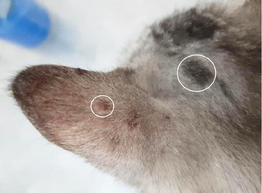
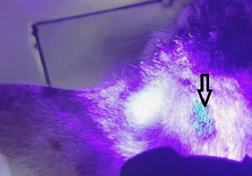
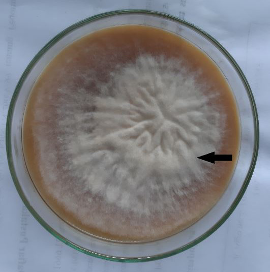
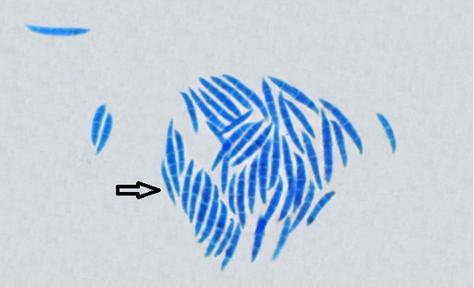
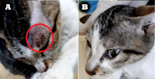
A total of 20 cats suspected of dermatophyte infection are shown to have clinical signs of alopecia ring, scale, and erythema with various degrees of severity (Figure 1). Wood’s lamp examination indicates (100%) positive results on all suspected cats with blue-green glow on skin lesions (Figure 2). Hyphae segments are also found in hair and skin scrapings examination under the microscope. The results of isolation and identification showed that they grew efficiently on Sabouraud’s dextrose agar. The macroscopic characteristics that can be observed are that they look like cotton, white in color, and become brownish-yellow with age (Figure 3). The results of the microscopic examination showed the presence of abundant macroaleuriospora, large, 6-15 cells, spindle-shaped, with curved or hooked ends and thick verrucose walls (Figure 4). Based on these characteristics, it can be concluded that the cat is infected with Microsporum canis. Macroscopic lesions progress (decrease in severity) on the last day (day 21st) of treatment using 2% Ketoconazole cream can be observed as shown in Figure 5. It is shown that the use of 2% Ketoconazole cream for cases of dermatophytosis in cats may reduce clinical lesions up to 85%.
It can be inferred that all 20 cats are infected with dermatophytes from examinations as shown in research results. Wood’s lamp examination will result in fluorescence on all dermatophytosis clinical lesions. Previous research explained that several dermatophyte species are known to produce phosphor as a by product of their development on hair and/or skin while some other fungi don’t have that quality. Thus, not all dermatophytes are able to exhibit fluorescence under wood’s lamp ; ; . Dermatophytes known for the ability to produce fluorescence are from the genus Microsporum . False positive results are often found in examinations using Wood’s lamp, so it becomes crucial to differentiate fluorescence from hair shaft or other things like crust, debris, or topical medication . Some researchers also recommend to do direct examination and fungal culture following a positive result by Wood’s lamp screening ; ; .
The results of microscopic examination of hair and skin scrapings shows that 20 cats in the study were positively infected with dermatophytes based on the discovery of hyphae segments (ectothrix). Watanabe et al. (2021) explained that direct microscopic examination of hair and skin scrapings is an early diagnosis method for dermatophytosis to detect the presence of arthroconidia and/or hyphae. The results of fungal culture in this study indicate that dermatophytosis cases in cats in Yogyakarta are dominated by M. canis species. The results of this study are similar to the findings of previous researchers who explained that three most common dermatophyte species infecting cats and dogs are M. canis, M. gypseum, and T. mentagrophytes ; ; ; ; . Research results by Moriello and Deboer (2012) and Koussidou-Eremondi et al. (2005) also showed that M. canis is a dermatophyte species that is commonly found in cats and is known to be the main dermatophyte found in humans.
Overall, dermatophytosis lesions found on the cats are round (circular) with varying sizes in each individual. This is similar to research results of da Cunha et al. (2019) which stated that the main clinical symptoms of dermatophyte infection in cats and dogs are alopecia, erythema, and scale with varying degrees of lesion severity. Moriello et al. (2017) and Bianchi et al. (2017) also stated that dermatophyte infection will manifest as superficial clinical signs, starting with the presence of erythema then followed by the appearance of scales and crusts, then alopecia. Paryuni et al. (2020) mentioned that dermatophytes are known to be unable to grow in the center of the lesion (an area that is experiencing inflammation), so the infection will spread centrifugally to the periphery of the lesion resulting in round lesions such as rings or ringworms.
Topical treatment of 2% Ketoconazole cream given for cases of cat dermatophytosis in this study brings out good outcomes based on the reduction of skin lesions. This is in accordance with the results of previous studies explaining that topical treatment for dermatophyte infections is known to prevent re-infection and is effective for carrier cats . A previous study by Putriningsih and Soedarmanto (2013) also reported that following topical administration of ketoconazole resulted in reducing the severity of clinical and pathological lesions in dogs infected with M. canis at day 21 of therapy. Indarjulianto et al. (2020) reported oral combination of antifungal ketoconazole for 35 days and itraconazole for 20 days respectively to cure dermatophytosis in cat. The side effects of giving this medication are also reported to be milder compared to peroral treatments . Ketoconazole is an azole antifungal known for its high effectiveness against dermatophytes . The mechanism of action of this drug is to inhibit cytochrome P450 14α-lanosterol demethylase . Ketoconazole treatment for 2-4 weeks is known to be effective in curing mild dermatophyte infections .
Conclusions and Recommendation
The main lesions of dermatophytosis in cats are alopecia, scale, and erythema. The clinical appearance of skin lesions cannot be relied upon as the sole criterion for diagnosing dermatophytosis. A Wood’s lamp examination can be helpful in some cases. Microscopic examination of hairs taken from suspected patients may be visualized for evidence of fungal hyphae or spores. Fungal culture on SDA media is a more sensitive diagnostic tool. Dermatophyte infections in cats in Yogyakarta region were dominated by M. canis species and topical application of 2% ketoconazole cream for 3 weeks was effective in reducing lesions caused by dermatophyte infections in cats.
Acknowledgements
Financial support was provided by PMDSU scholarship from KEMENRISTEK DIKTI NO. 3136/UNI/DITLIT/DIT-LIT/PT/2020 BATCH IV.
Novelty Statement
The prevalence of dermatophytosis in cats in Yogyakarta region is high, but reports on this case are still very limited. Therefore, in this study, we want to identify dermatophytosis in cats in Yogyakarta region and see the efficacy of antifungal Ketoconazole topical in cats infected with dermatophytes. Microsporum canis is the major species that infects cats in Yogyakarta region. The single therapy of antifungal ketoconazole topical has an effective effect against clinical features of Microsporum canis infection in cats.
Author’s Contribution
SW and SI: conceptualised the study, SW: drafted the experimental design, prepared, and edited the manuscript according to the title. TU and ADP: collected the data experiment and literature, edited the manuscript, and finalised the manuscript.
All authors read and approved the final manuscript.
Conflict of interest
The author have declared no conflict of interest.
Figures
Figure 1:.
Lesions from dermatophyte infection in body part of cat with alopecia, erythema, and scale in cat dermatophytosis (white ring).
Figure 2:.
Ear of cat with fluorescence (apple blue-green color), under Wood’s lamp examination (black arrow).
Figure 3:.
Fungal colony of Microsporum canis; Cottony, white to buff in colour; with increasing age becomes orange-brown (black arrow).
Figure 4:.
Microscopic structure of Microsporum canis with Lactophenol cotton blue stain (black arrow).
Figure 5:.
Macroscopic lesions progress (decrease in severity) on treatment with 2% Ketoconazole cream: A. Before treatment (red circle), B. The last day (day 21st) of treatment.
References
Asawanonda P, Taylor CR (1999). Wood’s light in dermatology. Int. J. Dermatol., 38: 801–807. https://doi.org/10.1046/j.1365-4362.1999.00794.x[CrossRef]
Behzadi P, Ranjbar R, Behzadi E (2014). Dermatophyte fungi: Infectious, diagnosis and treatment. SMU Med. J., 1: 50-61.
Bseiso EA, Nasr M, Sammour O, El-Gawad NAA (2015). Recent advances in topical formulation carriers of antifungal agents. Indian J. Dermatol., 81(5): 457-463. https://doi.org/10.4103/0378-6323.162328[CrossRef]
Colombo S, Scarampella F, Ordeix L (2012). Dermatophytosis and papular eosinophilic/mastocytic dermatitis (urticaria pigmentosa like dermatitis) in three Devon Rex cats. J. Feline Med. Surg., 14: 498–502. https://doi.org/10.1177/1098612X12440761[CrossRef]
Deboer DJ, Moriello KA (2006). Cutaneous fungal infections. In: Infectious disease of the dog and cat (ed. C.E. Greene). 3rd ed. St. Louis, Mo: Saunders/Elsevier. pp. 530-538.
Dong C, Angus J, Scarampella F (2016). Evaluation of dermoscopy in the diagnosis of naturally occurring dermatophytosis in cats. Vet. Dermatol., 27: 275–e265. https://doi.org/10.1111/vde.12333[CrossRef]
da Cunha MM, Capote-Bonato F, Capoci IRG, Bonato DV, Ghizzi LG, Paiva-Lima P, Baeza LC and Svidzinski TIE (2019). Epidemiological investigation and molecular typing of dermatophytosis caused by Microsporum canis in dogs and cats. PVM. 167: 39-45.
Fehr M (2015). Zoonotic potential of dermatophytosis in small mammals. J. Exotic. Pet. Med., 24: 308-316. https://doi.org/10.1053/j.jepm.2015.06.015[CrossRef]
Grable S (2019). Small animal dermatology for technicians and nurse. Wiley-Blackwell. pp. 65-84. https://doi.org/10.1002/9781119108641.ch5[CrossRef]
Graser Y, Monod M, Bouchara JP, Dukik K, Nenoff P, Kargl A (2018). New insights in dermatophyte research. Med. Mycol., 56: 2–9. https://doi.org/10.1093/mmy/myx141[CrossRef]
Gupta AK, Cooper EA (2008). Update in antifungal therapy of dermatophytosis. Mycopathologia, 166: 353-367. https://doi.org/10.1007/s11046-008-9109-0[CrossRef]
Haggag YN, Samaha HA, Nossair MA, Mohammad RMH (2017). Prevalence of dermatophytosis in some animals and human in Bahera Province, Egypt. Alex. J. Vet. Sci., 53(2): 64-71. https://doi.org/10.5455/ajvs.203688[CrossRef]
Indarjulianto S, Yanuartono, Purnamaningsih H, Wikansari P, Sakan GYI (2014). Isolation and identification of Microsporum canis from dermatophytosis dogs in Yogyakarta. J. Vet., 15(2): 212-216.
Indarjulianto S, Yanuartono, Widyarini S, Raharjo S, Purnamaningsih H, Nururrozi A, Hariwibowo N, Jainudin HA (2017). Microsporum canis infection in dermatitis cats. J. Vet., 18(2): 207-210. https://doi.org/10.19087/jveteriner.2017.18.2.207[CrossRef]
Koussidou-Eremondi T, Devliotou-Panagiotidou D, Mourellou-Tsatsou O, Minas A (2005). Epidemiology of dermatomycoses in children living in Northern Greece 1996-2000. Blackwell Publ. Ltd. Mycoses, 48: 11-16. https://doi.org/10.1111/j.1439-0507.2004.01067.x[CrossRef]
Lopes AI, Tavaria FK, Pintado ME (2017). Application of chitosan in the control of fungal infections by dermatophytes. Ann. Appl. Microbiol. Biotechnol. J., 1: 1006–1011. https://doi.org/10.36876/aamb.1006[CrossRef]
Marshall KL (2003). Fungal diseases in small mammals: Thera-peutic trends and zoonotic consideration. Vet. Clin. North. Am. Exotic. Anim. Pract., 6: 415-427. https://doi.org/10.1016/S1094-9194(03)00002-1[CrossRef]
Miller WH, Griffin CE, Campbell KL (2013). Fungal and algal skin diseases. In: Muller and Kirk’s Small Animal Dermatology, 7e (eds. W. Miller, C. Griffin and K. Campbell), St. Louis: Elsevier Mosby. pp. 223–283.
Moriello, KA, Stuntebeck R and Mullen L (2019). Trycophyton species and Microsporum gypseum infection and fomite carriage in cats from there animal shelter: a retrospective case series. J. Fel. Med. Surg. 1-4.
Moriello KA (2001). Diagnostic techniques for dermatophytosis. Clin. Tech. In. Small. Anim. Pract., 16(4): 219-224. https://doi.org/10.1053/svms.2001.27597[CrossRef]
Moriello KA (2014). Feline dermatophytosis aspects pertinent to disease management in single and multiple cat situations. J Feline Med. Surg., 16: 419-431. https://doi.org/10.1177/1098612X14530215[CrossRef]
Moriello KA, DeBoer DJ (2012). Dermatophytosis. In: Infectious diseases of the dog and cat, 4e (ed. C.E. Greene), St. Louis: Elsevier Saunders. pp. 558–602.
Moriello KA, Coyner K, Peterson S, Mignon B (2017). Diagnosis and treatment of dermatophytosis in dogs and cats: clinical consensud guidelines of the world association for veterinary dermatology. Vet. Dermatol., 28: 266-268. https://doi.org/10.1111/vde.12440[CrossRef]
Paryuni AD, Indarjulianto S, Widyarini S (2020). Dermatophytosis in companion animals: A review. Vet. World, 13(22): 1174-1181. https://doi.org/10.14202/vetworld.2020.1174-1181[CrossRef]
Pathania S, Rudramurthy SM, Narang T, Saikia UM, Dogra S (2018). A prospective study of the epidemiological and clinical patterns of recurrent dermatophytosis at a tertiary care hospital in India. Indian J. Dermatol. Ven. Lepro., 84(6): 678-684. https://doi.org/10.4103/ijdvl.IJDVL_645_17[CrossRef]
Putriningsih PAS and Soedarmanto I (2013). Clinical and pathological evaluation of Microsporum canis infection in dogs treated with ketoconazole, griseofulvin, and a combination of both. in: Thesis, Repository Universitas Gadjah Mada, Indonesia. https://repository.ugm.ac.id/id/eprint/121021
Rotta I, Ziegelmann PK, Otuki MF, Riveros BS, Bernardo NLMC, Correr CJ (2013). Efficacy of topical antifungal in the treatment of dermatophytosis: A mixed-treatment comparison meta-analysis involving 14 treatments. JAMA Dermatol., 149(3): 341-349. https://doi.org/10.1001/jamadermatol.2013.1721[CrossRef]
Scott DW, Miller WH, Griffin CE (2001). Fungal skin diseases. Muller and Kirk’s small animal dermatology. 6th ed. Philadelpia, Pa: WB Saunders. pp. 339-357. https://doi.org/10.1016/B978-0-7216-7618-0.50009-2[CrossRef]
Soedarmanto I, Yanuartono, Raharjo S, Nururrozi A, Guna JCA (2020). Combination of systemic and topical treatment for feline dermatophytosis: A case report. Acta Vet. Indo., 8(1): 18-23. https://doi.org/10.29244/avi.8.1.18-23[CrossRef]
Vandis M, Knoll JS (2007). Peer-reviewed: Canine dermatophyte infection. Vet. Med., pp. 771-774.
Watanabe R, Furuta H, Ueno Y, Nukada T, Niwa H, Shinyashiki N, Kano R (2021). First isolation of Tricophyton bullosum from a horse with dermatophytosis in Japan. Med. Mycol. Case Rep., 32: 81-83. https://doi.org/10.1016/j.mmcr.2021.04.004[CrossRef]
Weese JS, Yu AA (2013). Infectious folliculitis and dermatophytossis. Vet. Clin. Equine, 29: 559-575. https://doi.org/10.1016/j.cveq.2013.09.004[CrossRef]
Yamada S, Anzawa K, Mochizuki T (2019). An epidemiological study of feline and canine dermatophytosis in Japan. Med. Mycol. J., 60: 39-44. https://doi.org/10.3314/mmj.19.001[CrossRef]