(Fig. 2) and clinically represented by dermatitis with secondary superficial pyoderma which grossly showed two pruritic circumscribed alopecic lesions with erythema and hair loss in the front of the head (Fig. 2a). Histologically the lesion was manifested by Eosinophilic dermatitis with infiltration of eosinophils in the dermal layer, keratolysis and spongiosis with swelling of epidermal cells with clear cytoplasm [Fig. 2(a1) & 2(a2)]. A clinical case of a dog suffering from traumatic dermatitis with secondary bacterial infection was recorded (Fig. 1b) histological lesions revealed Lymphocytic dermatitis with infiltration of lymphocytes in the dermal layer [Fig. 2(b1)] together with Keratolysis, acantholysis, lysis of keratin and spongiosa cell layer reflected in severely atrophied epidermis [Fig. 2(b2)]. A dog with superficial pyoderma and ulceration (Fig. 2C) denoted lymphocytic dermatitis with infiltration of lymphocytes in the dermal layer [Fig. 2(c1) & 2(c2)]. Erythematous lesions with hair loss alopecic areas were another reported clinical cases (Fig. 3), some of them reported in the right elbow (Fig 3a) and showed acanthosis with increased thickness of spongiosa cell layer and spongiosis noted by the swelling in epidermal cells with clear cytoplasm [Fig 3(a1)]. Perivascular lymphocytic dermatitis with accumulation of lymphocytes around the congested blood vessel in the dermal layer were denoted [Fig. 3(a2)]. Another oval area of non-pruritic hair loss was detected in the back (Fig. 3b), of which skin biopsy showed ulceration with deep destruction in the epidermal layer together with dermal necrosis [Fig. 3(b1)], keratolysis and acantholysis with atrophied epidermis were also reported [Fig. 3(b2)].
Figure 1: Mites and dermatophytes showing; 10 (a), (a1) and (a2) showed Demodex, (b) showed Microsporum and (b1), (b2) showed Sarcoptic mites.
Parasitic infestations were recorded in many other clinical cases (Fig. 4 & 5). Three cases of flea allergic dermatitis (FAD) wisth pruritic lesion on the left trunk was reported (Fig. 4a &4b & 5a). Microscopic examination of skin biopsies showed lymphocytic dermatitis with accumulation of lymphocytes in the dermal layer [Fig. 4(a1)], ulceration together with dermal necrosis were also reported [Fig. 4(a2) & 4(b1)], necrotic hair follicle [Fig. 4(b2)], keratolysis with complete destruction in keratin layer and spongiosis
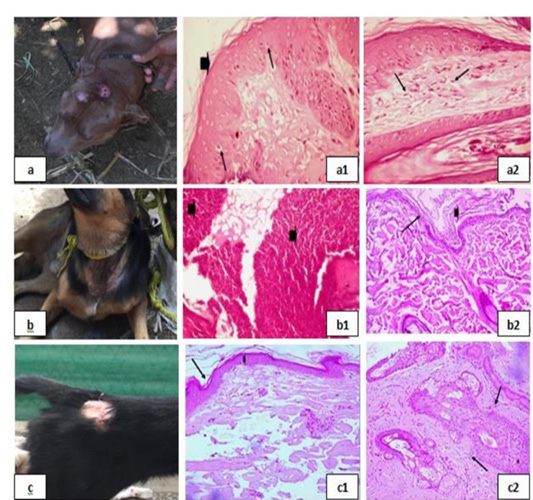
Figure 2: Bacterial infection dermopathies showing; 2a: Erythema and hair loss in the front of the head. 2(a1): keratolysis (arrow head) and spongiosis (arrows), (H&E X 200). 2(a2): Eosinophilic dermatitis with accumulation of eosinophils in the dermal layer (arrows), (H&E X 200). 2b: Traumatic dermatitis with secondary bacterial infection. 2(b1): Lymphocytic dermatitis (arrows), (H&E X 200). 2(b2): Keratolysis (arrow head) and acantholysis (arrow), (H&E X 200). 2c: Superficial pyoderma and ulcer formation. 2(c1), (c2): Lymphocytic dermatitis (arrows), (H&E X 200).
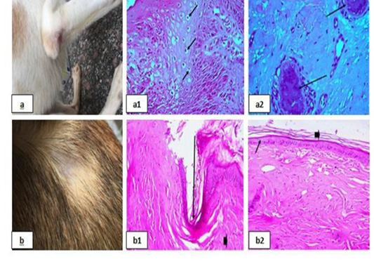
Figure 3: Erythematous lesions with hair loss alopecic areas showing; 3a: Erythematous lesions in the right elbow. 3(a1): Acanthosis and spongios (arrows), (H&E X 200). 3(a2): Perivascular Lymphocytic dermatitis (arrows), (H&E X 200). 3b: Erythematous lesions in the back. 3(b1): Ulceration (arrows), (H&E X 200). 3(b2): keratolysis (arrow head) and acantholysis (arrows), (H&E X 200).
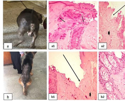
Figure 4: Parasitic infestations 1 showing; 4a, 4b: Flea allergic dermatitis (FAD) with pruritic lesion on the left trunk. 4(a1): Lymphocytic dermatitis (arrows), (H&E X 200). 4(a2), (b1): Ulceration (arrows), (H&E X 200). 4(b2): Necrotic hair follicle, (H&E X 200).
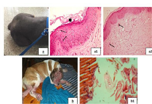
Figure 5: Parasitic infestations 2 showing; 5a: Flea allergic dermatitis (FAD) with pruritic lesion on the left trunk. 5(a1): Keratolysis (arrow head) and spongiosis (arrows), (H&E X 200). 5(a2): Eosinophilic dermatitis (arrows), (H&E X 200). 5b: Pyodemodicosis 5(b1): Demodex, (H&E X 200).
with swelling in epidermal cells and clear cytoplasm [Fig. 5(a1)], and eosinophilic dermatitis with accumulation of eosinophils in the dermal layer [Fig. 5(a2)] were recorded. A case of pyodemodicosis was recorded [Fig. 5b] and demodex was appeared by microscopic examination of biopsy [Fig. 5(b1)].
Dermatophytosis was also detected in many clinical cases (Fig. 8). Dermatophytosis with surface pyoderma and non-pruritic alopecia in the dorsal base of the neck was recorded (Fig. 8a), skin biopsy showed lymphocytic dermatitis with infiltration of lymphocytes in the dermal layer together with increased number of adipose cells [Fig. 8 (a1)]. Dermatophytosis with surface pyoderma and circumscribed non pruritic white patches was distributed on frontal region, dorsal plantar, ear pinna, and left thigh (Fig. 8b), skin biopsy showed necrotic dermatitis with sever necrosis in the dermal layer and leucocytic cells infiltrations [Fig. 8 (b1)].
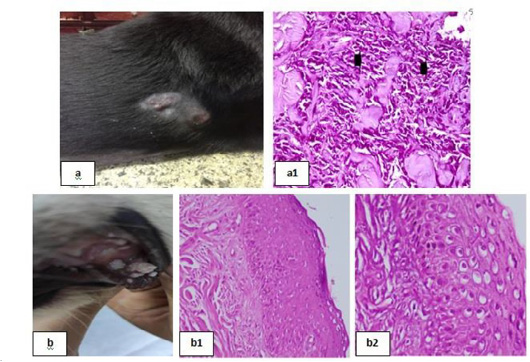
Figure 6: Skin tumors 1 showing; 6a: Soft subcutaneous lumps. 6(a1): Sarcoma cell tumor in the dermal layer with sheets of malignant cells, note the pleomorphism, hyperchromacia, increase nucleous : cytoplasmic ratio (arrow head), (H&E X 200). 6b: Cutaneous papillomatosis in the mouth 6(b1): Hyperplastic epidermis supported by fibrovascular tissue in the dermis, (H&E X 200). 6(b2): Irregular intracytoplasmic keratohyaline granules, (H&E X 200).
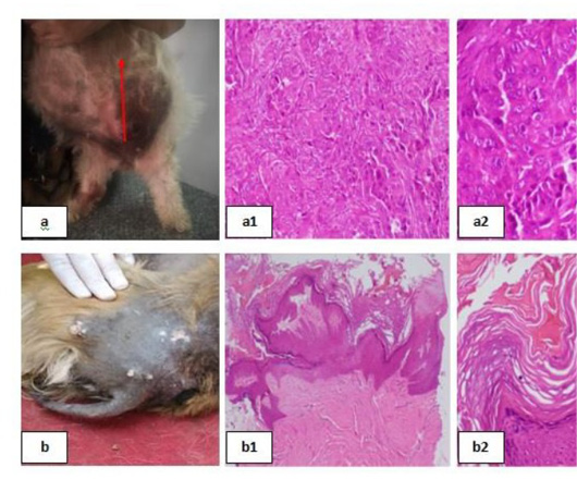
Figure 7: Skin tumors 2 showing; 7a: Mammary solid adenocarcinoma 7(a1): Carcinoma, solid type, (H&E X 200). 7(a2): Pleomorphic cells, hyperchromatic nuclei, central prominent nucleolus, and frequently detected mitosis, (H&E X 200). 7b: Canine papilloma, 7(b1): Hyperplastic epidermis , (H&E X 200). 7(b2): Prominent hyperkeratosis , (H&E X 200).
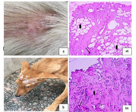
Figure 8: Dermatophytosis showing; 8a: Dermatophytosis with surface pyoderma and non-pruritic alopecia 8(a1): Lymphocytic dermatitis (arrow), with increased number of adipose cells (arrow head), (H&E X 200). 8b: Dermatophytosis with surface pyoderma and circumscribed white patches 8(b1): Necrotic dermatitis with sever necrosis in the dermal layer (arrow head) and leucocytic cells infiltrations (arrow), (H&E X 200).
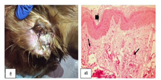
Figure 9: Chronic Otitis externa showing; 9a: Red inflamed ear. 9(a1): Dyskeratosis (arrow head) and eosinophilic dermatitis (arrows), (H&E X 200).
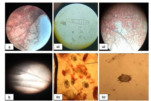
Figure 10: Mites and dermatophytes showing; 10 (a), (a1) and (a2) showed Demodex, (b) showed Microsporum and (b1), (b2) showed Sarcoptic mites.
Chronic Otitis externa was detected (Fig. 9a), skin biopsy showed dyskeratosis with disorganized keratin layer and eosinophilic dermatitis denoted by the infiltration of eosinophils in the dermal layer [Fig. 9(a1)].
Mites and dermatophytes were detected in many clinical cases (Fig. 10) including Demodex canis, Microsporum canis, and Sarcoptic scabiei adult mites [Fig. 10 (a), (a1), (a2), (b), (b1), and (b2)].
Skin tumors were detected in many clinical cases (Fig. 6 & 7). Haemangiosarcoma with soft subcutaneous lumps all over the body which were centrally depressed (Fig. 6a) was reported, microscopic examination of skin biopsies revealed sarcoma cell tumor in the dermal layer with sheets of malignant cells noted by pleomorphism, hyperchromacia and increase nucleus cytoplasmic ratio [Fig. 6(a1)]. Cutaneous papillomatosis in the mouth was detected (Fig. 6b), microscopically, there were hyperplastic epidermis supported by fibrovascular tissue in the dermis [Fig. 6(b1)] and irregular intracytoplasmic keratohyaline granules [Fig. 6(b2)]. Mammary solid adenocarcinoma with solid mass in the mammary gland and ulceration of the skin was detected (Fig. 7a), microscopic examination revealed carcinoma, solid type [Fig. 7(a1)] and pleomorphic cells showed hyperchromatic nuclei, central prominent nucleolus, and frequently detected mitosis [Fig. 7(a2)]. Canine papilloma, Nodular masses on the lateral side of the thigh with alopecia in groin and ventral abdomen with black discoloration were recorded (Fig. 7b), skin biopsy showed hyperplastic epidermis projects ventrally into the dermis [Fig. 7(b1)] together with prominent hyperkeratosis [Fig. 7(b2)].

