Research Article · Volume 11, Issue 4, 3 March 2023 · Open Access
Dermatophytosis in Cats: Clinical Signs and Identification of Etiological Agent
· · ·
Affiliations & Notes ⌄
1Department of Internal Medicine, Faculty of Veterinary Medicine, Universitas Gadjah Mada, 55281, Yogyakarta, Indonesia
2Department of Microbiology, Faculty of Veterinary Medicine, Universitas Gadjah Mada, 55281, Yogyakarta, Indonesia
3Department of Pathology, Faculty of Veterinary Medicine, Universitas Gadjah Mada, 55281, Yogyakarta, Indonesia. *Correspondence | Sitarina Widyarini, Department of Pathology, Faculty of Veterinary Medicine, Universitas Gadjah Mada, 55281, Yogyakarta, Indonesia
This study demonstrated that 55 cats from a total of 69 cats (79.7%) were positive to fungal infection by using direct Wood’s lamp examination and microscopic examination.
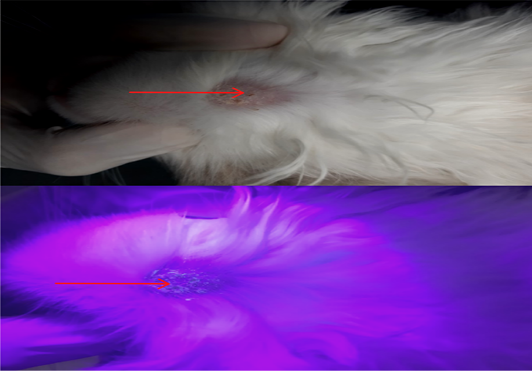
Physical examination demonstrated that 69 suspected cats showed clinical lesions such as alopecia, crust, and erythema (Figure 1).
Fungal culture demonstrated that M. canis (84%) was the most common agent of dermatophytosis in cats in special region of Yogyakarta followed by T. mentagrophytes (14%).
This preliminary examination could be used as a reference for the direct treatment of dermatophytosis in cats considering the test time and efficiency.
Selected from the article's own text.
Abstract
Fungal infection by dermatophytosis is a zoonotic disease distributed globally and well concern in public health. This study aimed to investigate clinical signs and the etiological agents of dermatophytosis in cats in Yogyakarta. A total of 69 cats were observed in this study. Physical examination, by using Wood’s lamp, and microscopic examination of hyphae on the skin was used for preliminary screening of dermatophytosis. Identification of fungi was conducted by scrapping area infection of the skin and then cultured on Dermatophyte Test Medium (DTM) for macroscopic and microscopic examination. The results of this study demonstrated that dermatophytosis in cats is characterized by the presence of multifocal alopecia, scale, pruritus, and erythema on the skin with a specific formation. The dominant clinical lesion was alopecia followed by crust and erythema, stated at 75.8%, 42.8%, and 1.86% respectively. Physical examination showed that 55 cats (79.7%) were positive fungal infections by using direct Wood’s lamp examination and skin scrappings. Moreover, the identification of agents demonstrated that 58 (84%) and 11 (16%) of cats were infected by M. canis and T. mentagrophytes, respectively. Besides clinical signs, Wood’s lamp, and microscopic examination, the gold standard for M. canis idenification is Dermatophyte Test Medium.
Keywords | Dermatophytosis, Cats, Zoonotic, M. canis, T. Mentagrophytes
INTRODUCTION
Dermatophytosis, or ringworm, is a potential zoonotic fungal infection. Dermatophytes are classified into the Trichophyton, Microsporum, and Epidermophyton genera based on the morphology of conidia and the secondary organs . Although the risk of ringworm is limited, the risk of occurrence, long-term illness, and the cost of treatment can be problematic due to ringworm ; ; . The number of cases of dermatophytosis in humans and animals, particularly dogs and cats, is increasing annually ; . It is observed that nearly 20 to 50 percent of human skin infections are caused mainly by zoonotic dermatophytes in pet animals (Microsporum canis and Trychophyton mentagrophytes) . A total of 50–70% of human mycotic infections occur in animal hosts, mainly pet animals ; .
In Indonesia, cases of dermatophytosis are reported to be more common in adult female cats and kittens than in male cats . A study conducted on dogs showed that 34 percent of Yogyakarta dogs had dermatophytosis . In Europe, dermatophytosis in dogs and cats is between 20 and 30% (Galuppi et al., 2013). Research shows that the main species of pet dermatophytosis are M. canis, which accounts for 81.8% to 97% . In many cats, dermatophytes cause mild and self-limited infection with hair loss, erythema, and scaling ; . Bad management of pets can increase the number of infected pets and lead to human infections.
The fact that dermatophytosis is infectious and contagious, dermatophytosis in cat should be monitor in order to prevent the transmission both in the animal and human. Methods of diagnostic play an important role in early detection of dematophyte infection both, in animal and human. Therefore, the aim of this study was to investigate clinical signs and etiological agent of dermatophytosis in cats in special region of Yogyakarta.
MATERIALS AND METHODS
This study was carried out at the Departement of Pathology, Departement of Internal Medicine and Department of Microbiology, Faculty of Veterinary Medicine, Universitas Gadjah Mada. Sixty nine (69) cats with suspected dermatophytosis from Special Region of Yogyakarta were used in this study. Data collection was conducted from April to August 2020. Physical examination and identification of etiological agent as previously reported . As brief, physical examination was performed by examining the skin lesions. Physical examination was followed by Wood’s lamp examination and confirmed with microscopic examination. Skin scrappings of cats suspected with dermatophytosis were collected. One part of each specimen was routinely subjected to direct microscopic examination for detection of fungal hyphae using potassium hydroxide (KOH) 10% solution. Other part of samples was inoculated onto DTM plates containing chloramphenicol (0,05 mg/mL; Kimia Farma, Indonesia) and cycloheximide (0,5 mg/mL; hplc cat 01820, Sigma Chemocal Co, USA), then the plates are incubated at 27-30°C, examined daily, and kept for at least 21 days . Following Frey et al. (1979) and Moriello (2001), the cultures of fungi were identified by examining macroscopic and microscopic fungal colonies. The reference of macroscopic and microscopic identification of the fungi was revealed in Table 1. The macromorphological examination is based on the reported consistency, surface color, reverse color, and change in the DTM color ; . Micromorphological examinations: Using colony in DTM, and then examined by a clear view window under the microscope for hyphae and conidia; in other media, the first subculture was done on dermal-agar, and then growth samples were placed in a slide with drops of lactophenol cotton blue (Hi-Media, Kennett Square, Pennsylvania, USA), covered with a cover and examined under the microscope for identification of hyphae, macroconidia, and microconidia . The procedures were approved by the Ethics Committee of Research and Testing Laboratory, Universitas Gadjah Mada No. 00057/04/LPPT/X/2019. The type of dermatophyte (species) and its correlation with variable sex, age, and clinical signs were statistically processed by the chi-square method. Statistical
Table 1: The gross and microscopic properties of common dermatophytes in DTM ; ; .
Dermatophyte species
Main animal involved
Wood’s lamp examination
Culture identification
Macroscopic characteristics
Microscopic characteristic
Micropsorum canis
Cats, dogs
Positive
Colony has more than one form: Most frequently cottony, white to buff in colour; with increasing age becomes brownish-yellow.
Macroaleuriospores are abundant, large, 6-15 celled, spindle-shaped, with curved or hooked ends and thick verrucose walls. Microaleuriospores are sparse, clavate, smooth-walled and usually sessile along the hypae.
Tricophyton mentagrophytes
Cats, dogs, rabbits, rodents
Negative
Colony present in two forms:
The downy type is floccose and white in colour. Older colonies may be cream-tan.
The granular form is either finely or coarsely, granular, cream to light buff in colour.
Macroaleuspores-rare on Sabouraud’s dextrose agar but may be present on enriched medium. When present, they are smooth, thin-walled, clavate and 3-4 celled. Microsleuriospores-abundant, subsperical, and borne along the hypae either singly or in clusters.
Mycrosporum gypseum
Soil (geophilic species), ringworm in animals
Negatif
Colony is initially white and floccose, later becoming powdery to granular and buff to cinnamon-brown in colour with central umbo and irregularly fringed border. Reverse is pale yellow to tan and occasionally red in some strains.
Macroaleuspores are abudant, spindle-shaped with rounded ends (not as pointed as M. canis), moderately thick-walled, enchinulate, 4-6 celled. Microaleuspores are clavate and usually sessile along the hyphae.
analysis was performed with IBM software Statistics SPSS® v.25 in the Microsoft Windows® 10 operating system.
RESULTS and Discussion
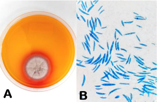
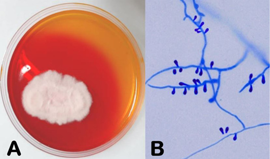
This study demonstrated that 55 cats from a total of 69 cats (79.7%) were positive to fungal infection by using direct Wood’s lamp examination and microscopic examination. Physical examination demonstrated that 69 suspected cats showed clinical lesions such as alopecia, crust, and erythema (Figure 1). Alopecia was the dominant clinical lesion, followed with crust and erythema, and stated at 75.8%, 42.8% and 1.86% respectively. Result of the fungal culture showed that all 69 skin scrapping samples grew on DTM, and M. canis was found to be the most common species (84%) followed by T. mentagrophytes (16%) (Figures 2-3). However, the type of dermatophyte (species) and its correlation with variable sex, age, and clinical signs results revealed no significance (P > 0,05).
Dermatophytes are the important superficial mycosis in cats especially in tropical countries, like Indonesia, the incidence of dermatophytosis in cats is significantly higher than in four seasons with constant humidity and temperature changes in the year ; . The increased prevalence of cat dermatophytosis is due to several factors, including the high number of cat strays in Indonesia . Dermatophyte are generally not classified further based on the location of the infection, but clinical signs usually include circular alopecic lesions and erythematous margins, and cats may be asymptomatic carriers of dermatophytes, leading to occult transmissions to other cats and human beings in close contact ; ; . Lesions in every cases of dermatophytosis are vary each species of animal, as suggested by Aktas and Yigit (2015) lesions caused by dermatophyte can be mild to severe depending on several factors including infected dermatophyte species, virulence factor, infection areas, secondary infection and environmental conditions. Other studies showed that dermatophyte infection in companion animals demonstrated lesions localized in face, legs, and/or tail . This study demonstatred that alopecia was the dominant clinical lesion, followed with crust and erythema. These observations were similar to those showed in previous studies .
Examination of clinical signs, screening test using Wood’s lamp and microscopic examination were performed in this study, as mention previously by ; . Examination of suspected dermatophytosis involves inspection of the skin for lesions in the area muzzle, lips, periocular, in and around the ear and ear margins, digits, medial aspect of the limb, axillary and tail . Physical examination is followed by a Wood’s lamp examination, which often identifies lesions (visualized as fluorescing hairs) that seen during light examination ; . A Wood’s lamp was a hand-held lamp that emits long-wave ultraviolet radiation through a nickel or cobalt glass filter ; and it was used for direct screening dermatophytosis. Under Wood’s lamp exposure active lesion of dermatophytosis resulted in glowing cat’s hair . However, it has been demonstrated that not all strains of dermatophyte glow under Wood’s lamp exposure . Direct microscopic examination of hairs aims to examine the appearance of fungal spores (arthroconidia) and hyphae. Several studies mentioned that direct microscopic examination was used to establish the diagnosis of fungal diseases .
Definitive diagnosis via fungal culture was considered the gold standard, including microscopic and macroscopic examination of the fungus following its cultivation in DTM (Frymus et al., 2017). Fungal culture demonstrated that M. canis (84%) was the most common agent of dermatophytosis in cats in special region of Yogyakarta followed by T. mentagrophytes (14%). Previous studies reported the same results that dermatophytes, such as M. canis, M. gypseum, T mentagrophytes, and M. nanum were the most predominant dermatophyte agents not only in cats but also in dogs in many areas of the world ; . A study by Neves et al. (2018) showed that, M. canis was isolated from 76.9% of the infected pets in Brazil. In this study, the number of dermatophyte positive cultures were quite high (100% from 69 samples), and these finding might be related with the climate during sample collection. It has been reported previously that the incidence of different dermatophyte species varies according to climate and natural reservoirs, therefore, the pattern of the species involved in dermatophytosis may be to some extent different in various geographical conditions in animals ; .
The descriptive analysis presented above revealed that dermatophytosis in cats can be detected early using clinical signs of primary lesions (such as alopecia, crust, and erythema), Wood’s lamp, and strip-tape examination. This preliminary examination could be used as a reference for the direct treatment of dermatophytosis in cats considering the test time and efficiency. All cats were juvenile and no older cats were taken for sample in our research. It is also more commonly reported that young age and old age are risk factors for acquiring dermatophytosis . Also, the research established that male cats have a higher risk of contracting the disease when compared with female cats since 69.5% of the recorded data were from male cats. A test of significance was carried out using the Chi-square method in order to establish whether the dermatophyte species, sex, age, and clinical signs of those who tested positive for the disease are independent or not. It was discovered from the Chi-square test of independence that both variables (dermatophyte species, sex, age, and clinical signs) are independent. It simply means there is no association between the variables when it comes to the spread of dermatophyes.
ACKNOWLEDGEMENTS
The author would like to thank the departments of pathology, internal medicine, and microbiology at the Faculty of Veterinary Medicine of the University of Gadjah Mada for their cooperation in conducting the research. Financial support was provided by PMDSU’s BATCH IV Scholarship. The authors thank the customers/cat owners for their good cooperation.
Novelty Statement
The incidence of dermatophytosis in Yogyakarta special region is high, however, very few published data regarding this matter. This study was conducted to identify cases of dermatophytosis in cats in Yogyakarta special region and to re-optimize the method for diagnosing dermatophytosis in cats.
AUTHOR’S CONTRIBUTION
SW and SI: Conceptualized the study.
SW: Developed experimental design, prepared manuscripts, and edited according to the title.
TU and ADP: Collect data experiments and literature, edit and finalize manuscripts.
All authors read and approved the final manuscript.
Conflict of interests
The authors have declared no conflict of interest.
Figures
Figure 1:.
Lesions from dermatophyte infection in body part of cat with crust and alopecia in the skin (red arrow); Skin lesions of cat with fluorescence (apple blue-green color) under Wood’s lamp examination (red arrow).
Figure 2:.
Fungal colony of M. canis in DTM (A); M. canis macroconidia (B).
Figure 3:.
Fungal colony of T. mentagrophytes in DTM (A); T. mentagrophytes microconidia (B).
References
Aktas E, Yigit N (2015). Hemolytic activity of dermatophytes species isolated from clinical specimens. J. Mycol. Med., 25(1): 25-30. https://doi.org/10.1016/j.mycmed.2014.10.014[CrossRef]
Aneke CI, Otranto D, Cafarchia C (2018). Theraphy and antifungal susceptibility profile of Microsporum canis. J. Fungi., 4(107): 1-14. https://doi.org/10.3390/jof4030107[CrossRef]
Bae JM, Lee RW (2020). 365-nm narrowband wood’s lamp for vitiligo and hypopigmentation disorders. J. Am. Acad. Dermatol., E283-e284. https://doi.org/10.1016/j.jaad.2019.08.064[CrossRef]
da Cunha MM, Capote-Bonato F, Capoci IRG, Bonato DV, Ghizzi LG, Paiva-Lima P, Baeza LC, Svidzinski TIE (2019). Epidemiological investigation and molecular typing of dermatophytosis caused by Microsporum canis in dogs and cats. Preventive Vet. Med., 167: 39-45. https://doi.org/10.1016/j.prevetmed.2019.03.019[CrossRef]
Day MJ, Breitschwerdt E, Cleaveland S, Karkare U, Khanna C, Kirpensteijn J, Kuiken T, Lappin MR, McQuiston J, Mumford E, Myers T, Palatnik-deSousa CB, Rubin C, Takashima G, Thiermann A (2012). Surveillance of zoonotic infectious disease transmitted by small companion animals. Emerg. Infect. Dis., 18(12). https://doi.org/10.3201/eid1812.120664[CrossRef]
Frey D, Oldfield, RJ, Bridger, RC (1979). Color atlas of pathogenic fungi. Year Book Medical Publishers.
Frymus T, Gruffydd-Jones T, Pennisi MG, Addie D, Belak S, Boucraut-Baralon C, Egberink H, Hartmann K, Hosie MJ, Lloret A, Lutz H, Marsilio F, Mostl K, Radford AD, Thiry E, Truyen U and Hornizek UMCDJ (2013). Dermatophytosis in cats:ABDC guidelines on prevention and management J. Feline. Med. Surg., 15(7): 598-604. https://doi.org/10.1177/1098612X13489222[CrossRef]
Galluppi R, Leveque JFC, Beghelli V, Bonoli C, Mattiolo M, Ostanello F, Tampieri MP, Accorsi PA (2013). Cortisol levels in cats, hair in presence or absence of Microsporum canis infection. Elsevier, 95: 1076-1080. https://doi.org/10.1016/j.rvsc.2013.07.023[CrossRef]
Haydar A, Erkhan K, Mitat G, Kiziltepe S (2012). Clinical effectiveness of ivermectin on bobine dermatophytosis makale kodu. Kafkas. Univ. Vet. Fak. Derg, 18(3): 523-526.
Hayette P, Sacheli R (2015). Dermatophytosis, trends in epidemiology and diagnostic approach. Curr. Fungal Infect. Rep., 9: 164–179. https://doi.org/10.1007/s12281-015-0231-4[CrossRef]
Hernandez-Bures A, Pieper JB, Bidot WA, O’Dell M, Sander WE, Maddox CW (2021). Survey of dermatophytes in stray dogs and cats with and without skin lesions in Puerto Rico and confirmed with MALDI-TOF MS. PLoS ONE 16(9): e0257514. https://doi.org/10.1371/journal.pone.0257514[CrossRef]
Indarjulianto S, Yanuartono, Purnamaningsih H, Wikansari P, Sakan GYI (2014). Isolation and identification of Microsporum canis from dermatophytosis dogs in Yogyakarta. J. Vet., 15(2): 212-216.
Indarjulianto S, Yanuartono, Widyarini S, Raharjo S, Purnamaningsih H, Nururrozi A, Hariwibowo N, Jainudin HA (2017). Microsporum canis infection in dermatitis cats. J. Vet., 18(2): 207-210. https://doi.org/10.19087/jveteriner.2017.18.2.207[CrossRef]
Jarjees KI, Issa NA (2022). First study on molecular epidemiology of dermatophytosis in cats,dogs, and their companions in the Kurdistan region of Iraq. Vet. World, 15: 2971-2978. https://doi.org/10.14202/vetworld.2022.2971-2978[CrossRef]
Lee JH, Kwon HS, Jung HM, Kim GM, Bae JM (2017). Wood’s lamp-induced fluorescence of milea. J. Am. Acad. Dermatol., E99-e100. https://doi.org/10.1016/j.jaad.2017.12.046[CrossRef]
Mahboubi M, Kazempour N (2015). The antidermatophyte activity of Allium hirtifolium Boiss aqueous extract. J. Mycol. Med., 25: e10-e14. https://doi.org/10.1016/j.mycmed.2014.10.010[CrossRef]
Moretti A, Agnetti F, Mancianti F, Nardoni S, Righi C, Moretta I, Morganti G, Papini M (2013). Dermatophytosis in animals: Epidemiological, clinical and zoonotic aspects. G. Ital. Dermatol. Venerol., 148(6): 563-572.
Moriello K (2014). Feline dermatophytosis. J. Feline. Med. Surg., 16(5): 419-431. https://doi.org/10.1177/1098612X14530215[CrossRef]
Moriello KA (2001). Diagnostic techniques for dermatophytosis W.B. Saunders Company. Clin. Tech. Small Anim. Pract., 16(4): 219-224. https://doi.org/10.1053/svms.2001.27597[CrossRef]
Moriello KA, Coyner K, Peterson S, Mignon B (2017). Diagnosis and treatment of dermatophytosis in dogs and cats: Clinical consensud guidelines of the world association for veterinary dermatology. Vet. Dermatol., 28: 266-268. https://doi.org/10.1111/vde.12440[CrossRef]
Murmu S, Debnath C, Pramanik AK, Mitra S, Jana S, Dey S, Banerjee S, Batabyal K (2015). Detection and characterization of zoonotic dermatophytes from dogs and cats in and around Kolkata. Vet. World, 8: 1078-1082. https://doi.org/10.14202/vetworld.2015.1078-1082[CrossRef]
Neves JJA, Paulino AO, Vieira EK, Nishida EK, Coutinho SDA (2018). The presence of dermatophytes in infected pets and their household environment. Arq. Bras. Med. Ved. Zootec., 70(6): 1747-1753. https://doi.org/10.1590/1678-4162-9660[CrossRef]
Paryuni AD, Indarjulianto S, Widyarini S (2020). Dermatophytosis in companion animals: A review. Vet. World, 13(22): 1174-1181. https://doi.org/10.14202/vetworld.2020.1174-1181[CrossRef]
Paryuni AD, Indarjulianto S, Untari T, Widyarini S (2022). Dermatophytosis in clinically infected cats: diagnoses and efficacy therapy. Adv. Anim. Vet. Sci., 10(8): 1713-1717. https://doi.org/10.17582/journal.aavs/2022/10.8.1713.1717[Full text][CrossRef]
Rashidian S, Falahati M, Kordbacheh P, Mahmoudi M, Safara M, Sadeghi TH, Mahmoudi S, Zaini F (2015). A study on etiologic agents and clinical manifestations of dermatophytosis in Yazd, Iran. Curr. Med. Mycol., 1(4): 20-25. https://doi.org/10.18869/acadpub.cmm.1.4.20[CrossRef]
Shokri H, Kosravi AR (2016). An epidemiological study of animals dermatomycoses in Iran. Mycmed, 622: 1-8. https://doi.org/10.1016/j.mycmed.2016.04.007[CrossRef]
Taha M, Elfangary M, Essa S, Younes A (2017). Species identification of dermatophytes isolated from human superficial fungal infections by conventional and molecular methods. J. Egypt. Womens. Dermatol., 14: 76-84. https://doi.org/10.1097/01.EWX.0000499598.84966.cb[CrossRef]
Weese JS, Fulford M (2010). In companion animal zoonoses. Wiley-Blackwell, New Jersey. pp. 278-279. https://doi.org/10.1002/9780470958957[CrossRef]
Zaki FHMD, Airin CM, Nururrozi A, Yanuartono, Indarjulianto S (2021). A review: The prevalence of dermatophytosis on cats in Indonesia and Turkey. Bio. Web Conf., 33: 1-5. https://doi.org/10.1051/bioconf/20213306004[CrossRef]