
Ras proto-oncogene encodes for small GTPases, downstream of epidermal growth factor receptor (EGFR) in the RAS/RAF/MAPK pathway. Wild type KRAS is associated with EGFR-signalling activation. In normal physiological conditions, it is activated by upstream signals when the GTP is exchanged with GDP. Playing an important role in regulation of differentiation and cell growth, RasGTPases behave as genetic switches. This transient process of GAP-mediated GTP hydrolysis becomes altered when the Kras gene is mutated. The most common Kras mutations are found in codon 12 and 13 and 61. Some other noncanonical mutations have been reported in codon 11, 14, 15, 17, 18, 19, 20, 22, 27, 30, 31, 117, 146 and 154. We aimed to demonstrate the conformational changes induced in two novel K RAS variants, p.E31K and p.G138V, identified in two CRC patients, which may account for transformative capacity by biochemical and signalling readouts in these patients. Dynamical implications and functional impact of variants were determined by in silico analysis and molecular docking of variants with GTP. MutationTaster was used for functional analysis of genetic variants and three-dimensional structure of mutant proteins were built by Swiss-Model and were further subjected to structural alignment and stability studies by I-Mutant Suite and DUET server. Both variants were predicted as ‘disease causing’ and protein stability analysis revealed p.G138V to be more destabilizing variant than p.E31K. When three-dimensional structures of variants were subjected to molecular docking with GTP, the mutated KRAS showed low binding affinity to the GTP as compared to the wild-type KRAS protein.
Research Article · Volume 51, Issue 4, 29 April 2019 · Open Access
Conformational Changes in Wild Type KRAS Induced by Two Novel Variants p.E31K and p.G138V
· · · · ·
Affiliations & Notes ⌄
- 1Department of Zoology, Kinnaird College for Women, 93-Jail Road, Lahore 54000, Pakistan 2Virtual University of Pakistan 3Benazir Bhutto Medical College, Mirpur, Azad Jammu and Kashmir, Pakistan 4Department of Biochemistry, Faculty of Science, King Abdulaziz University, Jeddah 21589, Saudi Arabia 5Department of Biochemistry, University of Central Punjab, 1 Kahayban-e-Jinnah, Johar Town, Lahore, Pakistan , * Corresponding author: arshaksbs@yahoo.com
- 2dr.arshakoori@ucp.edu.pk
- 3arshakoori.sbs@pu.edu
Article Info ⌄
- Journal:
- Pakistan Journal of Zoology
- Published:
- 29 April 2019

From the article
- KRAS has three sensitive sites which participate in the GTP hydrolysis; γ-phosphate of GTP binds to P-loop i.e., phosphate binding loop (10-16aa) of KRAS.
- After the KRAS-GTP complex is formed, a conformational change occurs in switch-I (30-38aa) and switch-II (59-67aa) regions.
- New close-range salt bridges observed in G12D variant were absent in wild type Kras.
- It was assumed that, negative charge of aspartate triggers several conformational and dynamical changes in Kras G12D which forms an electrostatic interactions with K16 and K88, furthermore, a salt bridge between K16 with D57 will be formed.
Selected from the article's own text.
Abstract
Introduction
Ras proteins (H, K ras4A, K ras4B, and N-ras) have 85% homology and amino acids 1-165 (G domain) are highly conserved between these four Ras proteins. Several motifs present in conserved domain are important for protein function including GTP binding, effector binding etc. ; . The human K ras contains an alternative fourth coding exon. Alternative RNA splicing specifies either of two isomorphic proteins differing by 25 amino acid residues at their carboxy-terminus . The first 85 amino acids have role in binding to GDP and GTP, this region also includes the phosphate binding loop (P loop, comprising of amino acids 10-16), Switch I region (amino acids 32-38) and Switch II region (amino acids 59-67). P loop binds to the γ phosphate of GTP and Switch I, II regulate binding to Ras regulators and effectors. At the carboxy terminal 25 amino acids show considerable variation, it is named as hypervariable domain (amino acids 165-168/169). In this region terminal-CAAX farnesylation motif is present which specify membrane localization of Ras (Fig. 1). Key cysteine residues responsible for palmitoylation in H ras, N ras and K ras4A are also present in this region (C in Fig. 1). A stretch of lysines, proximal to CVIM motif is responsible for localization of K ras4B (KKKKKK in Fig. 1) . Deletion of the CAAX motif leads to the interruption of the post translational modification, thereby preventing the trafficking of Ras to plasma membrane . Fluctuations in the switch I and II regions or P-loop regions can promote instability in these protein regions leading to the hampering of the GTP binding ; . As a consequence of mutations, the KRAS remains in an active GTP binding state and results in the continuous promotion of pro-proliferative signals downstream and tumorogenesis ; . More than one third of all human cancers are found associated with Kras gene mutations resulting in one million deaths per year ; ; ; . High frequency of Krasgene mutations are found in colorectal, lung and pancreatic cancers which are three of the four leading death cause by cancer in the world ; ; .
The most common Kras mutations are found in codon 12 and 13, which contribute about 95% of all mutation types, about 80% in codon 12 and 15% found in codon 13. The most common mutations in these two codons include G12C, G12V, G12D, G12A and G13D, which are found among 12% to 40% cases of cancers. Some other point mutations have been identified at codon 11 , 15, 18 , 19, 20 , 27, 30 31 , 61 (Enomoto et al., 1992), 117, 146 ; , 154 ; ; but with less frequency. Certain conformational changes in mutant protein can result in its constitutive activation. Tumors harbouring mutated isoforms may have achilles heel . In advance CRC, positive mutational status of K ras is associated with worse prognosis of the disease. KRAS mutation status should be considered as an important variable at the time of selection of therapy (Lièvre et al., 2008). To target the mutant K RAS pharmacologically, many in vitro and in vivo trials along with the computational metrics and MD simulation data analysis approach are being used ; ; ; ; ; and few of them are found promising. By using recent bioinformatics tools, we have analysed the possible functional impacts of two novel heterozygous mutations E31K and G138V in two colorectal cancer patients in silico.
Materials and methods
Two novel variants E31K and G138V of K RAS were selected for analysis. An heterozygous mutation at codon 31, substituting glutamic acid (GAA) to lysine AAA, was previously identified in a male patient (45 years) with moderately differentiated adenocarcinoma of mucinous type in T3 N0 M0 stage, invaded to the muscularis propria of colon with metastatic involvement in four or more regional lymph nodes. G138V was identified in a male patient (40 years) diagnosed with adenocarcinoma. Located in transverse colon, tumor was moderately differentiated infiltrative with vascular invasion classified in stage B. No family history of cancer was present in any of the studied subject.
Functional analysis by genomic tools
To predict the deleterious nature of the identified genetic variants (c.91G>A and c.413G>T corresponding to p.E31K and p.G138V) in KRAS gene, we employed MutationTaster as in-silico genomic prediction tools . MutationTaster is conservation/ evolutionary based algorithm, which demands the query in the form of Ensembl gene ID and nucleotide change with few flanking nucleotides of the variation ; .
Protein modelling and structural deviation analysis
The 3D structure of wild type KRAS protein, developed by X-Diffraction method, was obtained from Protein Data Bank. The crystal structure ‘4DSN’ was selected as a template to build further mutant protein models using Swiss-Model , as it covered both mutation positions (4DSN residue coverage: 2-164). The ligands, water molecules and other Het atoms present in the crystal structure were removed manually to avoid errors in building mutant models .
Protein stability analysis
The structural stability of the mutant KRAS proteins was analysed by I-Mutant Suite and DUET server ; . I-Mutant is a support vector machine (SVM)-based tool for the prediction of protein stability changes, upon single point mutations. After processing the protein sequence and the amino acid change, I-Mutant produces the prediction in the form of change in the Gibbs free energy (ΔG) value. The PDB structure 4-letter code, chain identifier as well as the mutation information such as residue position, wild-type and mutant residues codes in one-letter format were provided as an input for this server. The output of DUET is also in the form of ΔΔG values, wherein negative values denote destabilizing mutations.
Molecular docking
The three-dimensional structures of wild-type and mutant proteins were subjected to molecular docking with GTP by Hex docking server . Hex is the only docking and superposition program to use spherical polar Fourier (SPF) correlations to accelerate the calculations. Binding sites of the GTP was also analysed in the wild-type and mutant KRAS protein using PyMol, displaying all the interacting residues around 8Å.
Results
Genomic evaluation report
The output of the MutationTaster reveals one of the four predictions: ‘disease causing’ (probably deleterious), ‘disease causing automatic’ (known to be deleterious by database records) ‘polymorphism’ (probably harmless), and ‘polymorphism automatic’ (known to be harmless by database records). MutationTaster reported both p.E31K and p.G138V to be ‘disease causing’.
Protein structural alignment and stability report
When genetic variants were structurally aligned with the wildtype protein for structural deviation analysis using PyMol-molecular graphic system, wild-type KRAS was superimposed with mutant models and the Root Mean Square Deviation (RMSD) of 0.01 Å was noticed in p.E31K and p.G138V mutant models (Fig. 2). As DUET is an integrated computational web server; it calculated the combined/consensus predictions of mutation Cutoff Scanning Matrix (mCSM) and Site Directed Mutator (SDM) methods in a non-linear regression fashion using SVMs. I-Mutant Suite and DUET scores suggest that both the genetic variants are deleterious in nature. Particularly p.G138V mutant model is predicted to be of extremely low stability when compared to p.E31K protein mutant model (Table I).
Table I.- Protein stability analysis score of different tools for p.E31K and p.G138V mutant KRAS proteins. Units for all scores are Kcal/mole, negative values denotes destabilized protein.
| Nucleotide variant | Amino acid variant | mCSM score | SDM score | DUET score | I-Mutant suite |
| c.91G>A | p.E31K | 0.375 | -0.36 | 0.674 | -0.79 |
| c.413G>T | p.G138V | -0.392 | -0.72 | -0.196 | -0.58 |
GTP binding analysis
Molecular docking is extensively employed computation tool to analyse the molecular recognition that aims to predict the binding affinity and mode of a protein. Figure 2 depicts the docked complexes of A) Wt-KRAS-GTP B) Mt-E31K-KRAS-GTP and C) Mt-G138V-KRAS-GTP. The docking results of wild-type KRAS and its mutants (i.e., p.E31K and p.G138V) to GTP have revealed the binding energies -277.60, -278.27 and -279.67 Kcal/mole. The mutated KRAS protein may have low binding affinity to the GTP when compared to the wild-type KRAS protein. Moreover, the mutant and wild-type KRAS-GTP complex revealed different binding sites as exhibited by the visualization software (Table I). Herein, conformational changes induced in p.E31K and p.G138V proteins may result in its constitutive activation of Kras which resulted in worse prognosis of the disease; however the detailed functional activity and association of these variants to CRC needs be clarified by further studies.
Discussion
KRAS has three sensitive sites which participate in the GTP hydrolysis; γ-phosphate of GTP binds to P-loop i.e., phosphate binding loop (10-16aa) of KRAS. After the KRAS-GTP complex is formed, a conformational change occurs in switch-I (30-38aa) and switch-II (59-67aa) regions. Any mutation in these sites might affect its regulatory function (Xu et al., 2017; . Structural implications caused p.G12D and p.G13D were analysed by Chen et al. (2013) by calculating free energy profiles of binding processes of GTP, interacting with mutant and wild type and it was observed that GTP-binding pocket in mutant with p.G12D is more open than that of wild type and p.G13D proteins. By using a new integrated MD simulation data analysis approach Vatansever et al. (2017) depicted the induction of negative correlations between the fluctuations of SII and those of the P-loop, Switch I (SI) and α3 regions in K ras G12D and it was postulated that the deviation of active site residues impairs the GTP hydrolysis and GAP binding.SII fluctuations display increased level of fluctuations and negative correlations . New close-range salt bridges observed in G12D variant were absent in wild type Kras. It was assumed that, negative charge of aspartate triggers several conformational and dynamical changes in Kras G12D which forms an electrostatic interactions with K16 and K88, furthermore, a salt bridge between K16 with D57 will be formed.
Herein, it was observed that, E31K is predictable to disrupt the formation of effector loop thus affecting the downstream transducers and G138 residue is among the residues involved in the interprotein crystal interaction comprising the λ9 loop of KRAS. G138V may also destabilize the protein as it was predicted by the Duet stability analysis. Present results bring further acquiesce to the notion that heterogeneity of clinical outcomes in patients with mutant K ras may depends on variability in copy number and nature of diversity of mutant isoforms.
Statement of conflict of interest
The authors declare no conflict of interest.
Figures
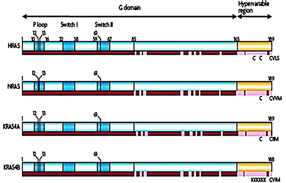
Isoforms of Ras (adopted from Schubbert et al., 2007).
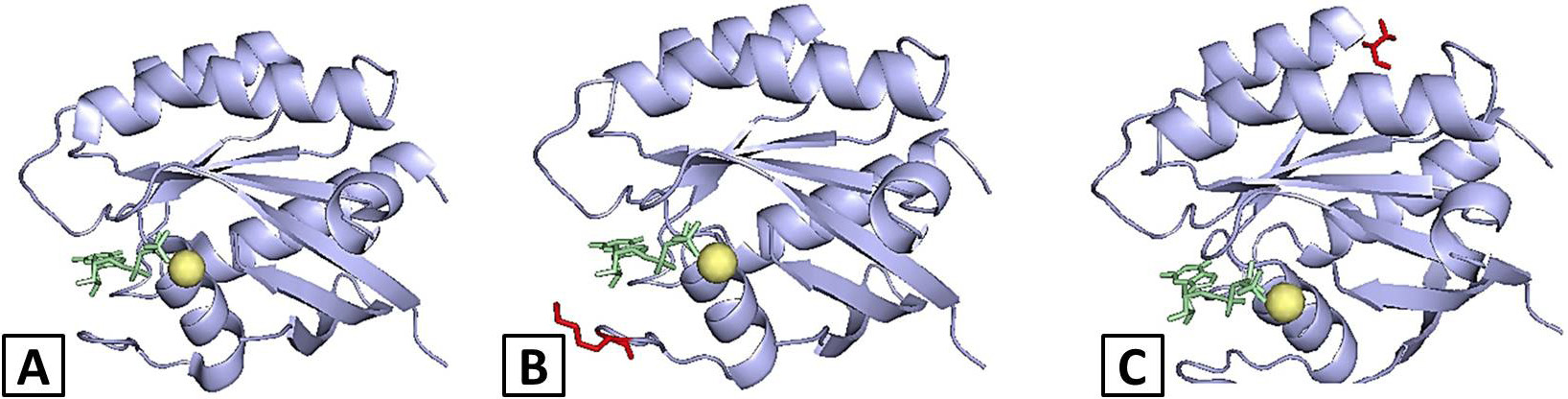
Docking analysis report: A, Wild-type KRAS-GTP complex; B, E31K-KRAS-GTP complex; C, G138V-KRAS-GTP complex. Colour coded as KRAS protein (paleblue cartoon), GTP (palegreen sticks), Magnesium ions (yellow spheres) and E31K and G138V mutations (red sticks).
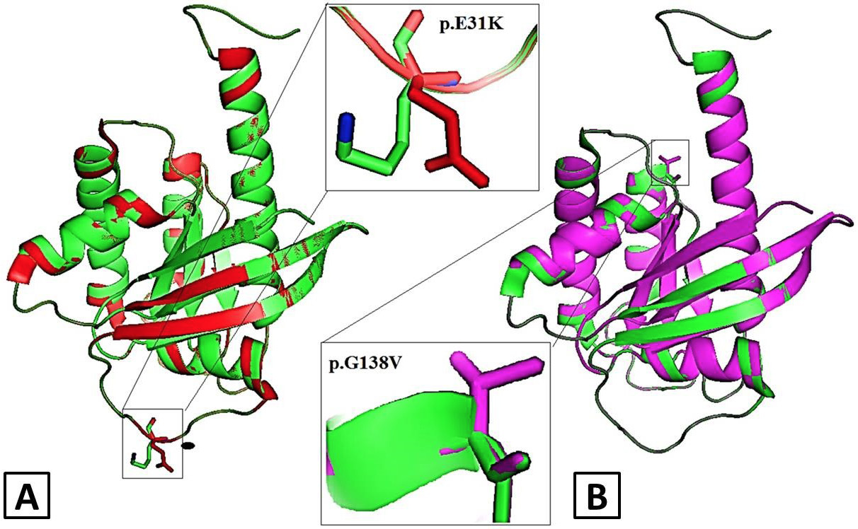
Structural alignment of wild-type and mutant models of KRAS in cartoon mode visualized by PyMol (A) p.E31K KRAS mutant model (red) superimposed with native KRAS protein (green) (B) p.G138V KRAS mutant model (magenta) superimposed with native KRAS protein (green) KRAS. Rectangle boxes depict the zoomed-in view of mutation sites.
References
- Adzhubei, I.A., Schmidt, S., Peshkin, L., Ramensky, V.E., Gerasimova, A., Bork, P., Kondrashov, A.S. and Sunyaev, S.R., 2010. A method and server for predicting damaging missense mutations. Nature Methods, 7: 248-249. https://doi.org/10.1038/nmeth0410-248[CrossRef]
- Buhrman, G., Casey, O., Zerbe, B., Kearney, B.M., Napoleon, R., Kovrigina, E.A., Vajda, S., Kozakov, D., Kovrigin, E.L. and Mattos, C., 2011. Analysis of binding site hot spots on the surface of Ras GTPase. J. mol. Biol., 413: 773-789. https://doi.org/10.1016/j.jmb.2011.09.011[CrossRef]
- Capriotti, E., Fariselli, P., Rossi, I. and Casadio, R., 2008. A three-state prediction of single point mutations on protein stability changes. BMC Bioinformatics, 9: S6. https://doi.org/10.1186/1471-2105-9-S2-S6[CrossRef]
- Chen, C.C., Er, T.K., Liu, Y.Y., Hwang, J.K., Barrio, M.J., Rodrigo, M., Garcia-Toro, E. and Herreros-Villanueva, M., 2013. Computational analysis of KRAS mutations: implications for different effects on the KRAS p. G12D and p. G13D mutations. PLoS One, 8: e55793. https://doi.org/10.1371/journal.pone.0055793[CrossRef]
- DeLano, W.L., 2002. Pymol: An open-source molecular graphics tool. CCP4 Newsl. Protein Crystallogr., 40: 82-92.
- Dogan, S., Shen, R., Ang, D.C., Johnson, M.L., D’Angelo, S.P., Paik, P.K., Brzostowski, E.B., Riely, G.J., Kris, M.G., Zakowski, M.F. and Ladanyi, M., 2012. Molecular epidemiology of EGFR and KRAS mutations in 3,026 lung adenocarcinomas: Higher susceptibility of women to smoking-related KRAS-mutant cancers. Clin. Cancer Res., 18: 6169-6177. https://doi.org/10.1158/1078-0432.CCR-11-3265[CrossRef]
- Enomoto, T., Weghorst, C.M., Inoue, M., Tanizawa, O. and Rice, J.M., 1991. K-ras activation occurs frequently in mucinous adenocarcinomas and rarely in other common epithelial tumors of the human ovary. Am. J. Pathol., 139: 777.
- Forbes, S., Clements, J., Dawson, E., Bamford, S., Webb, T., Dogan, A., Flanagan, A., Teague, J., Wooster, R., Futreal, P.A. and Stratton, M.R., 2006. COSMIC 2005. Br. J. Cancer, 94: 318-322. https://doi.org/10.1038/sj.bjc.6602928[CrossRef]
- Goitre, L., Trapani, E., Trabalzini, L. and Retta, S.F., 2014. The Ras superfamily of small GTPases: the unlocked secrets. In: Ras Signaling: Methods and protocols (eds. L. Trabalzini and S.F. Retta). Part of the Methods in Molecular Biology Book Series, Vol. 1120. Springer. Available at: https://link.springer.com/book/10.1007/978-1-62703-791-4 (Accessed on Feb 23, 2019).
- Guex, N., Peitsch, M.C. and Schwede, T., 2009. Automated comparative protein structure modeling with SWISS-MODEL and Swiss-PdbViewer: A historical perspective. Electrophoresis, 30(S1): S162-S173. https://doi.org/10.1002/elps.200900140[CrossRef]
- Habeck, M., 2002. Cancers with mutated Ras may have Achilles heel. Lancet Oncol., 3: 586. https://doi.org/10.1016/S1470-2045(02)00887-2[CrossRef]
- Hongyo, T., Buzard, G.S., Palli, D., Weghorst, C.M., Amorosi, A., Galli, M., Caporaso, N.E., Fraumeni, J.F. and Rice, J.M., 1995. Mutations of the K-ras and p53 genes in gastric adenocarcinomas from a high-incidence region around Florence, Italy. Cancer Res., 55: 2665-2672.
- Hunter, J.C., Gurbani, D., Ficarro, S.B., Carrasco, M.A., Lim, S.M., Choi, H.G., Xie, T., Marto, J.A., Chen, Z., Gray, N.S. and Westover, K.D., 2014. In situ selectivity profiling and crystal structure of SML-8-73-1, an active site inhibitor of oncogenic K-Ras G12C. Proc. natl. Acad. Sci., 111: 8895-900. https://doi.org/10.1073/pnas.1404639111[CrossRef]
- Hutchins, G., Southward, K., Handley, K., Magill, L., Beaumont, C., Stahlschmidt, J., Richman, S., Chambers, P., Seymour, M., Kerr, D. and Gray, R., 2011. Value of mismatch repair, KRAS, and BRAF mutations in predicting recurrence and benefits from chemotherapy in colorectal cancer. J. Clin. Oncol., 29: 1261-1270. https://doi.org/10.1200/JCO.2010.30.1366[CrossRef]
- Inoue, Y., Saigusa, S., Iwata, T., Okugawa, Y., Toiyama, Y., Tanaka, K., Uchida, K., Mohri, Y. and Kusunoki, M., 2012. The prognostic value of KRAS mutations in patients with colorectal cancer. Oncol. Rep., 28: 1579-1584. https://doi.org/10.3892/or.2012.1974[CrossRef]
- Jamal-Hanjani, M., Wilson, G.A., McGranahan, N., Birkbak, N.J., Watkins, T.B., Veeriah, S., Shafi, S., Johnson, D.H., Mitter, R., Rosenthal, R. and Salm, M., 2017. Tracking the evolution of non–small-cell lung cancer. New Engl. J. Med., 376: 2109-2121. https://doi.org/10.1056/NEJMoa1616288[CrossRef]
- Janakiraman, M., Vakiani, E., Zeng, Z., Pratilas, C.A., Taylor, B.S., Chitale, D., Halilovic, E., Wilson, M., Huberman, K., Ricarte-Filho, J.C. and Persaud, Y., 2010. Genomic and biological characterization of exon 4 KRAS mutations in human cancer. Cancer Res., 70: 5901-5911. https://doi.org/10.1158/0008-5472.CAN-10-0192[CrossRef]
- Janes, M.R., Zhang, J., Li, L.S., Hansen, R., Peters, U., Guo, X., Chen, Y., Babbar, A., Firdaus, S.J., Darjania, L. and Feng, J., 2018. Targeting KRAS mutant cancers with a covalent G12C-specific inhibitor. Cell, 172: 578-589. https://doi.org/10.1016/j.cell.2018.01.006[CrossRef]
- Lievre, A., Bachet, J.B., Boige, V., Cayre, A., Le Corre, D., Buc, E., Ychou, M., Bouché, O., Landi, B., Louvet, C. and André, T., 2008. KRAS mutations as an independent prognostic factor in patients with advanced colorectal cancer treated with cetuximab. J. clin. Oncol., 26: 374-379. https://doi.org/10.1200/JCO.2007.12.5906[CrossRef]
- Macindoe, G., Mavridis, L., Venkatraman, V., Devignes, M.D. and Ritchie, D.W., 2010. HexServer: An FFT-based protein docking server powered by graphics processors. Nucl. Acids Res., 38(Suppl-2): W445-449. https://doi.org/10.1093/nar/gkq311[CrossRef]
- Maurer, T., Garrenton, L.S., Oh, A., Pitts, K., Anderson, D.J., Skelton, N.J., Fauber, B.P., Pan, B., Malek, S., Stokoe, D. and Ludlam, M.J., 2012. Small-molecule ligands bind to a distinct pocket in Ras and inhibit SOS-mediated nucleotide exchange activity. Proc. natl. Acad. Sci., 109: 5299-5304. https://doi.org/10.1073/pnas.1116510109[CrossRef]
- McCormick, F., 2016. K-Ras protein as a drug target. J. mol. Med., 94: 253-258. https://doi.org/10.1007/s00109-016-1382-7[CrossRef]
- Misale, S., Fatherree, J.P., Cortez, E., Li, C., Bilton, S., Timonina, D., Myers, D.T., Lee, D., Gomez-Caraballo, M., Greenberg, M. and Nangia, V., 2019. KRAS G12C NSCLC models are sensitive to direct targeting of KRAS in combination with PI3K inhibition. Clin. Cancer Res., 25: 796-807. https://doi.org/10.1158/1078-0432.CCR-18-0368[CrossRef]
- Murtaza, B.N., Bibi, A., Nadeem, M.S., Chaudri, M.S. and Shakoori, A.R., 2012. Identification of novel mutation in codon 31 of Kirstein rat sarcoma viral oncogene homologue in colon cancer: Another evidence of non-canonical mutational pathwa. Pakistan J. Zool., 44: 1671-1676.
- Naguib, A., Wilson, C.H., Adams, D.J. and Arends, M.J., 2011. Activation of K-RAS by co-mutation of codons 19 and 20 is transforming. J. mol. Signal., 6: 2. https://doi.org/10.1186/1750-2187-6-2[CrossRef]
- Neumann, J., Zeindl-Eberhart, E., Kirchner, T. and Jung, A., 2009. Frequency and type of KRAS mutations in routine diagnostic analysis of metastatic colorectal cancer. Pathol. Res. Pract., 205: 858-862. https://doi.org/10.1016/j.prp.2009.07.010[CrossRef]
- Ostrem, J.M., Peters, U., Sos, M.L., Wells, J.A. and Shokat, K.M., 2013. K-Ras (G12C) inhibitors allosterically control GTP affinity and effector interactions. Nature, 503: 548-551. https://doi.org/10.1038/nature12796[CrossRef]
- Pantsar, T., Rissanen, S., Dauch, D., Laitinen, T., Vattulainen, I. and Poso, A., 2018. Assessment of mutation probabilities of KRAS G12 missense mutants and their long-timescale dynamics by atomistic molecular simulations and Markov state modelling. PLoS Comput. Biol., 14: e1006458. https://doi.org/10.1371/journal.pcbi.1006458[CrossRef]
- Patricelli, M.P., Janes, M.R., Li, L.S., Hansen, R., Peters, U., Kessler, L.V., Chen, Y., Kucharski, J.M., Feng, J., Ely, T. and Chen, J.H., 2016. Selective inhibition of oncogenic KRAS output with small molecules targeting the inactive state. Cancer Discov., 6: 316-329. https://doi.org/10.1158/2159-8290.CD-15-1105[CrossRef]
- Pires, DE., Ascher, D.B. and Blundell, T.L., 2014. DUET: A server for predicting effects of mutations on protein stability using an integrated computational approach. Nucl. Acids Res., 42(W1): W314-319. https://doi.org/10.1093/nar/gku411[CrossRef]
- Schubbert, S., Shannon, K. and Bollag, G., 2007. Hyperactive Ras in developmental disorders and cancer. Nat. Rev. Cancer, 7: 295-308. https://doi.org/10.1038/nrc2175[CrossRef]
- Schwarz, J.M., Rödelsperger, C., Schuelke, M. and Seelow, D., 2010. MutationTaster evaluates disease-causing potential of sequence alterations. Nature Methods, 7: 575-576. https://doi.org/10.1038/nmeth0810-575[CrossRef]
- Siegel, R., Ma, J., Zou, Z. and Jemal, A., 2014. Cancer statistics, 2014. CA: Cancer J. Clinic., 64: 9-29. https://doi.org/10.3322/caac.21208[CrossRef]
- Thompson, H., 2013. US National Cancer Institute’s new Ras project targets an old foe. Nat. Med., 19: 949-950. https://doi.org/10.1038/nm0813-949[CrossRef]
- Vatansever, S., Erman, B. and Gumus, Z., 2017. Oncogenic G12D mutation alters local conformations and dynamics of K-Ras. bioRxiv, 1: 178483. https://doi.org/10.1101/178483[CrossRef]
- Wang, J.Y., Hsieh, J.S., Chen, F.M., Yeh, C.S., Alexandersen, K., Huang, T.J., Chen, D.C. and Lin, S.R., 2003. High frequency of activated K-ras codon 15 mutant in colorectal carcinomas from Taiwanese patients. Int. J. Cancer, 107: 387-393. https://doi.org/10.1002/ijc.11417[CrossRef]
- Wang, J., Hu, K., Guo, J., Cheng, F., Lv, J., Jiang, W., Lu, W., Liu, J., Pang, X. and Liu, M., 2016. Suppression of KRas-mutant cancer through the combined inhibition of KRAS with PLK1 and ROCK. Nat. Commun., 7: 11363. https://doi.org/10.1038/ncomms11363[CrossRef]
- Wittinghofer, A. and Vetter, I.R., 2011. Structure-function relationships of the G domain, a canonical switch motif. Annu. Rev. Biochem., 80: 943-971. https://doi.org/10.1146/annurev-biochem-062708-134043[CrossRef]
- Wright, L.P. and Philips, M.R., 2006. Thematic review series: Lipid posttranslational modifications CAAX modification and membrane targeting of Ras. J. Lipid Res., 47: 883-891. https://doi.org/10.1194/jlr.R600004-JLR200[CrossRef]
- Xu, S., Long, B.N., Boris, G.H., Chen, A., Ni, S. and Kennedy, M.A., 2017. Structural insight into the rearrangement of the switch I region in GTP-bound G12A K-Ras. Acta Crystallogr. Section D: Struct. Biol., 73: 970-984. https://doi.org/10.1107/S2059798317015418[CrossRef]
Services
Article usage statistics
Loading…