
Achondroplasia is an autosomal dominant disorder of rhizomelic dwarfism. It is predominantly caused by de novo mutations in FGFR3. This study was aimed to determine the common variants of FGFR3 in one inherited, and eighteen sporadic cases of achondroplasia from Pakistan. Sanger sequencing analysis of FGFR3 exon 9 revealed that more than 90%cases had the c.1138G>A p.(Gly380Arg) variant. Our results suggest that c.1138G>A variant is the most common cause of achondroplasia in Pakistan, a finding which is similar to that reported for achondroplasia patients from other countries.
Short Communication · Volume 53, Issue 6, 17 October 2021 · Open Access
The c.1138G>A Variant of Fibroblast Growth Factor Receptor 3 is a Common Cause of Achondroplasia in Pakistan
· · · · ·
Affiliations & Notes ⌄
- 1School of Biological Sciences, University of the Punjab, Quaid-e-Azam Campus, Lahore 2 Institute of Process Engineering, University of the Chinese Academy of Sciences, Beijing 100190, China Niaz Muhammad and Samina Yasinshould be considered joint first author. , * Corresponding author: naz.sbs@pu.edu.pk
Article Info ⌄
- Journal:
- Pakistan Journal of Zoology
- Published:
- 17 October 2021

From the article
- Seventeen of the eighteen non-familial cases and all four affected members of the recruited family exhibited the c.1138G>A;p.(Gly380Arg) or the c.1138G>C; p.(Gly380Arg) rs28931614 variant in FGFR3 (Fig. 1b).
- The same variant c.1138G>A was found in fifteen of seventeen sporadic cases of achondroplasia.
- Thus 97% of achondroplasia patients had a transition of c.1138G>A while 2% achondroplasia cases were heterozygous for the transversion variant c.1138 G >C. These two FGFR3 variants were not detected for one affected individual ACH-NA10.
- It can be concluded that achondroplasia in Pakistan is most commonly caused by the substitution of p.(Gly380Arg) in FGFR3.
Selected from the article's own text.
Abstract
Material and methods
In this study a family ACH8 with four affected members (Fig. 1a), and a group of eighteen non-familial cases (ACH1, ACH3-7, ACH9-13, ACH19-23, HCP5, ACH-NA10) with a diagnosis of achondroplasia were studied. Patients were recruited from Sindh, Khyber Pakhtunkhwa and Punjab provinces of Pakistan. Ethical approval to conduct this study was obtained from Institutional Review Board, School of Biological Sciences, University of the Punjab, Lahore. Written informed consent was obtained from all participants and blood samples were collected. Ages of the participants varied from 5 to 40 years.
All affected individuals had rhizomelic dwarfism, normal sized trunks, prominent large foreheads and short extremities. They had heights less than the third percentile for their age. Their fingers were short and pointed away from each other. Few of the patients had large sized heads with prominent foreheads.
Genomic DNA was extracted from leukocytes by a standard protocol including sucrose lysis and salting out. Two sets of primers were designed using Primer 3 (http://bioinfo.ut.ee/primer3-0.4.0/) based on genomic sequence of FGFR3 (NM_000142.4) to amplify exons 9 c.1138G>A or c.1138G>C variants. PCR products were sequenced using Big Dye Terminator v.3.1 (ABI Thermo Fisher).
Results
Eighteen sporadic patients and all four affected members of the recruited family had typical manifestations of achondroplasia. Seventeen of the eighteen non-familial cases and all four affected members of the recruited family exhibited the c.1138G>A;p.(Gly380Arg) or the c.1138G>C; p.(Gly380Arg) rs28931614 variant in FGFR3 (Fig. 1b). Four affected members of the recruited family had the common c.1138G>A variant. The same variant c.1138G>A was found in fifteen of seventeen sporadic cases of achondroplasia. However, two patients had the c.1138G>C transversion of FGFR3. Thus 97% of achondroplasia patients had a transition of c.1138G>A while 2% achondroplasia cases were heterozygous for the transversion variant c.1138 G >C. These two FGFR3 variants were not detected for one affected individual ACH-NA10.
Discussion
Health issues of achondroplasia patients include cardiac diseases, neurological problems, spinal problems, leg abnormalities, obesity, respiratory issues and hearing impairments . The finding of the variants detected in sporadic cases of patients with achondroplasia is consistent with occurrence of de novo mutations in FGFR3. About 80 % cases of achondroplasia occur as de novo mutations . One of the main reasons hypothesized for prevalence of achondroplasia is increased paternal age. Offspring of fathers of older age have a higher rate of this disorder . Most of the affected participants collected in this study had fathers in the age range of 35-40 at the time of conception. In the single familial case of achondroplasia presented here, the phenotype was inherited dominantly as expected.
Rare genetic skeletal dysplasia is not registered or documented in a developing country like Pakistan. Public facility for appropriate genetic testing is also not available . Previously, a familial case of achondroplasia with three affected individuals (one parent and two offspring) was reported in Pakistan with p.(Gly380Arg) variant in FGFR3 . The present study is the first report from Pakistan in which a large number of subjects with achondroplasia were studied. It can be concluded that achondroplasia in Pakistan is most commonly caused by the substitution of p.(Gly380Arg) in FGFR3. Our results agree with the world-wide distribution of p.(Gly380Arg) mutation among achondroplasia cases. However, we did not identify the two variants resulting in the p.(Gly380Arg) substitution for one patient with achondroplasia. Future studies are warranted by sequencing of the entire FGFR3 gene in order to characterize the genetic underpinnings of his disorder.
Acknowledgements
We thank all the patients and their family members for participation.
Statement of conflicts of interest
The authors have declared no conflict of interest.
Figures
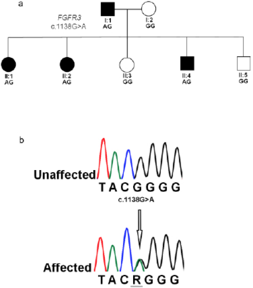
(a) Pedigree of the family ACH8 in which members exhibited dominantly inherited achondroplasia. (b) Chromatogram for FGFR3 selected region showing the most common transition c.G1138>A variant. The arrow indicates the point of mutation.
References
- Ahmad, F., Bilal, M. Khan, A. and Umair, M., 2019. Meta Gene, pp. 100559.
- Ajmal, M., Mir, A., Shoaib, M., Malik, S.A. and Nasir, M., 2017. Diagn. Pathol., 12: 47. https://doi.org/10.1186/s13000-017-0642-3[CrossRef]
- Fredwall, S.O., Maanum, G., Johansen, H., Snekkevik, H., Savarirayan, R. and Lidal, I.B., 2020. Clin. Genet., 97: 179-197. https://doi.org/10.1111/cge.13542[CrossRef]
- Katsumata, N., Mikami, S., Nagashima-Miyokawa, A., Nimura, A., Sato, N., Horikawa, R., Tanae, A. and Tanaka, T., 2000. Endocr. J., 47: S121-S124. https://doi.org/10.1507/endocrj.47.SupplMarch_S121[CrossRef]
- Nahar, R., Saxena, R., Kohli, S., Puri, R. and Verma, I.C., 2009. Indian J. Orthop., 43: 194. https://doi.org/10.4103/0019-5413.50856[CrossRef]
- Pauli, R.M., 2019. Orphanet J. Rare Dis., 14: 1. https://doi.org/10.1186/s13023-018-0972-6[CrossRef]
- Pehlivan, S., Ozkinay, F., Okutman, O., Cogulu, O., Ozcan, A., Çankaya, T. and Ulgenalp, A., 2003. Turk. J. Pediatr., 45: 99-101.
- Rousseau, F., Bonaventure, J., Legeai-Mallet, L., Pelet, A., Rozet, J-M., Maroteaux, P., Le-Merrer, M. and Munnich, A., 1994. Nature, 371: 252. https://doi.org/10.1038/371252a0[CrossRef]
- Shiang, R., Thompson, L.M., Zhu, Y.Z., Church, D.M., Fielder, T.J., Bocian, M., Winokur, S.T. and Wasmuth, J.J., 1994. Cell, 78:335-342. https://doi.org/10.1016/0092-8674(94)90302-6[CrossRef]
Services
Article usage statistics
Loading…