
Two hundred and seventy-eight clinical samples from hospital patients including pus, blood, nasal swabs, ear swabs, urethral swabs, wounds and sputum were processed for isolation of S. aureus. From these samples 50 S. aureus isolates were recovered, which were subjected to sensitivity testing against 18 antibiotics. The isolates were also screened for antibiotic resistance genes through PCRs. In S. aureus isolates highest resistance rates were observed against co-trimoxazole (70%). The isolates also showed high resistance against erythromycin, sparofloxacin and ofloxacin to which 52%, 48% and 46% of the isolates were found to be resistant. Multiple drug resistance (MDR) was noted in 78% of the S. aureus isolates. Out of the 50 isolates, 28 (56%) were found to be methicillin resistant S. aureus (MRSA). In the MRSA isolates highest resistance rate was detected against co-trimoxazole (75.0%), followed by piperacillin-tazobactam (64.2%), ofloxacin (60.7%) and sparofloxacin (60.7%). All the 28 MRSA were found to possess the mecA gene and no mecC variant was detected. Thirty two percent of the S. aureus isolates possessed no antibiotic resistance gene. The resistance genes detected in the isolates were aacA-D (50%), tetK (38%), ermC (30%), tetM (8%) and ermA (6%), while ermB, dfrA and cfr were not found. Additional studies are needed to get a better picture of situation of antibiotic resistance and antibiotic resistance genes in the local S. aureus strains.
Research Article · Volume 58, Issue 1, 27 December 2025 · Open Access
Antibiotic Resistance and Antibiotic Resistance Genes in Staphylococcus aureus Isolates from Hospital Patients in Islamabad, Pakistan
· · ·
Affiliations & Notes ⌄
- 1Department of Zoology, Faculty of Biological Sciences, Quaid-i-Azam University, Islamabad, Pakistan 2Islamabad Diagnostic Center, Islamabad, Pakistan * Corresponding author: saeedkhan@qau.edu.pk
Article Info ⌄
- Journal:
- Pakistan Journal of Zoology
- Published:
- 27 December 2025

From the article
- High levels of resistance were observed in the S. aureus isolates and multiple drug resistance (MDR) defined as resistance to ≥3 classes of antibiotics was noted in 39 (78%) isolates.
- The next common phenotype was resistance to group of 7 or 9 antibiotics observed in 4 (8%) isolates each for both groups.
- The antibiotic resistance gene aacA-D had the highest occurrence and tetK and ermC were also detected in a significant number of isolates.
- Further studies are required to know better the prevalence of antibiotic resistance and antibiotic resistance genes in Pakistani S. aureus strains.
Selected from the article's own text.
Abstract
INTRODUCTION
Staphylococcus aureus occurs as a commensal organism on the nasal passages, skin and mucous membranes of humans and animals. At the same time, it is also the leading cause of bacteremia, infective endocarditis and osteoarticular, skin, soft tissue, pleuropulmonary, and device-related infections. S. aureus is also one of the major organisms responsible for nosocomial infections in humans and it can cause infections after surgery or from implanted medical devices . S. aureus has been recovered from a wide variety of animals .
Resistance against several classes of antibiotics such as penicillins, glycopeptides, daptomycin, tetracyclines, aminoglycosides, linezolid, phenicols, lincosamides, pleuromutilins, macrolides, streptogramins, fusidic acid, mupirocin, fluoroquinolones, sulfonamides, trimethoprim and rifampicin has been observed in S. aureus. Resistance in S. aureus can occur by several mechanisms such as enzymatic modification or inactivation of antibiotic, alteration of antibiotic binding site, antibiotic efflux, acquisition of novel antibiotic-resistance target, change in the structure and composition of the bacterial cell wall and/or membrane to reduce the entry of the antibiotic into the bacterial cell .
In 1942, resistance to penicillin was reported in S. aureus that instigated the development of semi-synthetic penicillins such as methicillin and oxacillin. However, in the 1960s, the first isolates of MRSA were detected . Methicillin resistance is due to mecA gene which codes for an altered Penicillin Binding Protein called PBP2a ; . A novel mecA homologue called mecC was reported later. It was initially found in S. aureus isolates recovered from cattle and humans. The mecC gene has only 70% nucleotide similarity with the mecA gene . After its initial discovery in the UK in humans and bovine population, the mecC homologue of the mecA has been noted in 13 European countries and Australia and has been observed in MRSA from a variety of different animal species. It was also reported in MRSA from river water and urban waste-water in Spain .
Many studies have been conducted on epidemiology S. aureus in Pakistan and high percentages of the S. aureus isolates have been found to be MRSA. In the cities of Rawalpindi and Peshawar 42% and 36.1% of the clinical S. aureus isolates were noted to be MRSA. High resistance rates against erythromycin (99.0%), moxifloxacin (85.1%), ciprofloxacin (80.2%) and gentamicin (56.4%) were observed in Pakistani MRSA isolates ; .
The aim of the present study was to investigate the antibiotic susceptibility of S. aureus isolated from hospital patients in the city of Islamabad. This work also aimed to detect some selected antibiotic resistance genes in these isolates and to find the mec gene type (A or C) in the MRSA.
MATERIALS AND METHODS
Clinical samples and isolation of S. aureus
The study was carried out at Islamabad Diagnostic Center (IDC), which is a private diagnostic laboratory in Islamabad and at the Department of Zoology, Quaid-i-Azam University, Islamabad. A total of 278 clinical samples including pus (97), blood (126), nasal swabs (18), ear swabs (5), urethral swabs (3), wounds (3) and sputum (26) possibly containing S. aureus were received at IDC. The samples had been collected by the hospital staff from patients that had been admitted in various hospitals of Islamabad and were suffering from abscesses, wounds, pneumonia, throat infection, ear infection or urinary tract infection (community-acquired infections). Each sample was from a single patient. The samples were collected before any antibiotic was administered to the patients. Isolation of bacteria was done initially on Nutrient agar. Bacterial colonies were further sub-cultures on Blood agar and then on Mannitol Salt agar. To confirm the isolates as S. aureus, Grams’ staining was performed, colonial morphology and hemolysis on blood agar were noted and coagulase, catalase, oxidase, DNase and other biochemical tests were performed following standard protocols . The isolates were also subjected to a nuc gene PCR for molecular confirmation. Each S. aureus isolate was grown overnight in 5 ml of nutrient broth and the bacterial genomic DNA was isolated using DNAzol reagent (Thermofisher Scientific, Cat. No. 10503027) following manufacturer’s protocol. A 25 µl PCR mix was prepared containing 5 µl template genomic DNA, IX PCR buffer (NH4SO4), 0.2 mM dNTPs, 1 µM of each of the two primers (given in Table I), 1.5 mM MgCl2 and 2.5 U of Taq DNA polymerase (Thermofisher Scientific, Cat. No. EP0402). Cycling conditions were initial denaturation at 95°C for 10 min, followed by 35 cycles of denaturation at 95°C for 1 min, annealing at 55°C for 30 sec, and extension at 72°C for 1 min. The final extension was at 72°C for 10 min.
Antibiotic susceptibility testing
All the isolates confirmed as S. aureus through staining, growth characteristics, biochemical and molecular tests were sub-cultured on Mueller-Hinton agar (Oxoid, UK) and subjected to antimicrobial susceptibility against 18 antibiotics using the disc diffusion method following standard procedures as described in the Clinical Laboratory Standards Institute Manual M100 . The concentration of the antibiotics in the antibiotic impregnated discs (Oxoid, UK) were: linezolid (LNZ): 30µg, chloramphenicol (C): 30µg, clindamycin (DA): 2µg, erythromycin (E): 15 µg, cefoxitin (FOX): 30 µg, vancomycin (VA): 30µg, fusidic acid (FA): 10µg, tigecycline (TGC): 15µg, minocycline (MH): 30µg, amikacin (AK): 30µg, meropenem (MEM): 10µg, Co-trimoxazole (SXT): 25µg, sparofloxacin (SPX): 5µg, ciprofloxacin (CIP): 5 µg, levofloxacin (LEV): 5µg, piperacillin-tazobactam (PT): 110µg, ofloxacin (OFX): 5µg, gentamicin (CN): 10 µg. The results were interpreted as sensitive (S), intermediate (I) or resistant (R). Those isolates that were found to be resistant to cefoxitin were declared as MRSA as per guidelines .
PCRs for mec gene and other antibiotic resistance genes
PCRs were performed to find the mec gene type (A or C) and to detect other antibiotic resistance genes in the confirmed as S. aureus using primers shown in Table I. A multiplex PCR to detect the mec gene type was done on DNA of isolates that were declared MRSA based on sensitivity to cefoxitin. A 25 µl PCR mix contained 5 µl template DNA, IX PCR buffer (NH4SO4), 0.2 mM dNTPs, 1 µM of each of the four primers, 1.5 mM MgCl2 and 2.5 U of Taq DNA polymerase. Amplification conditions were 94°C for 5 min, followed by 30 cycles at 94°C for 30 sec, 59°C for 1 min and 72°C for 1 min. The final extension was at 72°C for 10 min.
PCRs were performed for the detection of the antibiotic resistance genes aacA-D, tetK, tetM, ermA, ermB, ermC, cfr and dfrA. For aacA-D, tetK, tetM, ermA and ermC genes, a multiplex PCR was performed ; . The reaction mix (25 µl) contained 10 µl template DNA, IX PCR buffer (KCl), 0.2 mM dNTPs, 0.5 µM of each of the 10 primers,
Table I. Names of genes, sequences of the primers and expected sizes of PCR products.
| Name of gene | Primer name | Sequence 5`→3` | Expected PCR product size (bp) |
| nuc | nuc-F | GCGATTGATGGT GATACGGTT | 270 |
| nuc-R | AGCCAAGCCTTGACGAACTAAAGC | ||
| mecA | mecA-F | TCCAGATTACAACTTCACCAGG | 162 |
| mecA-R | CCACTTCATATCTTGTAACG | ||
| mecC | mecC-F | GAAAAAAAGGCTTAGAACGCCTC | 138 |
| mecC-R | GAAGATCTTTTCCGTTTTCAGC | ||
| AacD-F | TAATCCAAGAGCAATAAGGGC | 227 | |
| AacD-R | GCCACACTATCATAACCACTA | ||
| ermA | ErmA-F | AAGCGGTAAACCCCTCTGA | 190 |
| ErmA-R | TTCGCAAATCCCTTCTCAAC | ||
| ermB | ErmB-F | CTATCTGATTGTTGAAGAAGGATT | 142 |
| ErmB-R | GTTTACTCTTGGTTTAGGATGAAA | ||
| ermC | ErmC-F | AATCGTCAATTCCTGCATGT | 299 |
| ErmC-R | TAATCGTGGAATACGGGTTTG | ||
| tetK | TetK-F | GTAGCGACAATAGGTAATAGT | 360 |
| TetK-R | GTAGTGACAATAAACCTCCTA | ||
| tetM | TetM-F | AGTGGAGCGATTACAGAA | 158 |
| TetM-R | CATATGTCCTGGCGTGTCTA | ||
| cfr | cfr-F | TGAAGTATAAAGCAGGTTGGGAGTCA | 746 |
| cfr-R | ACCATATAATTGACCACAAGCAGC | ||
| dfrA | dfrA-F | CTCACGATAAACAAAGAGTCA | 288 |
| dfrA-R | CAATCATTGCTTCGTATAACG |
2 mM MgCl2 and 2.5 U of Taq DNA polymerase. The cycling conditions were 94°C for 5 min, followed by 30 cycles at 95°C for 1 min, 55°C for 1 min, 72°C for 1 min. This was followed by final extension at 72°C for 90 sec. The PCR mixture (25 µl) for the ermB gene incorporated 5 µl template DNA, IX PCR buffer (KCl), 0.2 mM dNTPs, 1 µM of each of the forward and reverse primers, 1.5 mM MgCl2 and 2.5 U of Taq DNA polymerase. The amplification was performed at 95°C for 3 min, followed by 30 cycles at 95°C for 30 sec, 54°C for 30 sec, 72°C for 30 sec. The final extension was at 72°C for 4 min . In case of the cfr gene a 25 µl PCR mixture consisted of 5 µl template DNA, IX PCR buffer (KCl), 0.2 mM dNTPs, 1 µM of each of the forward and reverse primers, 1.5 mM MgCl2 and 2.5 U of Taq DNA polymerase. The cycling conditions were 94°C for 1 min, followed by 34 cycles at 94°C for 1 min, 48°C for 30 sec, 72°C for 3 min. The final polymerization step was 72°C for 7 min ; . The PCR mix (25 µl) for the dfrA gene comprised of 5 µl template DNA, IX PCR buffer (KCl), 0.2 mM dNTPs, 1 µM of each of the forward and reverse primers, 1.5 mM MgCl2 and 2.5 U of Taq DNA polymerase. The amplification protocol was 95°C for 2 min, followed by 30 cycles at 94°C for 30 sec, 50°C for 30 sec, 72°C for 30 sec. The final extension was at 72°C for 4 min .
RESULTS
S. aureus isolates
Fifty isolates of S. aureus were recovered from the 278 clinical samples that were processed for bacterial isolation, staining and biochemical tests. The distribution of S. aureus and MRSA isolates obtained from different clinical samples is shown in Table II. Highest number (28/97) of S. aureus isolates were from pus samples. The nuc gene PCR done for molecular confirmation of the 50 isolates produced an amplicon of expected size (270 bp).
Table II. Number of S. aureus and MRSA isolates recovered from various clinical samples.
| Pus | Blood | Nasal swab | Ear swab | Urethral swab | Wound | Sputum | Total | |
| No. of samples | 97 | 126 | 18 | 5 | 3 | 3 | 26 | 278 |
| S. aureus positive | 28 | 4 | 3 | 5 | 3 | 4 | 3 | 50 |
| MRSA positive | 18 | 2 | 1 | 3 | 2 | 1 | 1 | 28 |
Table III. Results of antibiotic sensitivity testing of the 50 S. aureus isolates.
| Antibiotic | R No. (%) | I No. (%) | S No. (%) |
| E | 26 (52%) | 9 (18%) | 15 (30%) |
| FOX | 28 (56%) | 7 (14%) | 15 (30%) |
| C | 4 (8%) | 11 (22%) | 35 (70%) |
| DA | 7 (14%) | 11 (22%) | 32 (64%) |
| VA | 0 (0%) | 10 (20%) | 40 (80%) |
| FA | 6 (12%) | 12 (24%) | 32 (64%) |
| LNZ | 2 (4%) | 0 (0%) | 48 (96%) |
| SXT | 35 (70%) | 7 (14%) | 8 (16%) |
| CN | 14 (28%) | 9 (18%) | 27 (54%) |
| AK | 9 (18%) | 9 (18%) | 32 (64%) |
| LEV | 22 (44%) | 17 (34%) | 11 (22%) |
| OFX | 23 (46%) | 14 (28%) | 13 (26%) |
| CIP | 20 (40%) | 11 (22%) | 19 (38%) |
| SPX | 24 (48%) | 13 (26%) | 13 (26%) |
| PT | 21 (42%) | 13 (26%) | 16 (32%) |
| TGC | 8 (16%) | 13 (26%) | 29 (58%) |
| MH | 4 (8%) | 14 (28%) | 32 (64%) |
| MEM | 9 (18%) | 13 (26%) | 28 (56%) |
LNZ, Linezolid; C, Chloramphenicol; DA, Clindamycin; E, Erythromycin; FOX, Cefoxitin; VA, Vancomycin; FA, Fusidic Acid; TGC, Tigecycline; MH, Minocycline; AK, Amikacin; MEM, Meropenem; SXT, Co-trimoxazole; SPX, Sparofloxacin; CIP, Ciprofloxacin; LEV, Levofloxacin; PT, Piperacillin-Tazobactam; OFX, Ofloxacin; CN, Gentamicin; R, Resistant; I, Intermediate; S, Sensitive.
Antibiotic susceptibility results
Results of antibiotic sensitivity testing are shown in Table III. Highest resistance rates were observed against co-trimoxazole (70%). This was followed by cefoxitin, erythromycin, sparofloxacin and ofloxacin to which 56%, 52%, 48% and 46% of the isolates, respectively were found to be resistant. Maximal sensitivity was observed to linezolid (96%), followed by vancomycin (80%) and chloramphenicol (70%). Twenty-eight (56%) isolates were found to be MRSA as determined by resistance to cefoxitin. The number of MRSA found in pus, blood, nasal swab, ear swabs, urethral swabs, wounds and sputum were 18, 2, 1, 3, 2, 1 and 1, respectively (Table II). The phenotypic antibiotic resistance combination patterns are shown in Table IV. A wide variation in antibiotic resistance phenotype was observed and 39 groups of antibiotic resistance patterns were seen. In general, the MRSA were resistant to larger number of antibiotics compared to the non-MRSA isolates. High levels of resistance were observed in the S. aureus isolates and multiple drug resistance (MDR) defined as resistance to ≥3 classes of antibiotics was noted in 39 (78%) isolates. Extensive drug resistance (XDR), determined as resistance to at least one antibiotic in all classes but susceptibility to at least two or fewer antimicrobial categories, was found in 2 (4%) isolates. Resistance to combinations of 4 or 5 antibiotics was the most common phenotype exhibited by 8 (16%) isolates each for both combinations. The next common phenotype was resistance to group of 7 or 9 antibiotics observed in 4 (8%) isolates each for both groups.
The antibiotic sensitivity of the 28 MRSA isolates is given in Table V. In MRSA highest resistance was against co-trimoxazole (75.0%). The resistance rates against piperacillin-tazobactam (64.2%), ofloxacin (60.7%) and sparofloxacin (60.7%) were also high. The antibiotics found to be most effective against MRSA were linezolid (92.8%), vancomycin (82.1%) and chloramphenicol (71.4%).
Antibiotic resistance genes
The PCR for mec gene type applied on all the 28 MRSA isolates produced a 162 bp band showing that all the MRSA had the mecA gene and none had the mecC gene. The findings on presence of antibiotic resistance genes in the S. aureus isolates are shown in Table VI. The gene aacA-D had the highest occurrence (50%), followed by tetK (38%) and ermC (30%). TetM (8%) and ermA (6%) had low frequencies, while ermB, dfrA and cfr were not found. A representative gel picture of resistance genes PCR is shown in Figure 1. The diversity in occurrence of antibiotic resistance genes in the isolates is shown in Table VII. No antibiotic resistance gene was found in 32% of the isolates. AacA-D occurred as the sole antibiotic resistance gene in 22% of the isolates. TetK was also found to exist as a single antibiotic resistance gene in 6% of the isolates. Six various combinations of antibiotic resistance genes were observed in 2% to 8% of the isolates.
Table IV. Antibiotic resistance patterns in the 50 S. aureus isolates.
| Antibiotic phenotype | No. of antibiotics | No. of resistant isolates | Source |
| SXT | 1 | 1 | Pus |
| SXT, E | 2 | 1 | Sputum |
| SXT, E, DA | 3 | 3 | Pus, Nasal Swab |
| SXT, DA, FOX | 3 | 1 | Pus |
| SXT, FOX, MEM | 3 | 1 | Pus |
| SXT, OFX, CIP, SPX | 4 | 3 | Wound |
| OFX, CIP, SPX, LEV | 4 | 1 | Sputum, Pus, Urethral swab |
| FOX, MEM, CIP, VA | 4 | 1 | Wound |
| FOX, MEM, CN, AK | 4 | 2 | Pus |
| FOX, CN, AK, FA | 4 | 1 | Pus |
| SXT, OFX, CIP, SPX, LEV | 5 | 1 | Blood |
| SXT, E, DA, MEM, MH | 5 | 3 | Ear Swab |
| SXT, FOX, OFX, CIP, CN | 5 | 2 | Wound |
| SXT, OFX, CIP, SPX, PT | 5 | 1 | Pus |
| E, FOX, OFX, CIP, SPX | 5 | 1 | Nasal Swab |
| E, FOX, CIP, SPX, CN, PT | 6 | 1 | Pus |
| SXT, FOX, OFX, CIP, SPX, LEV | 6 | 1 | Pus |
| SXT, MEM, OFX, CIP, SPX, LEV | 6 | 1 | Pus |
| SXT, OFX, VA, CN, AK, FA, LNZ | 7 | 1 | Blood |
| SXT, FOX, OFX, CIP, SPX, LEV, FA | 7 | 1 | Urethral Swab |
| SXT, FOX, OFX, CIP, LEV, CN, FA | 7 | 1 | Ear swab |
| E, FOX, OFX, CIP, SPX, LEV, CN | 7 | 1 | Pus |
| FOX, OFX, CIP, SPX, LEV, CN, AK | 7 | 1 | Blood |
| SXT, E, DA, OFX, CIP, SPX, LEV, FA | 8 | 1 | Pus |
| SXT, FOX, OFX, CIP, SPX, LEV, CN, AK | 8 | 1 | Ear Swab |
| SXT, FOX, OFX, CIP, LEV, CN, AK, FA | 8 | 1 | Blood |
| SXT, E, DA, FOX, OFX, CIP, SPX, LEV, FA | 9 | 1 | Pus |
| SXT, E, DA, FOX, VA, CN, AK, FA, LNZ | 9 | 1 | Sputum |
| SXT, E, FOX, OFX, CIP, SPX, LEV, CN, AK | 9 | 1 | Ear Swab, Urethral Swab |
| SXT, FOX, OFX, CIP, SPX, LEV, AK, TGC, MH | 9 | 1 | Pus |
| SXT, E, FOX, OFX, CIP, SPX, LEV, CN, AK, PT | 10 | 1 | Pus |
| SXT, E, FOX, OFX, CIP, SPX, LEV, CN, AK, TGC, C | 11 | 1 | Pus |
| SXT, E, FOX, OFX, CIP, SPX, LEV, CN, AK, PT, MH | 11 | 1 | Pus |
| SXT, E, FOX, MEM, OFX, CIP, SPX, LEV, CN, AK, C, MH | 12 | 1 | Pus |
| SXT, FOX, MEM, OFX, CIP, SPX, LEV, CN, AK, FA, PT, MH | 12 | 1 | Pus |
| SXT, DA, FOX, MEM, OFX, CIP, SPX, LEV, CN, AK, FA, PT, MH | 13 | 1 | Pus |
| SXT, E, FOX, MEM, OFX, CIP, SPX, LEV, CN, FA, LNZ, PT, TGC, MH | 14 | 1 | Pus |
| SXT, E, DA, FOX, OFX, CIP, SPX, LEV, VA, CN, AK, FA, LNZ, PT, TGC, C | 16 | 1 | Wound |
| SXT, E, DA, FOX, MEM, OFX, CIP, SPX, LEV, CN, AK, FA, LNZ, PT, TGC, C, MH | 17 | 1 | Pus |
Table V. Antibiotic sensitivity of the 28 MRSA isolates.
| Antibiotic | R No. (%) | I No. (%) | S No. (%) |
| E | 15 (53.5%) | 5 (17.8 %) | 8 (28.5%) |
| C | 2 (7.1%) | 6 (21.4%) | 20 (71.4%) |
| DA | 3 (10.7%) | 6 (21.4%) | 19 (67.8%) |
| VA | 0 (0%) | 5 (17.8%) | 23 (82.1%) |
| FA | 4 (14.2%) | 7 (25.0%) | 17 (60.7%) |
| LNZ | 2 (7.1%) | 0 (0%) | 26 (92.8%) |
| SXT | 21 (75.0%) | 4 (14.2%) | 3 (10.7%) |
| CN | 14 (50.0%) | 7 (25.0%) | 7 (25.0%) |
| AK | 7 (25.0%) | 5 (17.8%) | 16 (57.1%) |
| LEV | 10 (35.7%) | 9 (32.1%) | 9 (32.1%) |
| OFX | 17 (60.7%) | 8 (28.5%) | 3 (10.7%) |
| CIP | 14 (50.0%) | 7 (25.0%) | 7 (25.0%) |
| SPX | 17 (60.7%) | 7 (25.0%) | 4 (14.2%) |
| PT | 18 (64.2%) | 5 (17.8%) | 5 (17.8%) |
| TGC | 8 (28.5%) | 6 (21.4%) | 14 (50.0%) |
| MH | 2 (7.1%) | 8 (28.5%) | 18 (64.2%) |
| MEM | 6 (21.4%) | 7 (25.0%) | 15 (53.5%) |
For abbreviations of antibiotics, see Table III.
Table VI. Prevalence of antibiotic resistance genes in the 50 S. aureus isolates.
| Name of gene | Isolates found positive No. (%age) |
| aacA-D | 25 (50%) |
| cfr | 0 (0%) |
| dfrA | 0 (0%) |
| ermA | 3 (6%) |
| ermB | 0 (0%) |
| ermC | 15 (30%) |
| tetK | 19 (38%) |
| tetM | 4 (8%) |
Table VII. Diversity of antibiotic resistance genes in the 50 S. aureus isolates.
| Antibiotic resistance genes combinations | No. of isolates (% age) |
| No gene | 16 (32%) |
| tetK (alone) | 3 (6%) |
| aacA-D (alone) | 11 (22%) |
| tetK, ermC | 4 (8%) |
| tetK, aacA-D | 2 (4%) |
| tetK, ermC, aacA-D | 3 (6%) |
| ermC, aacA-D, tetM | 1 (2%) |
| tetK, ermC, aacA-D, ermA | 3 (6%) |
| tetK, ermC, aacA-D, tetM | 2 (4%) |
DISCUSSION
S. aureus is one of the most common causes of both community and hospital acquired infections. Although S. aureus causes a wide variety of clinical infections, the most common are those that affect skin and soft tissues and often lead to the formation of abscesses . In our study also most isolations of S. aureus were from pus samples. We found a high percentage of MRSA (56%) in our study and this is in line with previous international reports. In general, there has been an increase in the prevalence of MRSA in the Asia-pacific and in various other regions of the world with few exceptions ; . We tested our MRSA isolates for the presence of mecA or mecC alleles by a multiplex PCR. However, we found no isolate harboring the mecC allele. The occurrence of the mecC has generally been found to be low in humans .
In the present study the highest resistance rates were observed against co-trimoxazole. High resistance against co-trimoxazole has been reported in previous studies also. In a study in Nigeria 72.1% of S. aureus clinical isolates were found to be resistant to co-trimoxazole . Ninety six percent of Staphylococci isolated from meat samples have been found to be resistant to co-trimoxazole . Other antibiotics against which high rates of resistance were observed in S. aureus and MRSA in this study were the quinolones sparofloxacin and ofloxacin and the macrolide erythromycin. A total of 107 out of 122 (87.7%) of milk S. aureus isolates from various locations in South Africa were found to be resistant to erythromycin . In Iran 89.1% of MRSA were found to be resistant to erythromycin . Another study in Egypt found clinical MRSA isolates to be highly insusceptible to quinolones. In this, study 58% of the isolates were observed to be resistant to ofloxacin .
In the present study highly diverse patterns of antibiotic resistance were detected against the 18 antibiotics tested and 39 antibiotic resistance phenotype groups were observed. Wide variation in antibiotic resistance patterns in S. aureus have been noted in previous studies. Ninety-seven S. aureus clinical isolates tested for susceptibility to 14 antibiotics showed high divergence in resistance phenotype and 36 antibiotic resistance groups were observed . Sixteen resistance patterns were observed against 9 antibiotics in 33 S. aureus isolates recovered from ear discharges . Thirty S. aureus isolates were collected from seawater and sand from beaches and were tested against 15 antibiotics. The isolates showed 23 antibiotic resistance phenotypic patterns . In our study a high percentage (78%) of S. aureus isolates were found to carry MDR. High rates of MDR in S. aureus are being increasingly reported. Very high rates of MDR S. aureus were reported from China (100%) and Ireland (84.3%) ; .
In the present study the drugs found to be most effective against S. aureus were linezolid (96% for S. aureus and 92.8% for MRSA), vancomycin (80% for S. aureus and 82.1% for MRSA) and chloramphenicol (70% for S. aureus and 71.4% for MRSA). In previous studies both linezolid and vancomycin have shown excellent results against S. aureus. In a study 1116 S. aureus isolates collected over six years time (2009-2014) were tested for antibiotic sensitivity. None of the isolate was found to be resistant to linezolid or vancomycin . In a study in India, out of 250 S. aureus isolates, none was found to be resistant to either linezolid or vancomycin . Chloramphenicol has also been found to be highly effective against S. aureus in previous investigations. In a previous study in Pakistan 132 out of 174 (75.86%) of the MRSA isolates were found to be sensitive to chloramphenicol and this drug was suggested to be a good substitute to the latest expensive antibiotics in resource constrained-countries . Five out of 30 (83.3%) S. aureus isolates from seawater and beaches sand were found to be susceptible to chloramphenicol . None of the 221 S. aureus isolates recovered from various animal species were found to be resistant to chloramphenicol .
We screened our isolates for presence of 8 selected antibiotic resistance genes. The highest occurrence was that of aacA-D and 50% of the isolates were positive for it. The aacA-D gene has been detected at high levels in S. aureus in many previous studies. In Palestine 74.5% of MRSA isolates were found to harbor aacA-D gene . Hospital MRSA isolates recovered in Poland contained aacA-D at a rate of 72.3% . The other genes detected at considerable levels in our S. aureus isolates were tetK (38%) and ermC (30%). Both these genes have been detected at high levels in S. aureus in some of the earlier studies. TetK and ermC were detected in S. aureus clinical isolates at rates of 43.7% and 91.9%, respectively . MRSA isolates contained tetK and ermC at rates of 76.4% and 74.5%, respectively . A high percentage (85.5%) of MDR S. aureus clinical isolates in China were found to contain ermC gene . The genes found in low percentages in our study were tetM (8%) and ermA (6%). TetM has been observed at low levels in S. aureus in former studies in Palestine (16.4%) and Iran (27%) ; . ErmA was noted in 19.4% of clinical S. aureus isolates in Turkey , while no ermA was found in S. aureus collected from seawater and sand from beaches . Cfr, dfrA and ermB genes were not detected in the present study. No cfr gene was detected in 23 Staphylococcus isolates collected from meat samples . The occurrence rate of ermB in S. aureus clinical isolates in Turkey was 6.5% . No dfrA gene was detected in any of the 49 co-trimoxazole resistant Nigerian S. aureus isolates .
Regarding the diversity of antibiotic resistance gene occurrence pattern, 32% of our S. aureus isolates contained no resistance gene, while aacA-D occurred as sole antibiotic resistance gene in 22% of the isolates. Wide variation in occurrence of combinations of resistance gene was observed. Such patterns have been observed in earlier studies also ; .
The main factors that have been attributed to the development of antimicrobial resistance in Pakistan are irrational use of antibiotics and their extensive use in animal farming . There is a dire need to contain rising antibiotic resistance in the country. A plan called the “National Action Plan for the Control of Antimicrobial Resistance” in the country was devised by the Government of Pakistan. In this regular surveillance of antibiotic resistance, raising public awareness on the issue, enforcement of regulations of antimicrobial use in human and veterinary practice, research on antibiotic resistance and vaccines and improved hygiene etc. were suggested as antimicrobial resistance control strategies .
CONCLUSION
This study is one of the very few studies that have addressed the situation of antibiotic resistance and antibiotic resistance genes in Pakistani S. aureus strains. Detailed analysis of the findings of this study have been presented in the results part. More than half (56%) of the isolates were found to be MRSA. High resistance rates against co-trimoxazole, erythromycin, ofloxacin, sparofloxacin and piperacillin-tazobactam were observed. MDR was detected in a high proportion (78%) of the isolates. All the MRSA isolates carried the mecA gene and the mecC variant was not detected. The antibiotic resistance gene aacA-D had the highest occurrence and tetK and ermC were also detected in a significant number of isolates. Further studies are required to know better the prevalence of antibiotic resistance and antibiotic resistance genes in Pakistani S. aureus strains.
Declarations
Acknowledgement
The authors are grateful to the management of IDC for supporting this work.
Funding
This research work was funded by Quaid-i-Azam University.
IRB approval
This study was approved by the Advanced Studies and Research Board of Quaid-i-Azam University, Islamabad, Pakistan.
Ethics statement
This study was approved by the Bioethics/Biosafety Committee of Quaid-i-Azam University, Islamabad, Pakistan.
Statement of conflict of interest
The authors have declared no conflict of interest.
Figures
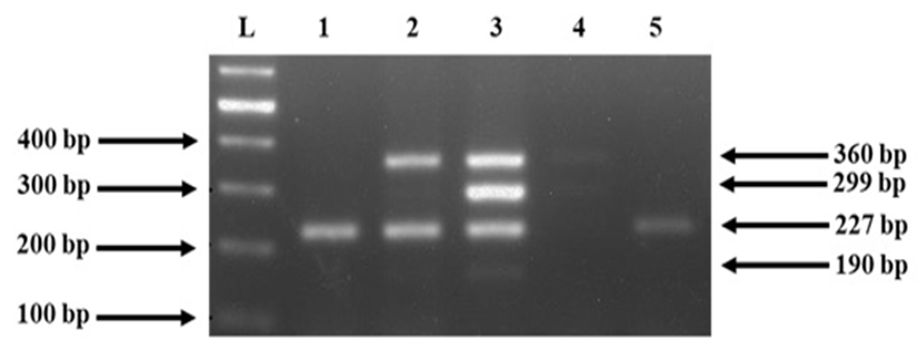
A representative gel showing results of PCR for detection of antibiotic resistance genes. M: molecular size marker. PCR product bands in the figure represent tetK (360 bp), ermC (299bp), aacA-D (227 bp) and ermA (190 bp).
References
- Adwan, G., Adwan, K., Jarrar, N. and Amleh, A., 2014. Molecular detection of nine antibiotic resistance genes in methicillin resistant Staphylococcus aureus isolates. Roum. Arch. Microbiol. Immunol., 73: 9-17.
- Aires-de-Sousa, M., 2017. Methicillin-resistant Staphylococcus aureus among animals: Current overview. Cl. Microbiol. Infect., 23: 373-380. https://doi.org/10.1016/j.cmi.2016.11.002[CrossRef]
- Akanbi, O.E., Njom, H.A., Fri, J., Otigbu, A.C. and Clarke, A.M., 2017. Antimicrobial susceptibility of Staphylococcus aureus isolated from recreational waters and beach sand in Eastern Cape Province of South Africa. Int. J. environ. Res. Publ. Hlth., 14: 1001. https://doi.org/10.3390/ijerph14091001[CrossRef]
- Akindolire, M.A., Babalola, O.O. and Ateba, C.N., 2015. Detection of antibiotic resistant Staphylococcus aureus from milk: A public health implication. Int. J. environ. Res. Publ. Hlth., 12: 10254-10275. https://doi.org/10.3390/ijerph120910254[CrossRef]
- Ali, A.M., Abbasi, S.A., Arif, S. and Mirza, I.A., 2007. Nosocomial infections due to methicillin resistant Staphylococcus aureus in hospitalized patients. Pak. J. med. Sci., 23: 593.
- Anonymus, 2011. Ministry of national health services, regulations and coordination Government of Pakistan National Institute of Health, Islamabad, Pakistan. J. Glob. Infect. Dis., 3: 285.
- Baba, T., Takeuchi, F., Kuroda, M., Yuzawa, H., Aoki, K., Oguchi, A., Nagai, Y., Iwama, N., Asano, K., Naimi, T., Kuroda, H., Cui, L., Yamamoto, K. and Hiramatsu, K., 2002. Genome and virulence determinants of high virulence community-acquired MRSA. Lancet, 359: 1819-1827. https://doi.org/10.1016/S0140-6736(02)08713-5[CrossRef]
- Balasubramanian, D., Harper, L., Shopsin, B. and Torres, V.J., 2017. Staphylococcus aureus pathogenesis in diverse host environments. Pathog. Dis., 75. https://doi.org/10.1093/femspd/ftx005[CrossRef]
- Brakstad, O.G., Aasbakk, K. and Maeland, J.A., 1992. Detection of Staphylococcus aureus by polymerase chain reaction amplification of the nuc gene. J. clin. Microbiol., 30: 1654-1660. https://doi.org/10.1128/jcm.30.7.1654-1660.1992[CrossRef]
- Dehkordi, F.S., Gandomi, H., Basti, A.A., Misaghi, A. and Rahimi, E., 2017. Phenotypic and genotypic characterization of antibiotic resistance of methicillin-resistant Staphylococcus aureus isolated from hospital food. Antimicrob. Resist. Infect. Contr., 6: 104. https://doi.org/10.1186/s13756-017-0257-1[CrossRef]
- Deyno, S., Toma, A., Worku, M., Bekele, M., 2017. Antimicrobial resistance profile of Staphylococcus aureus isolates isolated from ear discharges of patients at University of Hawassa comprehensive specialized hospital. BMC Pharmacol. Toxicol., 18: 35. https://doi.org/10.1186/s40360-017-0141-x[CrossRef]
- Duran, N., Ozer, B., Duran, G.G., Onlen, Y. and Demir, C., 2012. Antibiotic resistance genes and susceptibility patterns in staphylococci. Indian J. med. Res., 135: 389-396.
- Earls, M.R., Kinnevey, P.M., Brennan, G.I., Lazaris, A., Skally, M., O’Connell, B., Humphreys, H., Shore, A.C., Coleman, D.C., 2017. The recent emergence in hospitals of multidrug-resistant community-associated sequence type 1 and spa type t127 methicillin-resistant Staphylococcus aureus investigated by whole-genome sequencing: Implications for screening. PLoS One, 12: e0175542. https://doi.org/10.1371/journal.pone.0175542[CrossRef]
- Egyir, B., Guardabassi, L., Monecke, S., Addo, K.K., Newman, M.J. and Larsen, A.R., 2015. Methicillin-resistant Staphylococcus aureus strains from Ghana include USA300. J. Glob. Antimicrob. Resist., 3: 26-30. https://doi.org/10.1016/j.jgar.2014.11.006[CrossRef]
- Fayyaz, M., Mirza, I.A., Ahmed, Z., Abbasi, S.A., Hussain, A. and Ali, S., 2013. In vitro susceptibility of chloramphenicol against methicillin-resistant Staphylococcus aureus. J. Coll. Phys. Surg. Pak., 23: 637-640.
- Foster, T.J., 2017. Antibiotic resistance in Staphylococcus aureus. Current status and future prospects. FEMS Microbiol. Rev., 41: 430-449. https://doi.org/10.1093/femsre/fux007[CrossRef]
- Gade, N.D. and Qazi, M.S., 2013. Fluoroquinolone therapy in Staphylococcus aureus infections: Where do we stand? J. Lab. Phys., 5: 109-112. https://doi.org/10.4103/0974-2727.119862[CrossRef]
- Garcia-Alvarez, L., Holden, M.T., Lindsay, H., Webb, C.R., Brown, D.F., Curran, M.D., Walpole, E., Brooks, K., Pickard, D.J., Teale, C., Parkhill, J., Bentley, S.D., Edwards, G.F., Girvan, E.K., Kearns, A.M., Pichon, B., Hill, R.L., Larsen, A.R., Skov, R.L., Peacock, S.J., Maskell, D.J. and Holmes, M.A., 2011. Meticillin-resistant Staphylococcus aureus with a novel mecA homologue in human and bovine populations in the UK and Denmark: A descriptive study. Lancet Infect. Dis., 11: 595-603. https://doi.org/10.1016/S1473-3099(11)70126-8[CrossRef]
- Hashem, R.A., Yassin, A.S., Zedan, H.H. and Amin, M.A., 2013. Fluoroquinolone resistant mechanisms in methicillin-resistant Staphylococcus aureus clinical isolates in Cairo, Egypt. J. Infect. Dev. Countr., 7: 796-803. https://doi.org/10.3855/jidc.3105[CrossRef]
- Jevons, M.P., 1961. Celbenin-resistant staphylococci. Br. med. J., 1: 124. https://doi.org/10.1136/bmj.1.5219.124-a[CrossRef]
- Kehrenberg, C. and Schwarz, S., 2006. Distribution of florfenicol resistance genes fexA and cfr among chloramphenicol-resistant Staphylococcus isolates. Antimicrob. Agents Chemother., 50: 1156-1163. https://doi.org/10.1128/AAC.50.4.1156-1163.2006[CrossRef]
- Kobayashi, S.D., Malachowa, N. and DeLeo, F.R., 2015. Pathogenesis of Staphylococcus aureus abscesses. Am. J. Pathol., 185: 1518-1527. https://doi.org/10.1016/j.ajpath.2014.11.030[CrossRef]
- Kumar, R., Yadav, B.R. and Singh, R.S., 2010. Genetic determinants of antibiotic resistance in Staphylococcus aureus isolates from milk of mastitic crossbred cattle. Curr. Microbiol., 60: 379-386. https://doi.org/10.1007/s00284-009-9553-1[CrossRef]
- Loewen, K., Schreiber, Y., Kirlew, M., Bocking, N. and Kelly, L., 2017. Community-associated methicillin-resistant Staphylococcus aureus infection: Literature review and clinical update. Can. Fam. Phys., 63: 512-520.
- Monecke, S., Gavier-Widen, D., Hotzel, H., Peters, M., Guenther, S., Lazaris, A., Loncaric, I., Muller, E., Reissig, A., Ruppelt-Lorz, A., Shore, A.C., Walter, B., Coleman, D.C. and Ehricht, R., 2016. Diversity of Staphylococcus aureus isolates in European Wildlife. PLoS One, 11: e0168433. https://doi.org/10.1371/journal.pone.0168433[CrossRef]
- Okuma, K., Iwakawa, K., Turnidge, J.D., Grubb, W.B., Bell, J.M., O’Brien, F.G., Coombs, G.W., Pearman, J.W., Tenover, F.C., Kapi, M., Tiensasitorn, C., Ito, T. and Hiramatsu, K., 2002. Dissemination of new methicillin-resistant Staphylococcus aureus clones in the community. J.clin. Microbiol., 40: 4289-4294. https://doi.org/10.1128/JCM.40.11.4289-4294.2002[CrossRef]
- Osman, K., Alvarez-Ordonez, A., Ruiz, L., Badr, J., ElHofy, F., Al-Maary, K.S., Moussa, I.M.I., Hessain, A.M., Orabi, A., Saad, A. and Elhadidy, M., 2017. Antimicrobial resistance and virulence characterization of Staphylococcus aureus and coagulase-negative staphylococci from imported beef meat. Annls clin. Microbiol. Antimicrob., 16: 35. https://doi.org/10.1186/s12941-017-0210-4[CrossRef]
- Osman, K., Badr, J., Al-Maary, K.S., Moussa, I.M., Hessain, A.M., Girah, Z.M., Abo-Shama, U.H., Orabi, A. and Saad, A., 2016. Prevalence of the antibiotic resistance genes in coagulase positive and negative Staphylococcus in chicken meat retailed to consumers. Front. Microbiol., 7: 1846. https://doi.org/10.3389/fmicb.2016.01846[CrossRef]
- Paterson, G.K., Harrison, E.M. and Holmes, M.A., 2014. The emergence of mecC methicillin-resistant Staphylococcus aureus. Trends Microbiol., 22: 42-47. https://doi.org/10.1016/j.tim.2013.11.003[CrossRef]
- Ragbetli, C., Parlak, M., Bayram, Y., Guducuoglu, H. and Ceylan, N., 2016. Evaluation of antimicrobial resistance in Staphylococcus aureus isolates by years. Interdiscip. Perspect. Infect. Dis., 2016: 9171395. https://doi.org/10.1155/2016/9171395[CrossRef]
- Rubin, J.E., Ball, K.R. and Chirino-Trejo, M., 2011. Antimicrobial susceptibility of Staphylococcus aureus and Staphylococcus pseudintermedius isolated from various animals. Can. Vet. J., 52: 153-157.
- Saleem, Z., Hassali, M.A. and Hashmi, F.K., 2018. Pakistan’s national action plan for antimicrobial resistance: Translating ideas into reality. Lancet Infect. Dis., 18: 1066-1067. https://doi.org/10.1016/S1473-3099(18)30516-4[CrossRef]
- Shittu, A.O., Okon, K., Adesida, S., Oyedara, O., Witte, W., Strommenger, B., Layer, F. and Nubel, U., 2011. Antibiotic resistance and molecular epidemiology of Staphylococcus aureus in Nigeria. BMC Microbiol., 11: 92. https://doi.org/10.1186/1471-2180-11-92[CrossRef]
- Stegger, M., Andersen, P.S., Kearns, A., Pichon, B., Holmes, M.A., Edwards, G., Laurent, F., Teale, C., Skov, R. and Larsen, A.R., 2012. Rapid detection, differentiation and typing of methicillin-resistant Staphylococcus aureus harbouring either mecA or the new mecA homologue mecA(LGA251). Clin. Microbiol. Infect., 18: 395-400. https://doi.org/10.1111/j.1469-0691.2011.03715.x[CrossRef]
- Strommenger, B., Kettlitz, C., Werner, G. and Witte, W., 2003. Multiplex PCR assay for simultaneous detection of nine clinically relevant antibiotic resistance genes in Staphylococcus aureus. J. clin. Microbiol., 41: 4089-4094. https://doi.org/10.1128/JCM.41.9.4089-4094.2003[CrossRef]
- Szymanek-Majchrzak, K., Mlynarczyk, A. and Mlynarczyk, G., 2018. Characteristics of glycopeptide-resistant Staphylococcus aureus strains isolated from inpatients of three teaching hospitals in Warsaw, Poland. Antimicrob. Resist. Infect. Contr., 7: 105. https://doi.org/10.1186/s13756-018-0397-y[CrossRef]
- Ullah, A., Qasim, M., Rahman, H., Khan, J., Haroon, M., Muhammad, N., Khan, A. and Muhammad, N., 2016. High frequency of methicillin-resistant Staphylococcus aureus in Peshawar Region of Pakistan. Springerplus, 5: 600. https://doi.org/10.1186/s40064-016-2277-3[CrossRef]
- Versalovic, J., Carroll, K., Funke, G., Jorgensen, J., Landry, M. and Warnock, D., 2011. Manual of clinical microbiology. ASM, Washington DC. https://doi.org/10.1128/9781555816728[CrossRef]
- Wayne, P., 2017. Clinical and laboratory standards institute: Performance standards for antimicrobial susceptibility testing: 27th informational supplement. CLSI document M100-S27. 2017.
- Wong, J.W., Ip, M., Tang, A., Wei, V.W., Wong, S.Y., Riley, S., Read, J.M. and Kwok, K.O., 2018. Prevalence and risk factors of community-associated methicillin-resistant Staphylococcus aureus carriage in Asia-Pacific region from 2000 to 2016: A systematic review and meta-analysis. Clin. Epidemiol., 10: 1489-1501. https://doi.org/10.2147/CLEP.S160595[CrossRef]
- Yang, Y., Hu, Z., Shang, W., Hu, Q., Zhu, J., Yang, J., Peng, H., Zhang, X., Liu, H., Cong, Y., Li, S., Hu, X., Zhou, R. and Rao, X., 2017. Molecular and phenotypic characterization revealed high prevalence of multidrug-resistant methicillin-susceptible Staphylococcus aureus in Chongqing, Southwestern China. Microb. Drug Resist., 23: 241-246. https://doi.org/10.1089/mdr.2016.0078[CrossRef]
- Yilmaz, E.S. and Aslantas, O., 2017. Antimicrobial resistance and underlying mechanisms in Staphylococcus aureus isolates. Asian Pac. J. trop. Med., 10: 1059-1064. https://doi.org/10.1016/j.apjtm.2017.10.003[CrossRef]
Services
Article usage statistics
Loading…