
The resistance of antibiotics to organisms is a matter of global concern. Methicillin-resistant Staphylococcus aureus (MRSA) infection is rapidly increasing in both humans and animals. A two and half years old indigenous calf brought with myiasis wound and bacteria associated with myiasis wound was studied and further molecular detection confirms the presence of methicillin-resistant Staphylococcus aureus from the outer part of the wound but not from the inner part. The study helps and aware the veterinarians, health-workers, and general people regarding the situation of antibiotic resistance. As maggot helps in the reduction of bacteria, this can be used as medical therapy in the case of a different wound.
Case Report · Volume 6, Issue 2, 22 July 2020 · Open Access
Molecular Detection of Methicillin-resistant Staphylococcus aureus (MRSA) from a Clinical Case of Myiasis Wound
· · · · ·
Affiliations & Notes ⌄
- 1Department of Surgery and Obstetrics, Bangladesh Agricultural University, Mymensingh 2202, Bangladesh
- 2Department of Microbiology and Hygiene, Bangladesh Agricultural University, Mymensingh 2202, Bangladesh
- 3Faculty of Veterinary Science, Bangladesh Agricultural University, Mymensingh 2202, Bangladesh. moinul.vet@bau.edu.bd
Article Info ⌄
- Journal:
- Veterinary Sciences: Research and Reviews
- Published:
- 22 July 2020

From the article
- Based on the PCR amplification result, it was further confirmed that the isolated bacteria are Staphylococcus aureus and methicillin-resistant Staphylococcus aureus by nuc gene and mecA gene respectively (Figures 4 and 5).
- In our study we reported that MRSA was detected from the outer part of the wound but not from the inner part, this might be because of the unhygienic environmental conditions and untreated condition of myiasis wound.
- So, maggot can be used as medical therapy to make the wound clean.
- As MRSA is a global concern, the veterinary practitioner should be careful during antibiotic treatment.
Selected from the article's own text.
Abstract
Introduction
Myiasis refers to the infestation of humans and/or animals by dipterous larvae (Maggot) . Maggot is the larva of a dipterous insect with legless soft-body. Overall, more than 100 species of dipteran flies are present to cause Myiasis . Myiasis wound mostly occurs when fly larvae infest open wounds but not limited to larvae only, this can be infested by all dipteran life stages . Major predisposing factors of this sort of myiasis are poor socioeconomic conditions, extremes of age, and negligence . Myiasis is a well-known condition to veterinarians or practitioner from underdeveloped regions and causes severe economic losses globally . The prevalence of myiasis are reported both in human and animals mostly in rural, tropical and subtropical regions but now commonly seen in the temperate zone also ; .
As with any sort of infestation of parasites, myiasis causes concerns for the possibility of secondary bacterial infection, since certain species of fly and their larvae harbor associated bacteria . Different gram-positive (Corynebacterium spp.) and gram-negative (Proteus spp., Stenotrophomonas spp., Brevundimonas spp.) bacteria are isolated in the case of maggot infestations . Of particular concern in our case, methicillin-resistant Staphylococcus aureus (MRSA) is a gram-positive round shaped, anaerobic bacteria which are genetically distinct from other strains of Staphylococcus aureus. MRSA is usually a bacterium of both humans and animals and often described as community-associated, healthcare-associated, or livestock- associated. Around 10% of the sporadic infections are due to livestock-associated MRSA . The MRSA is only susceptible to Vancomycin but it has a carcinogenic effect. Therefore, we report a case of methicillin-resistant Staphylococcus aureus isolated from a calf with myiasis wound in Bangladesh, as this is of global concern.
Case presentation
Two and the half-year-old indigenous male bovine calf was brought to Veterinary Teaching Hospital, Bangladesh Agricultural University, Mymensingh having body weight 158 kg with the complaint of a wound. Visual examination revealed the case of myiasis leading to wound.
Materials and Methods
The dressing was performed, swab sample from the wound before dressing was collected using sterile cotton bud both from the outer and inner part of the wound and transferred to the nutrient broth for molecular study (Figure 1). The collected broth samples were incubated at 37 ºC for 2 hours for the enrichment and inoculated into selective media i.e. Mannitol salt agar (MSA) and incubated at 37 ºC for overnight. The next day the growth of bacteria was observed and the pure culture of each bacteria was obtained by repeated culture of a single colony. The pure cultures of isolated bacteria were subjected to Gram´s staining for observation of bacterial morphology, arrangement, and staining characteristics under the light microscope at 10X magnification, as per the method described by Jaman et al. (2018). Bacteria from pure culture subjected to DNA extraction by boiling method, as per described by Hussain et al. (2016) and PCR was done using S. aureus specific nuc gene and MRSA specific mecA gene primers, with expected product size 279 bp and 533 bp respectively. PCR amplification was done by initial denaturation at 95 °C for 5 min, followed by 30 cycles of denaturation at 95 °C for 1 min, annealing temperature of primers was 55°C for 45 sec and extension at 72 °C for 1 min. The final extension was conducted at 72 °C for 10 min. Forward and Reverse primers used to detect the nuc gene and mecA gene were mentioned in Table 1.
Table 1: Primers used in PCR for nuc gene and mecA gene.
| Primers | Primer’s sequence (5’-3’) | Product size | Reference |
| nuc F | 5'-GCG ATT GAT GGT GAT ACG GTD-3' | 279bp |
|
| nuc R | 5'-AGC CAA GCC TTG ACG AAC TAA AGC-3' | ||
| mecA F | 5'-AAA ATC GAT GGT AAA GGT TGGC-3' | 533bp | |
| mecA R | 5'-AGT TCT GGC ACT ACC GGA TTT TGC-3' |
Results and Discussion
Based on cultural characteristics and staining properties Staphylococcus aureus was identified from the outer part of the wound as on Mannitol Salt Agar (MSA), Staphylococcus aureus produces smooth and convex golden yellow colony (Figure 2) and on Gram´s staining, it shows grape-like cluster which is the characteristics of Gram-positive bacteria (Figure 3).
Based on the PCR amplification result, it was further confirmed that the isolated bacteria are Staphylococcus aureus and methicillin-resistant Staphylococcus aureus by nuc gene and mecA gene respectively (Figures 4 and 5).
S. aureus is well known causing agents of skin and soft tissue infections as well as food poisoning . Methicillin-resistant Staphylococcus aureus (MRSA) has emerged worldwide as a significant public health problem both in humans and animals and got zoonotic importance when scientists suggested the possibility of animals serving as reservoirs for human MRSA infection .
In our study we reported that MRSA was detected from the outer part of the wound but not from the inner part, this might be because of the unhygienic environmental conditions and untreated condition of myiasis wound. This result is also supported by Islam et al. (2015). The reason for no detection of bacteria from the inner part is maggot itself because maggot does not allow to grow any bacteria and eat dead tissues ; .
In conclusion, maggot inhibits the presence of MRSA. So, maggot can be used as medical therapy to make the wound clean. As MRSA is a global concern, the veterinary practitioner should be careful during antibiotic treatment.
Authors Contribution
PM and MMM performed clinical diagnosis and collected samples. PM, MMM, MAHA, AH and VKY performed the laboratory testing for the diagnosis. PM wrote and elaborates the manuscript. MH supervised the study. All authors read the manuscript and agree to be responsible for any aspect of the manuscript.
Conflict of interest
The authors have declared no conflict of interest.
Figures
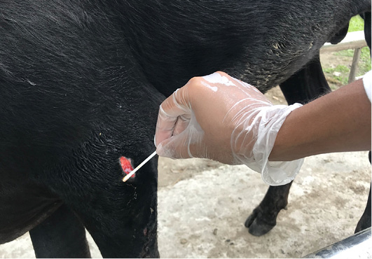
Collection of swab sample from myiasis wound.
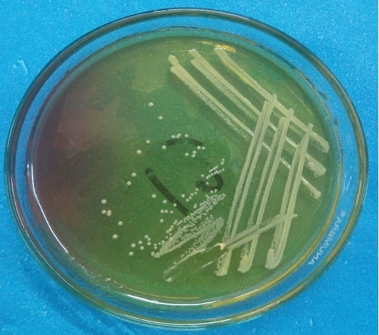
Fermentation of MSA by Staphylococcus aureus indicated by Smooth and convex formation of a yellowish colony.
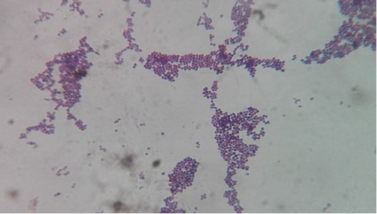
Staining characteristics of bacteria: Grape like clusters were found which are the characteristics of Staphylococcus aureus.
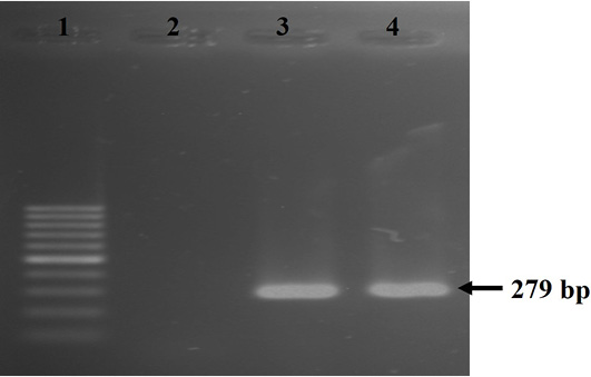
Results of PCR of Staphylococcus aureus specific nuc gene (size=279 bp). Here, Lane 1: 100 bp ladder, Lane 2: negative control, Lane 3: positive control, Lane 4- amplified nuc gene of S. aureus.
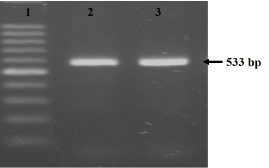
Amplification of the mecA gene (size=533 bp). Here, Lane 1: 100 bp ladder, Lane 2: positive control, Lane 3: amplified mecA gene of Staphylococcus aureus.
References
- Arnold, C., 2013. New science shows how maggots heal wounds. Sci. Am., 308(4). https://doi.org/10.1038/scientificamerican0413-19a[CrossRef]
- Cuny, C., Wieler, L.H. and Witte, W., 2015. Livestock-associated MRSA: The impact on humans. Antibiotics, 4(4): 521-543. https://doi.org/10.3390/antibiotics4040521[CrossRef]
- Garoy, E.Y., Gebreab, Y.B., Achila, O.O., Tekeste, D.G., Kesete, R., Ghirmay, R., Kiflay, R. and Tesfu, T., 2019. Methicillin-resistant Staphylococcus aureus (MRSA): prevalence and antimicrobial sensitivity pattern among patients, a multicenter study in Asmara, Eritrea. J. Can. J. Infect. Dis. Med. Microbiol., https://doi.org/10.1155/2019/8321834[CrossRef]
- Huntington, T.E., Voigt, D.W. and Higley, L., 2008. Not the usual suspects: human wound myiasis by phorids. J. Med. Entomol., 45(1): 157-159. https://doi.org/10.1093/jmedent/45.1.157[CrossRef]
- Hussain, K., Rahman, M., Nazir, K.H.M.N.H., Rahman, H. and Khair, A., 2016. Methicillin resistant Staphylococcus aureus (MRSA) in patients of community based medical College Hospital, Mymensingh, Bangladesh. Am. J. Biomed. Life Sci., 4(3): 26-29. https://doi.org/10.11648/j.ajbls.20160403.11[CrossRef]
- Islam, M.T., Al-Maruf, A., Mannan, M.A., Rahman, H.M.R., Tarafder, M.M., Samad, M.A., Al Noman, A., Hossain, M.B. and Rahman, M.M., 2015. Isolation and identification of associated bacteria and maggots from myiasis affected wounds of cattle and goats in Bangladesh. J. Adv. Vet. Anim. Res., 2(2): 95-100. https://doi.org/10.5455/javar.2015.b55[CrossRef]
- Jaman, M.M., Mishra, P., Rahman, M. and Alam, M.M., 2018. Clinical and laboratory investigation on the recurrence of the umbilical hernia after herniorrhaphy in bovine calves. J. Bangladesh Agric. Univ., 16(3): 464-470. https://doi.org/10.3329/jbau.v16i3.39418[CrossRef]
- Juyena, N., Tapon, M., Ferdousy, R., Paul, S. and Alam, M., 2013. A Retrospective study on occurrence of myiasis in ruminants. Progr. Agric., 24(1-2): 101-106. https://doi.org/10.3329/pa.v24i1-2.19110[CrossRef]
- Kalorey, D.R., Shanmugam, Y.K., Nitin V.C., Kapil K.B. and Sukhadeo B., 2007. PCR-based detection of genes encoding virulence determinants in Staphylococcus aureus from bovine subclinical mastitis cases. J. Vet. Sci., 8(2): 151-154. https://doi.org/10.4142/jvs.2007.8.2.151[CrossRef]
- Lwanga, A., Anis, M., Ayoubi, M., Sharma, J. and Khosla, P., 2018. Two cases of myiasis associated with malignancies in patients living in the continental United States. Cureus, 10(1): e2049. https://doi.org/10.7759/cureus.2049[CrossRef]
- Noutsis, C. and Millikan, L.E., 1994. Myiasis. Dermatol. Clin., 12(4): 729-736. https://doi.org/10.1016/S0733-8635(18)30136-0[CrossRef]
- Otranto, D., 2001. Myiasis: Devils in the flesh of humans and animals. Hosp. Pract., 53: 98-102.
- Penner, L.R., 1958. Concerning a rabbit cuterebrid, the larvae of which may penetrate the human skin (Diptera, Cuterebridae). J. Kansas Entomol. Soc., 31(2): 67-71.
- Pezzi, M., Bonacci, T., Leis, M., Mamolini, E., Marchetti, M.G., Krčmar, S., Chicca, M., Del Zingaro, C.N.F., Faucheux, M.J. and Scapoli, C., 2019. Myiasis in domestic cats: A global review. Parasites Vectors, 12(1). https://doi.org/10.1186/s13071-019-3618-1[CrossRef]
- Toth, E., Hell, E., Kovacs, G., Borsodi, A. and Marialigeti, K., 2006. Bacteria isolated from the different developmental stages and larval organs of the obligate parasitic fly, Wohlfahrtia magnifica (Diptera: Sarcophagidae). Microb. Ecol., 51(1): 13-21. https://doi.org/10.1007/s00248-005-0090-6[CrossRef]
Services
Article usage statistics
Loading…