Research Article · Volume 14, Issue 4, 13 September 2026 · Open Access
Anti-Inflammatory Effects of Low-Level Laser Therapy on Serum IL-6 and TNF-α Levels in an Egg-White Induced Rat Model of Acute Inflammation
· ·
Affiliations & Notes ⌄
1Department of Surgery and Obstetrics, College of Veterinary Medicine, University of Al-Qadisiyah, Iraq
2Department of Medical Biotechnology, College of Biotechnology, University of Al-Qadisiyah, Iraq
3Department of Pharmaceutical Chemistry, College of Pharmacy, University of Al-Qadisiyah, Iraq. *Correspondence | Muslim F. Diwan, Department of Surgery and Obstetrics, College of Veterinary Medicine, University of Al-Qadisiyah, Iraq
Within the context of available evidence, the present study supports the use of 8 J/cm² at 660 nm as an effective parameter for reducing inflammation in acute sterile inflammatory models.
We observed a time-dependent decrease in IL-6 and TNF-α levels at 1, 3, and 24 hours, respectively.
Compared with the positive control (G1) and negative control (G2) groups, cytokine levels in the low-level laser therapy (LLLT)–treated groups became significantly closer to baseline values at 24 hours.
Overall, the results indicate that LLLT reduces pro-inflammatory cytokine levels and may represent a potential therapeutic approach for the management of inflammatory processes.
Selected from the article's own text.
Abstract
Although essential for immune defense and tissue repair, inflammation remains a complex biological process. The pro-inflammatory cytokines interleukin-6 (IL-6) and tumor necrosis factor-alpha (TNF-α) play key roles in this response. Low-level laser therapy (LLLT) is intended as a non-invasive therapeutic alternative for modulating inflammatory processes. Therefore, the current study aimed to investigat the anti-inflammatory effects of low-level laser therapy (LLLT) on serum IL-6 and TNF-α levels in a rat model of inflammation induced by subcutaneous egg-white injection into the left hind paw. A total of thirty-six male rats (300 ± 1.65 g) were randomly allocated into four groups: G1 (negative control, no inflammation and no treatment), G2 (positive control, induced inflammation without treatment), G3 (LLLT at 6 J/cm²), and G4 (LLLT at 8 J/cm²). Treatments were applied for three consecutive days subsequently before inflammation. Serum was collected at 1, 3 and 24 hours post-inflammation to measure the resultant concentrations of cytokines through ELISA. In G1, the IL-6 level was 30.64 ± 6.83 pg/mL. In contrast, IL-6 levels significantly decreased (p < 0.05) in G3 and G4 to 15.21 ± 2.99 pg/mL and 11.36 ± 3.56 pg/mL, respectively. Similarly, the TNF-α concentration in G1 was 24.01 ± 3.58 pg/mL, whereas in G3 and G4 it significantly decreased (p < 0.05) to 18.82 ± 0.89 pg/mL and 16.82 ± 0.16 pg/mL, respectively. These results indicate that low-level laser therapy (LLLT) effectively attenuated the acute inflammatory cytokine response.
Inflammation is a biological response of the immune system that can be induce by a variety of factors; toxic compounds, damaged cells and pathogens like bacteria, and virus . Acute inflammation is crucial for healing and recovery while chronic inflammation can lead to a variety of disease and autoimmune conditions . Cytokines are small proteins released by cells, have a specific effect on the interactions and communications between cells. Cytokines are a broad group of signaling molecules that include lymphokines, monokines, chemokines, tumor necrosis factors (TNFs), interferons (IFNs), and interleukins, classified according to their source of secretion and biological function (Jung et al., 2007). Studies support the use of photobiomodulation (PBM) as a way to influence tissue remodeling by mitigating tissue inflammation while preserving the signaling necessary for tissue remodeling .
Beyond the skeletal muscles, PBM shows intestinal anti-inflammatory action in models of ulcerative colitis (UC). In the case of acetic acid–induced ulcerative colitis, PBM applied locally resulted in reduced disease activity indices, neutrophil-associated myeloperoxidase activity, and histopathology scores, while down-modulating TNF-α and IL-1. Sex-specific analyses of IL-6 (interleukin-6) in males, along with increases in counter-regulatory cytokines, indicated immunological rebalancing . A comparable study comparing local abdominal PBM to transcutaneous vascular PBM in UC showed that local treatment improved the mucosal architecture and the expression of mucosal mRNA of IL-6, TNF-α, and IFN-γ, demonstrating the dose geometry of PBM and cytokine control . Collectively, these observations demonstrate that tissue-proximal PBM suppress acute mucosal inflammation and restore epithelial immune crosstalk.
On the molecular level, PBM inhibits the inflammatory gene and protein expression induced by canonical stimuli. For instance, in human primary chondrocytes challenged with IL-1β, high-frequency near-infrared diode irradiation suppressed IL-6 and TNF-α and inhibited the various stages of NF-κB activation phosphorylation, nuclear translocation, and DNA binding which mechanistically integrates photonic dosing with the repression of cytokine-encoding genes . Analogous studies with periodontal fibroblasts showed distinctive IL-6 output patterns according to the wavelength used, 810 nm protocols consistently blunted stimulated outputs of IL-6 (and other chemokines), whereas 660 nm could be pro-inflammatory under specific conditions; mixed-wavelength regimens often resembled the anti-inflammatory profile typical of the 810 nm ; Papadelli et al., 2020). These in vitro results correspond with findings from animals and illustrate how wavelength and fluence PBM’s cytokine signature.
All models support PBM’s effectiveness in lowering TNF-α and, intermittently, IL-6, subject to timing, tissue, and load variants. In an osteoarthritic rat model, PBM was associated with reductions in TNF-α and MMP-13, while IL-6 had mixed effects consistent with IL-6’s complex roles in injury signaling and resolution . Broader disease settings, such as experimental spinal cord injury, also indicate PBM’s ability to decrease pro-inflammatory cytokines while enhancing neuroprotection and functional recovery, although heterogeneity warrants parameter standardization. These shared patterns suggest PBM can be fine-tuned to reduce pathological inflammation while maintaining reparation cytokine flux.
The systems-level review during the COVID-19 pandemic examined PBM as an adjunct to reduce cytokine-driven pathology. Clinical evidence is still in its infancy; however, during preliminary reports, PBM alongside standard care was associated with improved ventilatory metrics and a reduction in inflammatory markers, bolstering the plausibility for systemic cytokine modulation in humans . Collectively, the aforementioned findings delineate photobiomodulation (PBM) as a dose-dependent immunomodulatory intervention, characterized by suppression of NF-κB activity and peripheral pro-inflammatory cytokines, particularly TNF-α, while IL-6 may also contribute to regulated pro-recovery and tissue repair responses. The essential next steps involve precise dose mapping, tissue-specific optimization, and standardized data reporting to translate mechanistic insights into therapeutic protocols . Therefore, the present study investigated the anti-inflammatory effects of low-level laser therapy (LLLT) on serum IL-6 and TNF-α levels in a rat model of inflammation induced by subcutaneous egg-white injection into the left hind paw.
Materials and Methods
Ethics certification
All animal procedures taken are in accordance to the Al-Qadisiyah Veterinary Medicine College scientific board regulation (October 12 2025, Reg. 4290) and all national regulation policies with the use and care of laboratory animals. The protocol characterized the experimental design, randomization, anesthesia and analgesia, endpoints of humanitarian criteria, and criteria for premature euthanization. Prior to the study, investigators were trained in handling rodents, injecting, and phlebotomy. The protocol illustrated sharp pain as well as suffering, which is required for monitoring during the initiation of inflammation and the drawing of blood.
Daily, a veterinarian assessed the animals for overall health and body condition, including food and water intake, as well as their ability to stand and walk normally. A standardized anesthesia protocol (intramuscular ketamine/xylazine) was used for all blood collections to minimize stress-induced cytokine elevation. Humane endpoints for euthanasia included ≥15% body weight loss, persistent abnormal behavior (such as continuous stretching), self-mutilation, and ulceration at the injection site, as described by Gaertner (2008). When such endpoints were reached, animals were humanely euthanized using an overdose of ketamine/xylazine, followed by a secondary method to ensure death.
Animals
The subjects of this study included thirty-six adult male Rattus norvegicus, with an average body weight of 300 g. All subjects were obtained from a certified facility. Prior to experimentation, all animals were given a 7 day acclimatization period. Animals were kept in cohorts of three in polycarbonate cages. Animals were kept in controlled conditions of a temperature between (°C) 22 to 24, a humidity level of 45 to 65, and a 12 hour light and dark period. All animals were given access to food and water during the entire acclimatization period. Environmental stressors were reduced to a minimum.
Within groups animals were further divided into 3 time points. These time points included 1 hour, 3 hours, and 24 hours. Each of these time points included 3 animals. It was also shown that the subjects were well distributed throughout the groups. ELISA is a biochemical technique for the detection of the presence and the quantification of the specific antigen from a sample. A computer-generated randomization sequence was used to assign the animals into four groups, with nine animals per group. Group 1 served as the positive control (inflammation without treatment), Group 2 as the negative control (no inflammation and no laser treatment), Group 3 received laser preconditioning/treatment at 6 J/cm², and Group 4 received laser preconditioning/treatment at 8 J/cm². Each group was further divided equally for blood sample collection .
The induction of acute inflammation
Acute paw inflammation was triggered via subcutaneous injection of 0.05 mL undiluted hen egg white into the plantar muscles of the left hind paw using a 0.5 cc syringe. Before the injection, the paw was disinfected with 70% ethanol and allowed to dry. Injections were performed with utmost care at an angle to the surface to avoid inflicting excess tissue damage unrelated to the model. The animals were then returned to the home cages and were monitored for the next 30 minutes for any sudden untoward reactions .
These models enable kinetic sampling of systemic IL-6 and TNF-α at 1, 3, and 24 hrs in tandem with the rapid edema and the site-specific cytokine release. For the sake of consistency, all inductions were performed by the same trained individual over a short time period during the first half of the day, and the volume of injections was drawn up in a 0.5 cc, which was checked before each event.
Laser device and irradiation protocol
Photobiomodulation was done with a GaAlAs multi-wavelength system (Omega Laser Systems Ltd., UK), red light at 660 nm with 50 mW output in 146 Hz pulsed mode. Before each session, the device was powered for 5 min and checked way before device was used for laser for stabilization, and then checked with an optical power meter. Between animals, the hand-piece was cleaned and disinfected. The target energy densities for each dose was 6 J/cm² (G3) and 8 J/cm² (G4) .
In the days leading up to the induction of the inflammation model (Days −2, −1, and 0), laser preconditioning was applied once daily for three consecutive days to stimulate the immune response. On Day 0, inflammation was induced by egg-white injection. During the inflammatory phase, animals were anesthetized using a ketamine–xylazine combination administered intramuscularly at a 5:1 ratio (50 mg/kg ketamine and 10 mg/kg xylazine, respectively) .
The medication was injected intramuscularly and the volume of the blood that was drawn was around `2 to 3 mL` which was extracted alive with a surgical procedure called a terminal cardiac puncture. The blood was stored in special test tubes to avoid blood clotting. These samples were processed within `30 minutes` of collection in order to maintain records of the cytokines that were inflamed . The samples were collected from rats by the same way to avoid bias.
The serum was separated by centrifugation for 15 minutes at a 3000 rpm speed at a temperature of 4 ℃. The supernatant was aliquoted pro rata without exceeding 200 microliters to minimize the number of freeze-thaw cycles in order to facilitate processing and transferred to pre-labeled 1.5 mL microtubes. Hemolyzed (Visually assessed) samples were excluded because of the risk of interfering with immunoassays. All items used in the sample were free of endotoxins and were classified as low binding items with the aim of preserving the integrity of the cytokines.
Determination of serum concentration of IL-6 and TNF-α
Rat IL-6 and TNF-α ELISA kits (Elabscience) were used for the quantitative determination of cytokine levels in serum samples, following the manufacturer’s instructions. In brief, a volume of 100 µL of each sample was added to the appropriate wells of a micro-ELISA plate. For the blank well, only reference standard and sample diluent were added. The plate was gently mixed, covered with a plate sealer, and incubated at 37 °C for 90 minutes. After incubation, the liquid was removed from each well. Immediately, 100 µL of biotinylated detection antibody working solution was added to each well. The plate was covered again and incubated at 37 °C for 60 minutes. All wells were aspirated and washed three times using wash buffer. Washing was performed by filling each well with approximately 350 µL of wash buffer. Next, 100 µL of HRP-conjugate working solution was added to each well. The plate was covered and incubated at 37 °C for 30 minutes. The washing step was then repeated five times. Subsequently, 90 µL of substrate solution was added to each well, and the plate was incubated at 37 °C for approximately 15 minutes in the dark. Then, 50 µL of stop solution was added to each well, resulting in an immediate color change to yellow. The optical density (OD) of each well was measured immediately using a microplate reader at a wavelength of 450 nm.
Statistical analysis
Primary outcomes were plasma samples of IL-6 and TNF-α at 1, 3, and 24 hours of each assay. Data were filtered for outliers using robust procedures and graphical checks, normalcy (Shapiro–Wilk) and homoscedasticity (Levene). The cytokine estimations were given as mean ± standard error. Differences between groups across time were analyzed using two-way analysis of variance (ANOVA), with factors including Group (G1–G4) and Time (1, 3, and 24 hours). Planned post-hoc comparisons were performed using Fisher’s Least Significant Difference (LSD) test, in accordance with the reported LSD thresholds.
In addition to p-values, all results were supplemented by effect sizes (η² for omnibus tests; Cohen’s d for pairwise) and 95% CIs. When assumptions were violated, data were Box-Cox transformed or were subjected to aligned-rank transform analysis as sensitivity checks; conclusions were cross-validated to ensure alignment. Controlled family wise α was 0.05. Analysis was performed using the current versions of R or SPSS; all code and outputs are available upon request. present means for each group at each timepoint with pooled SEs and LSD values, as appropriate for the study design .
Results
Interleukin-6 (IL-6)
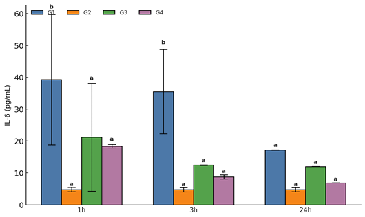
Serum IL-6 concentrations showed a marked time-dependent response following egg-white–induced inflammation in the plantar region of the paw. Levels increased sharply after induction and subsequently declined over time. Photobiomodulation (PBM) attenuated this IL-6 response in a dose-dependent manner, with a more pronounced effect observed at 8 J/cm².
Overall, the highest IL-6 concentration was recorded in G1 (positive control) (30.64 ± 6.83 pg/mL), whereas the negative control (G2) exhibited baseline non-inflamed levels (4.78 ± 0.01 pg/mL). Treatment groups showed significant reductions in IL-6 levels compared with G1 (p < 0.05), with G3 (6 J/cm²) showing a mean concentration of 15.21 ± 2.99 pg/mL and G4 (8 J/cm²) showing the lowest level among treated groups (11.36 ± 3.56 pg/mL).
Across time points, IL-6 levels peaked at 1 hour (20.92 ± 7.08 pg/mL) and then declined significantly at 3 hours (15.38 ± 6.89 pg/mL) and 24 hours (10.21 ± 2.76 pg/mL) (p < 0.05). These temporal changes were significantly influenced by both treatment and time factors, as confirmed by two-way ANOVA and Fisher’s LSD post-hoc analysis. Group and time comparisons exceeded the predefined LSD thresholds (Group LSD= 12.51, Time LSD= 14.45, Interaction LSD= 25.02), confirming statistically significant differences between inflamed and treated groups (p < 0.05), particularly in the 8 J/cm² group.
At individual time points, G1 exhibited the highest IL-6 values at 1 hour (39.26 ± 20.4 pg/mL), which were significantly higher than both G3 (21.20 ± 16.9 pg/mL) and G4 (18.42 ± 0.57 pg/mL) (p < 0.05). At 3 hours, IL-6 levels in G1 decreased to 35.51 ± 13.2 pg/mL, while G3 and G4 further decreased to 12.46 ± 0.11 pg/mL and 8.76 ± 0.66 pg/mL, respectively (p < 0.05). By 24 hours, IL-6 levels approached baseline values, with G1 at 7.15 ± 0.09 pg/mL, G3 at 11.99 ± 0.04 pg/mL, and G4 at 6.91 ± 0.04 pg/mL, nearing the negative control level (~4.78 pg/mL).
The differences between G1 and G4 at early time points (1–3 hours) exceeded the LSD thresholds (p < 0.05), and this effect persisted at 24 hours, suggesting sustained modulation of cytokine activity by PBM, particularly at 8 J/cm². Overall, these findings indicate that IL-6 dynamics are both time- and dose-dependent, with photobiomodulation effectively attenuating the acute inflammatory peak and promoting a faster return toward baseline levels (Figure 1).
TNF-α
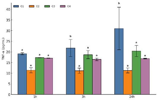
TNF-α (tumor necrosis factor-alpha) exhibited a complementary but not identical pattern to IL-6 (interleukin-6). The inflamed control group (G1) showed the highest overall TNF-α concentration (24.01 ± 3.58 pg/mL), whereas the negative control (G2) remained near baseline levels (11.30 ± 0.05 pg/mL) (p < 0.05 vs. G1). Photobiomodulation (PBM) significantly reduced TNF-α levels in treated groups compared with G1 (p < 0.05), with G3 (6 J/cm²) showing 18.82 ± 0.89 pg/mL and G4 (8 J/cm²) showing the lowest level among treated groups (16.82 ± 0.16 pg/mL).
Across time points, TNF-α showed a slower and more gradual response compared to IL-6. In the negative control (G2), TNF-α increased modestly from 16.24 ± 1.68 pg/mL at 1 hour to 17.06 ± 2.24 pg/mL at 3 hours, reaching a peak of 19.91 ± 4.14 pg/mL at 24 hours (p < 0.05). Statistical analysis using two-way ANOVA followed by Fisher’s LSD test confirmed significant differences between groups and time points, exceeding the predefined thresholds (Group LSD = 4.83, Time LSD = 5.58, Interaction LSD = 9.66), particularly in the 8 J/cm² group (p < 0.05).
At 1 hour, TNF-α levels in G1 were 17.32 ± 0.10 pg/mL, which were significantly higher than in G3 and G4 (p < 0.05). At 3 hours, G1 showed an increase to 21.85 ± 4.03 pg/mL, whereas G3 and G4 exhibited lower levels of 18.72 ± 1.93 pg/mL and 16.51 ± 0.62 pg/mL, respectively (p < 0.05). At 24 hours, TNF-α peaked in G1 (31.01 ± 9.99 pg/mL), while laser-treated groups showed significantly reduced levels (20.43 ± 2.62 pg/mL in G3 and 16.90 ± 0.13 pg/mL in G4), approaching the negative control level (11.32 ± 1.19 pg/mL) (p < 0.05).
Overall, the 24-hour time point demonstrated the most pronounced differences, supporting that photobiomodulation exerts a sustained anti-inflammatory effect on TNF-α. The higher fluence (8 J/cm²) consistently produced greater suppression of late-phase cytokine escalation compared to 6 J/cm² (Figure 2).
Discussion
Inflammation is the body’s initial response to injury or bacterial infection, during which pro-inflammatory cytokines such as IL-6 and TNF-α are among the first mediators released. The present data demonstrate that IL-6 levels were suppressed following 660 nm photobiomodulation (PBM) at energy densities of 6 and 8 J/cm². Furthermore, both IL-6 and TNF-α levels measured 24 hours post-treatment were lower than those of the negative control group (G1). These findings are consistent with previous studies. For example, Rodrigues et al. reported that low-level laser therapy (LLLT) in rats promoted vascular responses, enhanced mucosal tissue repair, and modulated IL-6 and TNF-α levels. Similarly, Bitencourt et al. observed reductions in IL-6 and TNF-α associated with decreased inflammatory activity.
In the present study, the reduction in IL-6 production was consistent with the observed decrease in TNF-α levels, particularly within the first 3 hours (Figure 2). Experimental evidence by Hempel et al. demonstrated that exposure to near-infrared irradiation at 8 J/cm² suppressed IL-6 and TNF-α activity in primary chondrocytes, along with inhibition of NF-κB signaling. Additional preclinical studies have shown that red and near-infrared PBM can reduce inflammatory mediators and influence macrophage polarization, supporting a shift toward anti-inflammatory responses.
At the clinical level, Zou et al. reviewed the inhibitory effects of PBM on TNF-α, IL-1β, and IL-6 in gingival and peri-surgical models, suggesting a systemic anti-inflammatory mechanism similar to that observed in this study. Collectively, evidence from cellular, tissue, and whole-organism studies supports the interpretation that cytokine modulation observed here is not unique to egg-white–induced edema but reflects a broader biological response associated with PBM pathways.
However, PBM effects are known to be context-dependent and may exhibit biphasic responses. For example, Hu et al. reported that PBM did not exacerbate inflammation in mouse wound models but, in some cases, slightly increased inflammatory cell aggregation and IL-6 expression. This suggests that treatment outcomes depend on factors such as timing, tissue condition, and dose parameters. In agreement with this concept, the present findings show that IL-6 levels were reduced below control levels at 8 J/cm², particularly between 3 and 24 hours post-treatment, indicating an optimal therapeutic window for anti-inflammatory effects without excessive signaling.
Broader analyses, variability in PBM outcomes due to differences in wavelength, fluence, and treatment timing. The structured pre-conditioning and post-induction protocol used in this study helps address this variability. While confirming the anti-inflammatory effects of PBM, the results also emphasize the need for standardized treatment parameters to avoid inconsistent cytokine responses.
The finding that 8 J/cm² was more effective than 6 J/cm² supports a dose-response relationship in PBM. Zhang and Qu (2023) demonstrated that cytokine secretion (IL-1β, IL-6, TNF-α) can vary depending on energy density and exposure conditions, suggesting the presence of multiple optimal therapeutic ranges rather than a single fixed setting. Additionally, studies by Rodrigues and Bitencourt indicate that factors such as treatment type (local vs. systemic) and biological variables (e.g., sex) may influence cytokine responses.
Within the context of available evidence, the present study supports the use of 8 J/cm² at 660 nm as an effective parameter for reducing inflammation in acute sterile inflammatory models. However, it is important to consider the fundamental principle of PBM: treatment outcomes are influenced by the interaction of dose, timing, and tissue characteristics, which together determine the extent of IL-6 and TNF-α modulation.
Conclusion
We observed a time-dependent decrease in IL-6 and TNF-α levels at 1, 3, and 24 hours, respectively. Compared with the positive control (G1) and negative control (G2) groups, cytokine levels in the low-level laser therapy (LLLT)–treated groups became significantly closer to baseline values at 24 hours. Overall, the results indicate that LLLT reduces pro-inflammatory cytokine levels and may represent a potential therapeutic approach for the management of inflammatory processes.
Acknowledgment
The author is grateful to the University of AL-Qadisiyha/ College of Veterinary Medicine for all the facilities to achieve this study.
Novelty Statement
The novelty of our study lies in evaluating the effect of different doses of low-level laser therapy (LLLT) on inflammation by measuring serum levels of IL-6 and TNF-α.
Author’s contribution
MFD conceptualized and designs the experiment. QZB and NTA carried out the experimental work. All authors analyzed the data and drafted the manuscript. All authors reviewed and approved the final version of the manuscript.
Generative AI and AI assisted technology statement
The authors declare that no generative AI and AI assisted technology was used in the creation of this manuscript.
Conflict of interest
The authors have declared no conflict of interest.
Figures
Figure 1:.
Serum IL-6 (interleukin-6) concentration (pg/ mL ± SE) in negative control (G2), positive control (G1), and laser-treated groups (G3: 6 J/cm² and G4: 8 J/cm²) at 1, 3, and 24 hours after induction of rat paw egg-white inflammation, showing the lowest concentration in G4 at 24 hours. a-b different superscripts on the bars indicate significant difference (p < 0.05).
Figure 2:.
Serum TNF-α (tumor necrosis factor-alpha) concentration (pg/mL ± SE) in negative control (G2), positive control (G1), and laser-treated groups (G3: 6 J/ cm² and G4: 8 J/cm²) at 1, 3, and 24 hours after induction of rat paw egg-white inflammation, showing the lowest concentration in G4 at 24 hours. a-b different superscripts on the bars indicate significant difference (p < 0.05).
References
Abidi AH, Mayall RE, Ruan CX, Ou KL, Walinski CJ (2021). Immunomodulatory activity seen as a result of photobiomodulation therapy in stimulated primary human fibroblasts. Arch. Oral Biol., 121: 104968. https://doi.org/10.1016/j.archoralbio.2020.104968[CrossRef]
Albertini AB, Villaverde F, Aimbire MAC, Salgado JM, Bjordal LP, Aves EM, Costa MS (2007). Anti-inflammatory effects of low-level laser therapy (LLLT) with two different red wavelengths (660nm and 684nm) in carrageenan-induced rat paw edema. J. Photochem. Photobiol. B: Biol., 89(1): 50-55. https://doi.org/10.1016/j.jphotobiol.2007.08.005[CrossRef]
Bitencourt KCQM, Schapochnik A, de Souza V, Rodrigues VMM, da Cruz MP, Damazo AS (2023). Effects of photobiomodulation in the experimental acetic acid-induced colitis: comparison between male and female. Lasers Med. Sci., 38(1): 271. https://doi.org/10.1007/s10103-023-03932-w[CrossRef]
da Cruz Tobelem D, Silva T, Araujo T, Andreo L, Malavazzi TCS, Horliana ACRT (2022). Effects of photobiomodulation in experimental spinal cord injury models: A systematic review. J. Biophotonics., 15(8): e202200059. https://doi.org/10.1002/jbio.202200059[CrossRef]
de Matos BTL, Buchaim DV, Pomini KT, Barbalho SM, Guiguer EL, Reis CHB (2021). Photobiomodulation therapy as a possible new approach in COVID-19: A systematic review. Life (Basel). 11(6): 580. https://doi.org/10.3390/life11060580[CrossRef]
Elisabeth NB, Jovie MD, Meilany FD, Donald EK (2021). Egg white-induced inflammation models: A study of edema profile and histological change of rat’s paw. J. Adv. Pharma. Technol. Res., 12(2): 109-112. https://doi.org/10.4103/japtr.JAPTR_262_20[CrossRef]
Gaertner DJ (2008). Anesthesia and analgesia for laboratory rodents. Anesthesia and analgesia in laboratory animals: pp. 240–297. https://doi.org/10.1016/B978-012373898-1.00010-4[CrossRef]
Gregio VA, Martinelli A, Malavazzi TCS, Andreo L, Terena SML, Bussadori SK (2025). Photobiomodulation and aquatic training reduce TNF-α expression and enhance muscle fiber area in Wistar rats with compensatory hypertrophy. Lasers Med. Sci., 40(1): 47. https://doi.org/10.1007/s10103-025-04290-5[CrossRef]
Harry NES, Abinawanto AB, Mukh S, Fadhillah F, Retno L, Teja K, Dwi R, Indra S, Iin KHB, Triningsih T, Bisma BP (2024). IL-6 and IL-10 levels in Rats blood plasma as immune responses post radioiodine (I-131) administration. Asian Pac. J. Cancer Prevent., 25(3): 1017-1023. https://doi.org/10.31557/APJCP.2024.25.3.1017[CrossRef]
Hempel L, Körholz D, Bönig H, Schneider M, Klein- Vehne A, Packeisen J, Mauz-körholz C, Burdach S (1995). Interleukin-10 directly inhibits the interleukin-6 production in t-cells. Scand. J. Immunol., 41(5): 462-466. https://doi.org/10.1111/j.1365-3083.1995.tb03593.x[CrossRef]
Hu J, Zhang Y, Peng X, Li X, Li S, Liu H, Dong H, Lv Y, Hu D (2024). Biphasic photobiomodulation of inflammation in mouse cutaneous wounds. Photonics Lasers Med., 13(2): e20240028.
Jahani-Sherafat S, Maroufi SF, Sedighimehr N, Salimi A, Moradi A, Ghavidel-Parsa B (2023). The effectiveness of photobiomodulation therapy in musculoskeletal pain: a narrative review. Pain Res. Manage. 2023: 6660834.
Jung-Ming Z, Jianxiong A (2007). Cytokines, inflammation and pain. Int. Anesthesiol. Clin., 45(2): 27-37. https://doi.org/10.1097/AIA.0b013e318034194e[CrossRef]
Martinelli A, Andreo L, Alves AN, Terena SML, Santos TC, Bussadori SK (2021). Photobiomodulation modulates the expression of inflammatory cytokines during the compensatory hypertrophy process in skeletal muscle. Lasers Med. Sci., 36(4): 791-802. https://doi.org/10.1007/s10103-020-03095-y[CrossRef]
Nambi G (2021). Does low level laser therapy has effects on inflammatory biomarkers IL-1β, IL-6, TNF-α, and MMP-13 in osteoarthritis of rat models-a systemic review and meta-analysis. Lasers Med. Sci., 36(3): 475-484. https://doi.org/10.1007/s10103-020-03124-w[CrossRef]
Papadelli A, Kyriakidou K, Kotsakis GA, Pepelassi E, Kallis A, Vrotsos IA (2021). Immunomodulatory effects of Nd:YAG (1064 nm) and diode laser (810 nm) wavelengths to LPS-challenged human gingival fibroblasts. Arch. Oral. Biol., 122: 104982. https://doi.org/10.1016/j.archoralbio.2020.104982[CrossRef]
Rishabh C, Ajay P, Vipul A, Mujeeba R, Anand K, Arjun SK, Siddhi S, Sukriti S, Vikas M (2023). Chronic stress predisposes to the aggravation of inflammation in autoimmune diseases with focus on rheumatoid arthritis and psoriasis. Int. Immunopharmacol., 125(A): 111-146. https://doi.org/10.1016/j.intimp.2023.111046[CrossRef]
Rodrigues VMM, Bitencourt KCQM, Schapochnik A, de Souza V, da Palma Cruz M, Damazo AS (2023). Comparison between local abdominal and transcutaneous tail vein photobiomodulation in experimental rat model of ulcerative colitis. Lasers Med. Sci., 38(1): 247. https://doi.org/10.1007/s10103-023-03910-2[CrossRef]
Sakata S, Kunimatsu R, Tsuka Y, Nakatani A, Gunji H, Yanoshita M, Kado I, Ito S, Putranti N, Prastya R, Hirose N, Tanimoto K (2022). High-frequency near-infrared diode laser irradiation suppresses IL-1β-induced inflammatory cytokine expression and NF-κB signaling pathways in human primary chondrocytes. Lasers Med. Sci., 37(2): 1193-1201. https://doi.org/10.1007/s10103-021-03371-5[CrossRef]
Weinberg SL, Sarah KA (2008). Statistics using SPSS: An integrative approach. Cambridge University Press.
Zhang R, Qu J (2023). The mechanisms and efficacy of photobiomodulation therapy for arthritis: A comprehensive review. Int. J. Mol. Sci., 24(18): 14293. https://doi.org/10.3390/ijms241814293[CrossRef]
Zou Q, Zhang P, Sun H, Wang Z, He J, Yu F (2024). Low-level laser therapy on soft tissue healing after periodontal and implant surgeries: A systematic review. Front. Oral Health, 5: 1387542. https://doi.org/10.1186/s12903-024-05258-7[CrossRef]
Anti-Inflammatory Effects of Low-Level Laser Therapy on Serum IL-6 and TNF-α Levels in an Egg-White Induced Rat Model of Acute Inflammation · ResearchersLinks