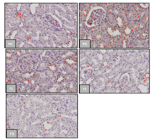
Based on Table 2. the positive control group had a significant difference of TNF–α expression mean (p<0,05) compared to the negative control group. Blueberry extract therapy groups on treatments 1 and 2 had no significant difference (p>0.05) compared to the positive and negative control group, while the treatment 3 group has a significant difference compared to the positive control group. These results showed that blueberry therapy at a dosage 1500mg/kg was able to provide a significant therapeutic effect on the incidence of acute kidney injury. TNF-α can appear in normal conditions in small amounts as an immune response produced by macrophages in the phagocytosis process of dead cells and antigens that enter the body (Chu, 2013).
The positive control group had the highest mean TNF-α expression which had 65.35%, it was due to acute kidney injury after 50% glycerol induction and not given any therapy to relieve the inflammation. 50% glycerol induction will cause rhabdomyolysis conditions thereby increasing the production of nephrotoxic myoglobin. Myoglobin induces oxidative stress and lipid peroxidation of proximal tubular cells, then inducing the release of a series of mediators, including the pro-inflammatory cytokine TNF-α. The more inflammation in the tissue, the higher the TNF-α level (Wu, et. al., 2017).
The Blueberry extract however only presented a slight decrease in TNF-α expression for T1 and T2 groups compared to T3 group marked with a different notation from positive control group (PC). The average TNF-α expression between treatment groups decreased in proportion to the increase in the therapeutic dose of blueberry extract. This is in line with the research of Nair et al. (2014) stated that there was a decrease in the production of proinflammatory cytokines TNF-α in the kidneys in animal models of metabolic syndrome that had been given blueberry extract therapy. The average value of TNF-α expression in treatment group 1 (dose 500 mg/kg) was 52.43%, treatment group 2 (dose 1000 mg/kg) was 48.28%, and treatment group 3 (dose 1500 mg/kg) was 33.46%. When the three therapy groups were compared to the effect of reducing TNF-α expression, the treatment group 3 (dose 1500 mg/kg) was the best dose of blueberry fruit extract in reducing TNF-α expression as an anti-inflammatory in rats model of acute kidney injury.
The decrease in TNF-α expression was due to the blueberry extract therapy containing high amounts of anthocyanins and flavonoids. Based on the results of phytochemical testers, our blueberry extract contains an anthocyanin content of 150.98 ppm. Blueberry extract anthocyanins are reported to act as anti-inflammatory and antioxidants in diabetic retinopathy (Huang et al., 2018). Anthocyanins are also reported to have antioxidant, anti-inflammatory, anti-carcinogenic, and protective effects from heart damage and improvement of the microcirculation (Ghosh, 2007). Flavonoids can reduce the expression of pro-inflammatory cytokines/chemokines including TNF-α and inhibit the activity of NF-Kß and stimulate cell regeneration in necrotic tubules. Anthocyanin acts as decreasing the lipid inflammatory perox enzyme so that it can suppress the production of pro-inflammatory cytokines TNF-α (Nicoletti et al., 2015).
The mechanism of action of blueberry compounds has been reported to vary in various cell systems. Blueberry polyphenols act as an anti-inflammatory in brain microglial cells by developing several inflammatory cytokines, TNF-a, LPS, nitric oxide, and IL1b (Ma H et al., 2018a; Ma L et al., 2018; Lau et al., 2007). Some of the compounds contained in the most abundant blueberries include the conjugates of delphinidin, malvidin, and cyanide, which exhibit strong anti-tumorigenic and anti-inflammatory effects (Pereira et al., 2017). Anthocyanin-delphinidin compounds work through various pathways such as caspase activation and cytokine inhibition (Haseeb et al., 2013), including Delphinidin inhibits NF-kB and COX-2 activation induced by IL-1b human chondrocyte expression. IL-1b and NF-kB are major mediators of inflammation and disease progression. The combination of cyanidin, malvidin, peonidin, petunidin, and delphinidin (chloride form) suppresses the growth of aggressive non-small cell lung cancer and reduces the expression of NO, PGE2, IL-8, iNOS, and COX-2 on the lower thrusters of NSAIDs, acids. 5-aminosalicylate (Serra et al., 2013).